Abstract (J. Kor. Oral Maxillofac. Surg. 2009;35:361-366)
Ⅰ.
서 론일반적으로 상악에서의 임플란트 성공율은 하악에 비해 낮으며, 상악 구치부에서 더욱 성공을 예견하기 힘들다
1-7). 이러한 결과는 일차적으로 좋지 못한 골질에 의한 치조골 과 임플란트와의 골접촉 면적 불량과 관련이 있다
8-10). 임플 란트와 골의 접촉 면적을 증가 시키기 위해 새로운 임플란 트는 표면처리 (티타늄 표면, HA 표면, sandblasting되거나 화학적 부식을 시킨 표면 등) 기술이 적용되었다
11-16). 골량 이 부족하고 골질이 불량한 곳에서 유리한 임플란트 골접 촉 면적을 얻을 수 있는 Ti-6Al-4V의 구형 입자(spherical particle)의 multilayed porous surface zone을 가지는 새로운
texture가 개발되었다
17-20). Sintered porous implants의 이런 3 차원적인 표면 형태는 골의 침투 후 3차원적 기계적 결합 을 일으킨다고 알려져 있다
11,21). Sintered porous surfaced implants는 골질이 좋지 않은 곳에서도 threaded implants보 다 짧은 길이로 사용 가능하다
11). 전치부 보다 구치부는 교 합력이 많이 작용하며
10), 악궁의 후방부는 전방부보다 endosseous root-form implants의 식립 후 예지성이 상대적 으로 낮다
3,7). 따라서 구치부 식립 시 일반적으로 임플란트 를 식립해야 하지만 식립 부위의 골이 부족해 긴 임플란트 를 식립 할 수 없는 상황이 자주 있게 된다
22). Sintered porous implants는 짧은 길이인 경우에도 다양한 경우에서 성공적으로 적용이 가능함이 보고되었다
22-24). 또한 최근 5mm길이의 엔도포어 임플란트에 관한 다기관의 연구에서 성공적인 성공율을 보이는 논문이 발표되었다
25).
이번 우리 연구에서는 상악 부분 무치악 부위에 식립된 sintered porous-surfaced dental implant(Endopore
�Dental Implant system, Innova Corporation, Toronto, ON, Canada)의 장기간의 기능 후의 여러 상황에 따른 임플란트 생존율을 분석하는데 그 목적이 있다.
손 동 석
705-718
대구광역시 남구 대명4
동3056-6
대구가톨릭대학병원 치과 구 강악안면외과학교실Dong-Seok Sohn
Dept. of Oral and Maxillofacial Surgery, Daegu Catholic University Hospital, 3056-6 Daemyung 4-Dong, Nam-Gu, Daegu, Republic of Korea. 705-718 Tel: 82-53-650-4291 Fax: 82-53-622-7067
E-mail: [email protected]
상악 구치부에 식립된 엔도포아 임플란트의 후향적 연구
김상수∙안미라∙이원혁,∙정희승*∙신임희**∙손동석
대구 가톨릭대학교 의과대학 치과 구강악안면외과학교실, *대구 서문치과, **대구 가톨릭대학교 의과대학 통계학교실
A RETROSPECTIVE STUDY OF SINTERED POROUS-SURFACED DENTAL IMPLANTS IN RESTORING THE POSTERIOR MAXILLA
Sang-Soo Kim, Mi-Ra Ahn, Won-hyuk Lee, Heui-Seung Jung*, Im-hee Shin**, Dong-Seok Sohn Dept. of Oral and Maxillofacial Surgery, Daegu Catholic University Hospital,
* Daegu Seomun Dental Clinic, ** Dept. of statistics, College of Medicine, Daegu Catholic University
Purpose: The purpose of this retrospective report was to analyze long-term survival rate of sintered porous-surfaced dental implant (Endopore
�Dental Implant system, Innova Corporation, Toronto, ON, Canada).
Methods: 61 partially edentulous patients were received a total of 127 Endopore dental implants in the maxilla. Of the 127 implants, 24 implants were restored with individual (ie, non-splinted) crowns, while 103 implants were splinted to other implants. Medical records and radiographs were evaluated and analyzed by the cumulative survival rate, location of implants, implants length and diameter, crown/implant ratio and whether the implant was splinted. Chi squire test was used statistically.
Result: Of the 127 implants, 8 implants (6.3%) were removed and and cumulative survival rate was 93.7%.
Conclusion: Endopore implants showed satisfactory results after up to 8 years function periods in the edentulous posterior maxilla.
Key words: Endopore
�, Sintered porous-surfaced implant, survival rate, edentulous posterior maxilla
[원고접수일 2009.6.2. / 1차수정일 2009.6.12. / 2차수정일 2009.6.27. / 게재확정일 2009.7.13.]
Ⅱ.
연구대상 및 방법대구가톨릭대학 병원과 1개의 개인 치과의원에서, 상악 구치부에 Endopore
�임플란트 식립 후 보철물이 올라간 61 명의 환자를 대상으로 조사를 하였다. 식립된 임플란트 직 경의 종류는 2종류(4.1mm, 5.0mm)이고 길이는 4종류 (5mm, 7mm, 9mm, 12mm)를 사용하였다. Smooth collar의 높이는 2종류(1mm, 2mm)를 사용하였다. 모든 임플란트는 고정성 보철물(연결고정 보철물 103개, 단일 보철물 24개) 로 수복되었다. 초진시, 임플란트 식립 후, 고정성 보철물 수복 후에 파노라마와 치근단 방사선 사진을 촬영하였고, 치근단 방사선 사진은 평행촬영법을 사용하였다.임플란트 식립 위치에 따른 임플란트의 생존율, 임플란트의 직경 및 길이에 따른 생존율, 최종 보철물의 연결고정 유무에 따른 생존율, 치관/임플란트 비율에 따른 생존율을 조사하였다.
치관/임플란트 비율 계산을 위해 치관의 길이는 임플란트 장축에 평행한 중심선을 따라 machined smooth collar의 상 연에서 보철물 교합면까지의 길이로 정하였고, 임플란트
의 길이는 임플란트 장축에 평행한 중심선을 따라 mach- ined smooth collar의 상연에서 임플란트 식립체의 최하방 까지의 길이로 정하였다(Fig. 1). 이 두 부분의 길이를 계측 하여 각 임플란트마다 치관/임플란트 비율을 계산하였다.
임플란트 실패의 기준은 임플란트를 구강내에서 제거하였 을 경우로 정하였다.
통계분석은 SPSS Win.Ver 14.0 프로그램을 이용하여 분 석하였다. 식립 위치에 따른 임플란트 생존율, 임플란트 직 경 및 길이에 따른 생존율, 최종 보철물의 연결고정 유무에 따른 생존율, 치관/임플란트 비율에 따른 분포 및 생존율에 대해 Chi-square test를 사용하였으며 P<0.05인 경우 통계학 적으로 유의하다고 판정하였다.
Ⅲ.
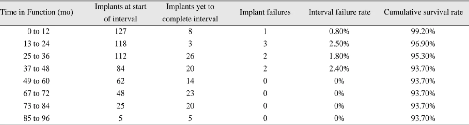
결 과상악 구치부에 식립한 총 127개의 중, 보철 기능 후 첫 1 년 동안의 생존율은 99.2%(1개 실패)였으며 기능 4년까지 의 전체 생존율은 93.7%(8개 실패)였다. 그 이후 실패한 증 례는 관찰되지 않았으며 전체 8년 기능 동안의 생존율은 93.7%로 변화가 없었다(Table 1).
1. 임플란트 식립 위치에 따른 임플란트 분포 및 생존율
전체 127개의 임플란트를 치아 위치에 따라 분포를 분류 해 보면 제1소구치에 9개(7.1%), 제2소구치에 19개(15.0%), 제1대구치에 55개(43.3%), 제2대구치에 44개(34.6%)로 나 타났다. 제1, 2대구치부위는 전체 임플란트 중 99개(77.9%) 가 식립되었다.
제1소구치 부위에 식립된 임플란트의 생존율은 77.8%(7 개 식립, 2개 실패), 제2소구치 부위에 식립된 임플란트의 생존율은 94.7%(18개 식립, 1개 실패), 제1대구치 부위에 식립된 임플란트의 생존율은 94.5%(52개 식립, 3개 실패), 제2대구치 부위에 식립된 임플란트의 생존율은 95.5%(42 개 식립, 2개 실패)로 각각 나타났고 치아위치에 따른 임플 란트 분포 및 생존율에 대한 통계학적으로 유의한 차이가 없었다(P=0.241)(Table 2).
Fig. 1. A:Length of crown, B:Length of implant.
Table 1.
Life Table AnalysisTime in Function (mo) Implants at start Implants yet to
Implant failures Interval failure rate Cumulative survival rate of interval complete interval
0 to 12 127 8 1 0.80% 99.20%
13 to 24 118 3 3 2.50% 96.90%
25 to 36 112 26 2 1.80% 95.30%
37 to 48 84 20 2 2.40% 93.70%
49 to 60 62 14 0 0% 93.70%
67 to 72 48 23 0 0% 93.70%
73 to 84 25 20 0 0% 93.70%
85 to 96 5 5 0 0% 93.70%
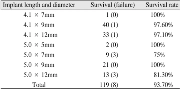
2.임플란트 직경 및 길이에 따른 생존율
직경은 4.1mm, 5.0mm으로 분류하였고 길이는 5mm, 7mm, 9mm, 12mm로 분류되었다. Smooth collar는 2종류 (1mm, 2mm)를 사용하였다. 직경 4.1mm 임플란트는 76개 가 식립되었고 총 식립 개수의 59.8%를 차지하였으며, 직 경 5.0mm 임플란트는 51개가 식립되었고 총 식립 개수의 41.2%를 차지하였다. 직경 4.1mm 임플란트의 생존율은 96.1%를 보였으며, 직경 5.0mm 임플란트의 생존율은 90.2%를 보였다.
길이는 5mm, 7mm, 9mm, 12mm로 분류하였고 길이 5mm 임플란트는 2개가 식립되었고 총 식립 갯수의 1.57%를 차 지하였으며, 길이 7mm 임플란트는 13개가 식립되었고 총 식립 개수의 10.2%를 차지하였다. 길이 9mm 임플란트는 62 개가 식립되었고 총 식립 개수의 48.8%를 차지하였으며 길 이 12mm 임플란트는 총 50개가 식립되었고 총 식립 개수의 39.4%를 차지하였다. 생존율은 5mm 임플란트는 100%, 7mm 임플란트는 76.9%, 9mm 임플란트는 98.4%, 12mm 임 플란트는 92%이었다. 상악 구치부에 식립된 전체 임플란트 의 생존율은 93.7%이었다. 직경 4.1mm 임플란트의 생존율 은 97.4%(76개 중 74개 생존)로 직경 5.0mm 임플란트의 생 존율 88.2%(51개 중 45개 생존)보다 높게 나타났으며, 두 임 플란트의 직경(4.1mm, 5.0mm)에 따른 생존율은 통계학적 으로 유의한 차이를 보였다(P=0.038). 식립된 수가 적은 길 이 5mm, 7mm 임플란트를 제외한 길이 9mm 임플란트의 생 존율은 98.4%(62개 중 61개 생존)로 길이 12mm 임플란트의 생존율 94%(50개 중 47개 생존)보다 높게 나타났으며 두 임 플란트 길이에(9mm, 12mm) 따른 생존율은 통계학적으로
유의한 차이를 보였다(P=0.022)(Table 3).
3. 최종 보철물의 연결고정 유무에 따른 생존율
최종 수복물 형태가 연결 고정 보철물인 경우는 103개로 전체의 81.1%를 차지하였으며 골유착 실패로 임플란트를 제거한 경우는 6개로 생존율은 94.2%이다. 연결고정을 하 지 않은 경우는 24개로 전체의 18.9%를 차지하였으며 골유 착 실패로 임플란트를 제거한 경우는 2개로 생존율은 91.7%로 나타났다. 최종 보철물의 연결고정 유무에 따른 생존율 에 대한 통계학적으로 유의한 차이가 없었다 (P=0.649)(Table 4).
4 .치관/임플란트 비율에 따른 분포 및 생존율
치관/임플란트 비율이 1.00이하가 4개(3.1%), 1.00 초과 ~ 1.50 이하가 54개(42.5%), 1.50 초과 ~ 2.00 이하가 46개 (36.2%), 2.00 초과 ~ 2.50 이하가 17개(13.4%), 2.51 초과 ~ 3.00 이하가 3개(2.4%), 3.00 초과가 3개(2.4%)로 나타났다.
전체 127개 중 123개(96.9%)가 1.00 초과의 치관/임플란트 비율에 포함 되었다.
치관/임플란트 비율이 1.00이하일 때 100%의 생존율, 1.00 초과 1.50 이하일 때 94.4%의 생존율, 1.50 초과 2.00 이 하일 때 93.5%의 생존율, 2.00 초과 2.50 이하일 때 88.2%의 생존율, 2.51 초과 3.00 이하일 때 100% 생존율, 3.00 초과일 때 100%의 생존율을 나타냈다. 치관/임플란트 비율에 따른 분포 및 생존율에 대한 통계학적으로 유의한 차이가 없었 다(P=0.903)(Table 5).
Table 2.
Implant Location by Tooth TypeTooth location Survival (failure) Survival rate
First premolars 7 (2) 77.80%
Second Premolars 18 (1) 94.70%
First molars 52 (3) 94.50%
Second molars 42 (2) 95.50%
Total 119 (8) 93.70%
Table 3.
Implants Length UsedImplant length and diameter Survival (failure) Survival rate
4.1 × 7mm 1 (0) 100%
4.1 × 9mm 40 (1) 97.60%
4.1 × 12mm 33 (1) 97.10%
5.0
× 5mm2 (0) 100%
5.0 × 7mm 9 (3) 75%
5.0 × 9mm 21 (0) 100%
5.0 × 12mm 13 (3) 81.30%
Total 119 (8) 93.70%
Table 4.
Splinted VS Non-SplintedSurvival failure Survival rate
Non-Splinted 22 2 91.70%
Splinted 97 6 94.20%
Table 5.
Crown/Implant RatioC/I ration Survival (Failure) Survival rate
<1.00 4(0) 100%
1.00 to 1.50 51(3) 94.40%
1.50 to 2.00 43(3) 93.50%
2.00 to 2.50 15(2) 88.20%
2.51 to 3.00 3(0) 100%
>3.00 3(0) 100%
Ⅳ.
고 찰Branemark에 의 해 골 유 착 의 개 념 이 알 려 진 이 후 machined smooth surfaced implant는 임상에서 많이 사용되 었으나 초기 골유착이 약하고 골질이 불량한 곳에서는 높 은 실패율이 보고되었다
2,26-28). Engquist 등은 Type IV골질에 서 브레네막 시스템 임플란트의 74%의 성공율을 보고하였 고
26), Johns 등은 브레네막 시스템을 이용한 overdenture의 multicenter study에서 50%의 실패를 보고하였다
27). Block은 174명에게 443개의 HA임플란트 식립하였고 이중 233개가 5년이상, 80개가 8년이상 기능하였음을 보고하였다. 또한 제2대구치에서 가장 높은 실패율 보고하였고 과도한 교합 압의 작용 때문이라고 설명하였다
16). 반면에 sintered porous surfaced implant는 여러 상황에서 짧은 길이임에도 성공적 으로 적용가능하다
24,25). Porous material은 임플란트 표면과 인접한 골조직 두 면간의 움직임을 최소화하고 골의 파괴 를 억제할 수 있다. Threaded machined smooth surfaced implant는 골과 임플란트 사이에 단순히 긴밀한 접촉을 하 고 있지만 porous surface implant는 서로 연결된 구멍속으 로 골이 성장하여 3차원적 결합을 하여 강한 골유착을 제 공한다
17). 초기고정이 상대적으로 빨라서 하악은 10주 상 악은 16주 정도면 초기고정이 가능하다
19). 4년간 경과관찰 한 결과 3차원적인 구조에 본 ingrowth는 골에 최적의 힘 전 달하고 crestal bone loss를 최소화하였다
19). 이러한 구조는 교합력에 의한 shear force, tensile forces에 효과적으로 저항 할 수 있고 결과적으로 주위골에 균등하게 스트레스를 전 달할 수 있으며
29), 따라서 스트레스와 관련된 골의 미세손 상(골흡수, 섬유조직의 함입)임플란트의 실패를 피할 수 있다
22). Deporter 등은 상악에 151개의 엔도포어 임플란트 를 식립하였으며 식립된 전체 임플란트의 76.8%가 상악 구 치부에 식립되었으며 전체 임플란트의 평균 34.6개월의 기 능 기간 동안의 임플란트 생존율을 97.3%로 보고하였다
30). 우리의 이번 연구에서는 상악 구치부에 식립한 총 127개의 중, 보철 기능 후 첫 1년 동안의 생존율은 99.2%(1개 실패) 였으며 기능 4년까지의 전체 생존율은 93.7%(8개 실패)였 다. 그 이후 실패한 증례는 관찰되지 않았으며 전체 8년 기 능 동안의 생존율은 93.7%로 변화가 없었다. 또한 상악 구 치부에서 식립된 부위에 따른 임플란트의 생존율의 통계 학적으로 유의한 차이는 관찰되지 않았다. 이는 엔도포어 임플란트가 골질이 불량한 상악 구치부에서도 식립 위치 에 큰 영향을 받지 않고 좋은 생존율을 얻을 수 있음을 의 미한다.
임플란트 길이가 하악은 10mm이하, 상악은 13mm이하일 때 상대적으로 높은 실패율을 보이기 때문에
1)짧은 임플란 트만 식립가능한 경우에는 이러한 점을 고려할 필요가 있 다. Bahat 등은 브레네막 시스템의 7mm길이의 임플란트에 서 9.5%의 실패율을 보고하였고
2), 또 다른 연구에서는 Wyatt와 Zarb는 브레네막 시스템의 7mm길이에서 25%의
실패율을 보고하였다
28). Artzi 등은 248개의 HA coated implants를 식립한 후 10년 경과 관찰한 결과, 13mm 길이는 97.9%, 15mm 길이는 96.5%, 10mm는 88.2%, 8mm는 75%의 생존율을 보고하였으며 길이가 짧을수록 불리하다고 하였 다
29). 반면에 Buser 등은 총 2,359개의 titanium plasma- sprayed 임플란트에서 8년 누적 생존율이 임플란트 길이 사 이에 통계학적으로 유의한 차이가 없음을 보고하였다
30). Ten Bruggenkate 등은 253개의 6mm 길이 titanium plasma- sprayed 임플란트가 1~7년 기능 동안 93.8%의 누적성공율 을 보임을 보고하였다
31). Fugazzoto 등은 상악 구치부에 식 립된 9mm 이하의 총 987개 ITI 임플란트의 최대 84개월 기 능까지(평균 29.3개월 기능)의 누적 성공율을 95.1%로 보 고하였다
32). Deporter등은 엔도포어 임플란트가 7mm 길이 에서도 만족스러운 결과를 나타냄을 보고하였고
22), 또한 최근 5mm길이의 엔도포어 임플란트에 관한 다기관의 연 구에서 성공적인 성공율을 보이는 논문을 발표하였다
25). 우리의 이번 연구에서 총 식립된 127개의 임플란트의 평균 길이는 9.9mm였으며 생존율은 93.7%로 나타났고, 식립된 수가 적은 길이 5mm, 7mm 임플란트를 제외한 길이 9mm 임플란트의 생존율은 98.4%(62개 중 61개 생존)로 길이
12mm 임플란트의 생존율 94%(50개 중 47개 생존)보다 높
게 나타났으며, 두 임플란트 길이에(9mm, 12mm) 따른 생 존율은 통계학적으로 유의한 차이를 보였다(P=0.022). 또 한 직경 4.1mm 임플란트의 생존율은 97.4%(76개 중 74개 생존)로 직경 5.0mm 임플란트의 생존율 88.2%(51개 중 45 개 생 존 )보 다 높 게 나 타 났 으 며, 두 임 플 란 트 의 직 경 (4.1mm, 5.0mm)에 따른 생존율은 통계학적으로 유의한 차 이를 보였다(P=0.038). 이와 같은 차이는 아마도 임플란트 식립 부위의 잔존골 높이가 부족하고 골질이 좋지 못한 불 리한 식립 환경에서 초기고정을 얻기 위해 더 긴 길이 및 더 큰 직경의 임플란트를 식립한 것이 그 원인으로 작용한 것으로 생각된다.
상악에서 연결고정하지 않은 임플란트 단일 보철물의 5 년 기능 후의 성공율 혹은 생존율은 48.8%~100%로 보고되
었다
33-42). Deporter 등은 상악에서 단일 보철물로 수복된 엔
도포어 임플란트의 성공율을 92.9%로 보고하였다
43). 또한 보철물의 연결 고정 유무가 임플란트 주변 치조정 골 흡수 차이에 영향을 주지 않았음을 보고 하였다
33). 또한 우리의 이번 연구에서도 상악 구치부에서 엔도포어 임플란트가 단일 보철물로 수복된 경우 Deporter 등의 연구와 유사한 91.7%(24개 중 2개 실패)의 생존율이 나타났으며 임플란트 보철의 연결고정 유무에 따른 생존율에는 통계학적 유의 한 차이가 없었다(P=0.649).
브레네막 시스템 임플란트가 소개되었을 때, 임플란트 주변 치조정 골에 과도한 응력이 생기는 것을 피하기 위해 긴 임플란트(작은 치관/임플란트 비, 즉 치관/임플란트
<0.5)가 필요하였고 이것은 표준으로 받아들여졌었다
28,44,45).
반면에 짧은 sintered porous-surfaced 임플란트는 주로 치관
/임플란트 비가 불리한 상황(즉 치관/임플란트 > 0.5)으로 주로 수복되었으며 그럼에도 불구하고 이러한 형태의 임 플란트는 임플란트 성공에 다소 불리한 상하악의 구치부 에서 조차도 임상적으로 만족할 만한 결과를 보였다
22,33,46,47). 우리의 이번 연구에서 전체 식립된 127개의 임플란트 중 123개(96.9%)가 임플란트와 보철물의 치관/임플란트 비율 이 1을 초과하였고, 치관/임플란트 비율에 따른 임플란트 의 생존율에서 통계학적으로 유의한 차이가 관찰되지 않 았다(P=0.903).
본 연구에서 실패한 임플란트는 모두 4명의 환자에서 각 2개씩 총 8개였다. 이 환자들은 모두 상악동 거상술과 임플 란트 식립을 동시에 받았다. 상악동 거상술 없이 잔존골 높 이에 맞추어 식립된 임플란트 중에 실패는 없었다. 이러한 결과는 임플란트 식립부위의 풍부한 잔존골의 존재가 임 플란트 성공에 관련 있음을 의미한다.
식립된 위치에 따른 임플란트의 생존율, 보철물의 연결 고정 유무에 따른 임플란트의 생존율, 치관/임플란트 비율 에 따른 임플란트의 생존율에서는 통계학적으로 유의한 차이를 보이지 않았다. 임플란트의 직경 및 길이에서 통계 학적으로 유의한 차이를 보였으나 오히려 직경이 더 작은 경우(4.1mm와 5.0mm 중)와 길이가 더 짧은 경우(9mm와 12mm 중)에서 더 높은 임플란트 생존율을 보였다. 이는 엔 도포어 임플란트의 생존율이 임플란트의 식립 부위, 임플 란트의 작은 직경 및 짧은 길이, 임플란트 보철물의 연결고정 유무, 치관/임플란트 비율에 큰 영향을 받지 않고 불리한 조건에서도 만족할 만한 생존율을 얻을 수 있음을 말해준 다.
Ⅴ.
결 론이 연구에는 총 61명의 환자에서 127개의 임플란트가 추 적 조사되었다. 전체 임플란트의 96.9%에서 임플란트와 보 철물의 치관/임플란트 비율이 1을 초과하였고, 식립된 위 치에 따른 임플란트의 생존율, 보철물의 연결고정 유무에 따른 임플란트의 생존율, 치관/임플란트 비율에 따른 임플 란트의 생존율에서는 통계학적으로 유의한 차이를 보이지 않았다. 임플란트의 누적된 생존율은 8년까지 93.7%로 나 타났다. 상악 구치부 무치악 환자에서 엔도포어 임플란트 보철물은 장기간 기능 후에 만족할 만한 결과를 보였다.
참고문헌