This is an open-access article distributed under the terms of the Creative Commons Attribution Non-Commercial License (http://creativecommons.org/
licenses/by-nc/4.0/), which permits unrestricted non-commercial use, distribution, and reproduction in any medium, provided the original work is properly cited.
CC
7-mm-long dental implants: retrospective clinical outcomes in medically compromised patients
Truc Thi Hoang Nguyen*, Mi Young Eo*, Yun Ju Cho, Hoon Myoung, Soung Min Kim
Department of Oral and Maxillofacial Surgery, Dental Research Institute, School of Dentistry, Seoul National University, Seoul, Korea
Abstract(J Korean Assoc Oral Maxillofac Surg 2019;45:260-266)
Objectives: Dental implants shorter than 8 mm, called short dental implants (SDIs), have been considered to have a lower success rate than standard length implants. But recent studies have shown that SDIs have a comparable success rate, and implant diameter was more important for implant sur- vival than implant length. Also, SDIs have many advantages, such as no need for sinus lifting or vertical bone grafting, which may limit use in medi- cally compromised patients.
Materials and Methods: In this study, 33 patients with 47 implants 7-mm long were examined over the last four years. All patients had special medical history and were categorized into 3 groups: systemic disorders, such as diabetes mellitus (controlled or uncontrolled), mental disability, and uncontrolled hypertension; oral cancer ablation with reconstruction, with or without radiotherapy; diverse osteomyelitis, such as osteoradionecrosis and bisphosphonate-related osteonecrosis of the jaw. Most of these patients have insufficient residual bone quality due to mandible atrophy or sinus pneu- matization.
Results: The implant diameters were 4.0 (n=38), 4.5 (n=8), and 5.0 mm (n=1). Among the 47 implants placed, 2 implants failed before the last follow- up. The survival rate of 7-mm SDIs was 95.74% from stage I surgery to the last follow-up. Survival rates did not differ according to implant diameter.
The mean marginal bone loss (MBL) at 3 months, 1 and 2 years was significantly higher than at implant installation, and the MBL at 1 year was also significantly higher than at 3 months. MBL at 1 and 2 years did not differ significantly.
Conclusion: Within the limitations of the present study, the results indicate that SDIs provide a reliable treatment, especially for medically compro- mised patients, to avoid sinus lifting or vertical bone grafting. Further, long-term follow-up is needed.
Key words: Dental implants, Survival rates, Alveolar bone loss
[paper submitted 2018. 6. 15 / revised 1st 2018. 8. 6, 2nd 2018. 8. 28 / accepted 2018. 8. 30]
Copyright © 2019 The Korean Association of Oral and Maxillofacial Surgeons. All rights reserved.
I. Introduction
Current literature defines dental implants shorter than 8 mm as short dental implants (SDIs)1-4. SDIs were considered to have a lower success rate than standard length implants4,5. However, no distinct linear relationship between implant length and survival rate has been identified4,6, and recent stud- ies have shown that SDIs have comparable success rates7-10.
In some situations, the mechanical stress on a shorter implant might be lower than that on a longer implant11-13.
In some patients, pathologic conditions lead to insufficient residual bone quality. Planning implant therapy in these pa- tients needs careful consideration to gain predictable results and avoid complications.(Fig. 1) In patients with insufficient bone volume, several procedures can be used such as maxil- lary sinus elevation, guided bone regeneration or edentulous ridge expansion; but they all involve prolonged healing time, higher morbidity, and high cost5. Recently, SDIs have been considered to be an alternative resolution to those conditions.
While the success and survival of SDIs has been widely investigated, studies on the survival rate of SDIs in medically compromised patients are limited. The purpose of this study was to determine the survival rate of SDIs in medically com- promised patients. There are several systemic disorders that were approved to have the significance influence on dental implant treatment success. Although some authors did not Soung Min Kim
Department of Oral and Maxillofacial Surgery, School of Dentistry, Seoul National University, 101 Daehak-ro, Jongno-gu, Seoul 03080, Korea TEL: +82-2-2072-0213 FAX: +82-2-766-4948
E-mail: [email protected]
ORCID: https://orcid.org/0000-0002-6916-0489
*These authors contributed equally to this work as first authors.
find the negative effect of diabetes mellitus to implantation outcomes14, other studies found statistically significant rela- tionship of implant failure and diabetes mellitus (controlled and uncontrolled)15. The compromised condition of gingival microvascular in diabetes patients may affect wound healing and increasing the risk of infection15.
The effect of uncontrolled hypertension condition on sur- vival rate of dental implant is under controversy16. The risk of cardiovascular complications and renal failure in uncon- trolled hypertension patients are well established17. These complications may affect the ossteointegration and change of alveolar bone level. Result of some studies show that patients with cardiovascular disease had increased peri-implant bone loss and peri-implantitis18.
In this retrospective study, patients who had special medi- cal histories were categorized into 3 groups: systemic dis- orders, such as uncontrolled or controlled diabetes mellitus, mental disability, and uncontrolled hypertension; oral cancer ablation with reconstruction, with or without radiotherapy;
diverse osteomyelitis, such as osteoradionecrosis and bisphosphonate-related osteonecrosis of the jaw (BRONJ).
Most of these patients have insufficient residual bone quality due to mandible atrophy or sinus pneumatization. Marginal bone loss (MBL) is a generally accepted parameter for evalu- ating bone response around a dental implant19. Therefore, we evaluated MBL of SDIs on panoramic radiographs taken at implant installation, 3 month, 1-year and 2-years follow-up visits.
Thirty-three patients with forty-seven implants that were 7-mm long were examined during the last 4 years. The im- plant diameters were 4.0 (n=38), 4.5 (n=8), and 5.0 mm (n=1).
We analyzed SDI survival rate in the 3 patient groups, evalu- ated MBL, and discussed clinical implications.
II. Materials and Methods
1. Patients data
Thirty-three patients with SDIs placed from January 2015 to March 2018 at the Department of Oral and Maxillofacial Surgery at Seoul National University Dental Hospital (Seoul, Korea) were evaluated in this study.
Sample was chosen according to the following inclusion criterions: (1) medically compromised patients that belong to at least one of 3 groups: systemic disorders, such as diabetes mellitus (controlled or uncontrolled), mental disability, and uncontrolled hypertension; oral cancer ablation with recon- struction that associated to implantation sites, with or without radiotherapy; and diverse osteomyelitis such as osteoradione- crosis or BRONJ (Table 1); (2) patients were treated with the installation of internal submerged tapered Luna (Shinhung, Seoul, Korea) and internal non-submerged Stella (Shinhung) sand blasted and acid etched (S&E) SDIs; and (3) patients didn’t receive any bone augmentation at the implantation site.
All the selected patients have insufficient residual bone qual- ity due to mandible atrophy or sinus pneumatization.
All implants were placed through 1- or 2-stage procedures with a 3- to 6-month interval. Under local anesthesia, im- plants were installed according to the Luna and Stella implant surgical protocol by a single maxillofacial implant surgeon.
A B
Fig. 1. Schematic drawing of short den- tal implant use in patients with insuffi- cient residual bone volume due to sinus pneumatization (A) or mandible atrophy (B).
Truc Thi Hoang Nguyen et al: 7-mm-long dental implants: retrospective clinical outcomes in medically compromised patients. J Korean Assoc Oral Maxillofac Surg 2019
Table 1. Distribution of patients in medical history groups Medical history groups No. of patients1 Systemic disorder (hypertension, diabetes, mental
disability, heart disease, etc.) 9
Oral cancer ablation with reconstruction (with or
without radiotherapy) 13
Osteomyelitis 16
1There are five patients had more than one special medical history including: osteomyelitis condition on a medical history of systemic disorder or previously oral cancer treatment.
Truc Thi Hoang Nguyen et al: 7-mm-long dental implants: retrospective clinical outcomes in medically compromised patients. J Korean Assoc Oral Maxillofac Surg 2019
All implants initially achieved good primary stability. A panoramic radiograph was taken of all cases after implant surgery. This retrospective data analysis was approved by the Institutional Review Board of Seoul National University (S- D20180022).
2. Marginal bone loss evaluation
MBL was determined from panoramic radiographs and ex- pressed as the distance from the implant shoulder to the most coronal bone-to-implant contact on the mesial and distal sides of the implant. The relationship between the implant shoulder and marginal bone was measured mesially and distally by using reference lines including a line along the longitudinal implant axis, a horizontal line at the most coronal level of the implant shoulder, and two horizontal lines at the most coronal level of bone-to-implant contact mesially and distally20. MBL was evaluated on panoramic radiographs taken at implant placement, and at 3 months, 1 year, and 2 years follow-up visit. The MBL was measured at the same magnification on all installation and follow-up radiographs. Each aspect was measured 3 times, and the average was recorded.(Fig. 2) The change in MBL from installation at follow-up visits and changes between consecutive visits were calculated. A failed implant was considered as a lost or mobile implant or severe
peri-implantitis that required prompt removal.
3. Statistical analysis
The collected data included descriptive and quantitative data. IBM SPSS Statistics software (ver. 25.0; IBM Corp., Armonk, NY, USA) was used for statistical analyses. De- scriptive statistics were used to analyze and calculate the dis- tributions of qualitative variables. For analyzing quantitative variables to assess MBL, mean and standard deviation were calculated. We evaluated MBL data using the Shapiro–Wilk normality test. Data review and statistical analysis were per- formed by a single researcher (T.T.H.N.).
III. Results
Among the 33 patients, 11 were male, and 22 were female.
Patient ages at installation ranged from 30 to 82 years and averaged 62 years. In total 47 implants were installed with diameters of 4.0 (n=38), 4.5 (n=8), and 5.0 mm (n=1). Of the 47 implants, 6 were Stella implants, and 41 were Luna implants. Nineteen implants were installed in the maxilla and twenty-eight in the mandible.(Table 2) The follow-up periods ranged from 7 to 36 months with an average of 15 months.
In total 45 success implants, there were 19 implants sup-
Table 2. Short dental implant installation locations
Maxilla Mandible Total
Anterior 2 6 8
Posterior 17 22 39
Total 19 28 47
Truc Thi Hoang Nguyen et al: 7-mm-long dental implants: retrospective clinical outcomes in medically compromised patients. J Korean Assoc Oral Maxillofac Surg 2019
Table 3. Prosthesis data of 45 success implants
Types of prosthesis No. of implants
Single crown 19
Multiple fixed prosthesis 22
Removable overdenture 4
Truc Thi Hoang Nguyen et al: 7-mm-long dental implants: retrospective clinical outcomes in medically compromised patients. J Korean Assoc Oral Maxillofac Surg 2019
A B
10 9 8 7 6 5 4 3 2 1 0 mm
2
3 4
10 9 8 7 6 5 4 3 2 1 0 mm
2
3 4
1 1
Fig. 2. Marginal bone loss (MBL) evalu- ation used in this study, reference lines were drawn to calculate bone loss on the mesial and distal sides of implant:
longitudinal implant axis (“1”), horizontal line at the most coronal level of the im- plant collar (“2”), horizontal lines at the most coronal level of bone-to-implant contact at the mesial and distal sites (“3”
and “4”). MBL measurement in a Stella implant (Shinhung; A) and Luna implant (Shinhung; B).
Truc Thi Hoang Nguyen et al: 7-mm-long dental implants: retrospective clinical outcomes in medically compromised patients. J Korean Assoc Oral Maxillofac Surg 2019
porting single crown restorations, 22 implants supporting multiple fixed prostheses, 4 implants supporting removable overdentures.(Table 3)
1. Survival rate
Among the 47 implants placed, 2 implants failed before the last follow-up. The survival rate of 7-mm SDIs was 95.74%
from stage I surgery to the last follow-up. The survival rates of 4.0-mm-diameter implants was 94.74%, 4.5-mm-diameter implants was 100%, and 5.0-mm-diameter implants was 100%.(Table 4) Both failed implants were 4.0 mm in diam- eter in a patient who had oral cancer and underwent recon- struction. Survival rates for the three diameters did not differ significantly (P=0.069; P>0.05).
2. Marginal bone loss
The mean MBL between implant installation and 1 month on the mesial and distal aspects was 0.34±0.47 mm and 0.53±0.57 mm, between installation and 1 year on the mesial and distal aspects was 0.53±0.58 mm and 0.67±0.56 mm, respectively. It was 0.58±0.60 mm and 0.71±0.60 mm, re- spectively, between implant installation and 2 years. Between 3 month and 1 year, the mean MBL on mesial and distal as- pects increased by 0.19±0.24 and 0.14±0.02; between 1 and 2 years, it was 0.05±0.12 and 0.04±0.05 mm, respectively. The mean MBL at 3 months and 1 and 2 years was significantly higher than at implant installation. The mean MBL at 1 year
was also significantly higher than at 3 months. The MBL at 1 and 2 years did not differ significantly (P<0.05).(Table 5)
IV. Discussion
Using the longest possible implants was considered con- ventional therapy based on the principle that longer implants would have higher survival rates and a more favorable prog- nosis21. However, in many clinical conditions, long implants were limited or unfavorable due to insufficient bone volume, maxillary sinus pneumatization, and inferior alveolar nerve canal position.
The indication for SDIs remains controversial because of challenges such as less bone-to-implant contact due to re- duced implant surface, more crestal bone resorption due to a smaller surface over which to distribute forces, and increased crown-to-implant (C/I) ratio11,19. The technique for installing SDIs involves some considerations. First, the direction is eas- ily distorted when drilling because the hole made for SDIs is shallower than for longer implants. Therefore, placing an SDI requires more skill. Second, the hole made by the counterbore should not be too deep because of the short fixture. Third, the implant-supported restoration should not be too large.
Despite these considerations, SDI has many advantages to both the patient and surgeon. Using SDI avoids bone graft- ing and nerve transposition, reduces donor site morbidity for autogenous bone grafting, reduces nerve damage for nerve transposition, and, therefore, reduces treatment time and cost and patient discomfort22. SDI can help decrease the possibil- ity of contact with adjacent tooth roots, lower the risk of sur- gical paresthesia, reduce bone overheating, and lower the risk of bone graft exposure, which brings significant advantages to implant therapy for medical compromised patients.
We present 3 cases of SDI placement, one from each medical condition group. Case 1 was a 76-year-old female who had squamous cell cancer and was treated with maxil- lary mass resection and radiotherapy on the left, and then underwent radical neck dissection and radiotherapy due to
Table 5. Marginal bone loss (MBL) evaluation on the mesial and distal aspects of short dental implants at 3 months, 1 year, and 2 years MBL from installation (mm)1 MBL from previous visit (mm)2
Mesial Distal Mesial Distal
3 months 0.34±0.47 0.53±0.57 0.34±0.47 0.53±0.57
1 year 0.53±0.58 0.67±0.56 0.19±0.24 0.14±0.02
2 years 0.58±0.60 0.71±0.60 0.05±0.12 0.04±0.05
1Mean MBL at 3 months, 1 year, and 2 years was significantly higher than at implant installation (P<0.05).
2Mean MBL at 1 year was significantly higher than at 3 months. MBL at 1 and 2 years did not differ significantly (P<0.05).
Truc Thi Hoang Nguyen et al: 7-mm-long dental implants: retrospective clinical outcomes in medically compromised patients. J Korean Assoc Oral Maxillofac Surg 2019 Table 4. Survival rate of the 47 short dental implants
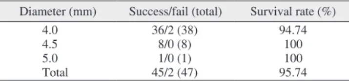
Diameter (mm) Success/fail (total) Survival rate (%)
4.0 36/2 (38) 94.74
4.5 8/0 (8) 100
5.0 1/0 (1) 100
Total 45/2 (47) 95.74
Survival rates did not differ significantly among the three diameter groups (P=0.069).
Truc Thi Hoang Nguyen et al: 7-mm-long dental implants: retrospective clinical outcomes in medically compromised patients. J Korean Assoc Oral Maxillofac Surg 2019
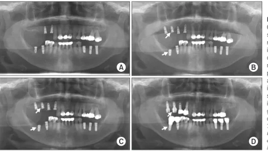
right neck metastasis. An implant was planned for the eden- tulous region of the right posterior mandible. After consider- ing mandible atrophy and proximity to the inferior alveolar nerve, a 4.5 mm×7 mm Stella implant was placed. The implant achieved good stability and bone integration after loading and showed acceptable MBL at 3 years.(Fig. 3) Case 2 is a 72-year-old male with osteomyelitis and a history of hypertension and diabetes. A 4 mm×7 mm Stella implant was installed in the 37 tooth site. The implant showed good stabil- ity and low MBL after loading and at 1 year.(Fig. 4) Case 3 is a 67-year-old female who had hypertension and osteomyelitis in the posterior right mandible. A previous implant installed in the 37 tooth position failed due to bone resorption. In ad- dition, the edentulous posterior of the right maxilla also had insufficient bone and sinus pneumatization. Therefore, in planning the implant therapy, a 4 mm×7 mm Luna implant
was chosen for the 16 position, and a 4.5 mm×7 mm Stella implant was chosen for the 47 tooth position after removal of the failed implant. The two implants showed good stability and acceptable MBL on follow-up examination.(Fig. 5)
Of the 47 implants, two failed, and the survival rate was 95.74%. The two failed implants belonged to a patient with oral cancer who was treated with mandibular resection and reconstruction. The postoperative bone had insufficient vol- ume and unfavorable quality. Dental implant treatment has few absolute contraindications, and the impact of health risks on implant outcome remains unclear due to the scarcity of prospective studies19. However, studies have shown a nega- tive impact of bisphosphonates on implant success19. In oral cancer patients, a lack of residual bone following resection makes placing implants in an ideal position difficult23,24. Considering that all patients were medically compromised,
A B
C D
Fig. 3. Panoramic radiograms of a 76-year-old female who had squamous cell cancer, treated with maxillary mass resection and radiotherapy on the left, radical neck dissection and radiotherapy due to neck metastasis on the right (A).
A 4.5 mm×7 mm Stella (Shinhung) was installed in the 47 tooth position (arrow;
B). The implant achieved good stability and bone integration, and showed ac- ceptable bone loss at 3 years (arrows;
C, D).
Truc Thi Hoang Nguyen et al: 7-mm-long dental implants: retrospective clinical outcomes in medically compromised patients. J Korean Assoc Oral Maxillofac Surg 2019
A B
C D
Fig. 4. Panoramic radiogram of a 72-year-old male with osteomyelitis and a history of hypertension and dia- betes. A. Preoperation radiogram. B. A 4 mm×7 mm Stella (Shinhung) implant was installed in the 37 tooth position (arrow). C, D. The implant achieved good stability and bone integration, and showed acceptable bone loss at 1 year (arrows).
Truc Thi Hoang Nguyen et al: 7-mm-long dental implants: retrospective clinical outcomes in medically compromised patients. J Korean Assoc Oral Maxillofac Surg 2019
including cancer and BRONJ, the survival rate of 7-mm- long implants in the general population would be higher than in this study. The failed implants in this study were placed adventurously in alveolar bone, which had insufficient height and had been involved in cancer treatment and reconstruction surgery. Most implant failures were reported early, during the healing phase at abutment connection25-29.
To guarantee long-term clinical results, maintaining stable marginal bone is more critical with SDIs18. MBL is a gener- ally accepted parameter to evaluate the bone response around a dental implant. Originally, a mean MBL of ≥1.5 mm in the first year and an MBL of ≥0.2 mm per year afterward was considered a threshold for implant success20,30. Randomized, controlled studies31 on SDIs in the posterior maxilla had an MBL from 1.02 to 0.1 mm. In this study, the MBL results on the mesial and distal aspects after 1 year were 0.53±0.58 mm and 0.67±0.56 mm, respectively, and 0.58±0.60 mm and 0.71±0.60 mm, respectively, after 2 years. These MBL results are within the success threshold20; however, long-term follow-up is needed.
V. Conclusion
The present study showed comparable survival rates of SDIs in medically compromised patients to the conventional implants in a healthy population. In addition, the stability of marginal bone around an SDI in these patients was accept- able in comparison with MBL in healthy patients. The results suggest that placing an SDI is a reliable treatment option, especially for medically compromised patients, and can be an alternative when sinus lifting or vertical bone grafting should
be avoided. Further, long-term follow-up and evaluation of SDIs in these patients is needed.
ORCID
Truc Thi Hoang Nguyen, https://orcid.org/0000-0002-8667-6698 Mi Young Eo, https://orcid.org/0000-0001-7055-9924 Yun Ju Cho, https://orcid.org/0000-0002-1818-5280 Hoon Myoung, https://orcid.org/0000-0002-9984-8479 Soung Min Kim, https://orcid.org/0000-0002-6916-0489
Authors’ Contributions
All authors read and approved the final manuscript.
T.T.H.N. read and wrote the manuscript, M.Y.E. prepared ret- rospective data and wrote the manuscript, Y.J.C. prepared all figures and references, H.M. revised and corrected the manu- script, and S.M.K. designed and wrote the entire article.
Acknowledgements
This study was supported by Basic Science Research Program through the National Research Foundation of Korea funded by the Ministry of Education (2017R1D- 1A1B03036054).
Ethics Approval and Consent to Participate
This retrospective data analysis was approved by the In- stitutional Review Board of Seoul National University (S- D20180022).
A B
C D
Fig. 5. Panoramic radiogram of a 67-year-old female who had hyperten- sion and osteomyelitis in the posterior right mandible. A. A previous implant installed in the 37 tooth position failed due to bone resorption. B. A 4 mm×7 mm Luna (Shinhung) implant was placed at the 16 tooth position (arrow), and a 4.5 mm×7 mm Stella (Shinhung) implant was placed at the 47 tooth po- sition (arrow) after removal of the failed implant. C, D. The two implants showed good stability and acceptable bone loss on follow-up examination (arrows).
Truc Thi Hoang Nguyen et al: 7-mm-long dental implants: retrospective clinical outcomes in medically compromised patients. J Korean Assoc Oral Maxillofac Surg 2019
Conflict of Interest
No potential conflict of interest relevant to this article was reported.
References
1. Renouard F, Nisand D. Impact of implant length and diameter on survival rates. Clin Oral Implants Res 2006;17 Suppl 2:35-51.
2. Jain N, Gulati M, Garg M, Pathak C. Short implants: new horizon in implant dentistry. J Clin Diagn Res 2016;10:ZE14-7.
3. Al-Hashedi AA, Taiyeb Ali TB, Yunus N. Short dental implants:
an emerging concept in implant treatment. Quintessence Int 2014;45:499-514.
4. Nisand D, Renouard F. Short implant in limited bone volume. Peri- odontol 2000 2014;66:72-96.
5. Sennerby L, Roos J. Surgical determinants of clinical success of osseointegrated oral implants: a review of the literature. Int J Prosthodont 1998;11:408-20.
6. Wyatt CC, Zarb GA. Treatment outcomes of patients with implant- supported fixed partial prostheses. Int J Oral Maxillofac Implants 1998;13:204-11.
7. Srinivasan M, Vazquez L, Rieder P, Moraguez O, Bernard JP, Belser UC. Efficacy and predictability of short dental implants (<8 mm): a critical appraisal of the recent literature. Int J Oral Maxil- lofac Implants 2012;27:1429-37.
8. Karthikeyan I, Desai SR, Singh R. Short implants: a systematic review. J Indian Soc Periodontol 2012;16:302-12.
9. Lemos CA, Ferro-Alves ML, Okamoto R, Mendonça MR, Pellizz- er EP. Short dental implants versus standard dental implants placed in the posterior jaws: a systematic review and meta-analysis. J Dent 2016;47:8-17.
10. Benlidayi ME, Ucar Y, Tatli U, Ekren O, Evlice B, Kisa HI, et al.
Short implants versus standard implants: midterm outcomes of a clinical study. Implant Dent 2018;27:95-100.
11. Esfahrood ZR, Ahmadi L, Karami E, Asghari S. Short dental im- plants in the posterior maxilla: a review of the literature. J Korean Assoc Oral Maxillofac Surg 2017;43:70-6.
12. Pierrisnard L, Renouard F, Renault P, Barquins M. Influence of im- plant length and bicortical anchorage on implant stress distribution.
Clin Implant Dent Relat Res 2003;5:254-62.
13. Anitua E, Tapia R, Luzuriaga F, Orive G. Influence of implant length, diameter, and geometry on stress distribution: a finite ele- ment analysis. Int J Periodontics Restorative Dent 2010;30:89-95.
14. Chen H, Liu N, Xu X, Qu X, Lu E. Smoking, radiotherapy, dia- betes and osteoporosis as risk factors for dental implant failure: a meta-analysis. PLoS One 2013;8:e71955.
15. Moy PK, Medina D, Shetty V, Aghaloo TL. Dental implant failure rates and associated risk factors. Int J Oral Maxillofac Implants 2005;20:569-77.
16. Guobis Z, Pacauskiene I, Astramskaite I. General diseases influ- ence on peri-implantitis development: a systematic review. J Oral
Maxillofac Res 2016;7:e5.
17. Liddelow G, Klineberg I. Patient-related risk factors for implant therapy. A critique of pertinent literature. Aust Dent J 2011;56:417- 26; quiz 441.
18. Renvert S, Aghazadeh A, Hallström H, Persson GR. Factors related to peri-implantitis - a retrospective study. Clin Oral Implants Res 2014;25:522-9.
19. Kasai T, Pogrel MA, Hossaini M. The prognosis for dental im- plants placed in patients taking oral bisphosphonates. J Calif Dent Assoc 2009;37:39-42.
20. Galindo-Moreno P, León-Cano A, Ortega-Oller I, Monje A, O Val- le F, Catena A. Marginal bone loss as success criterion in implant dentistry: beyond 2 mm. Clin Oral Implants Res 2015;26:e28-34.
21. Diz P, Scully C, Sanz M. Dental implants in the medically compro- mised patient. J Dent 2013;41:195-206.
22. Grant BT, Pancko FX, Kraut RA. Outcomes of placing short dental implants in the posterior mandible: a retrospective study of 124 cases. J Oral Maxillofac Surg 2009;67:713-7.
23. Barrowman RA, Wilson PR, Wiesenfeld D. Oral rehabilitation with dental implants after cancer treatment. Aust Dent J 2011;56:160-5.
24. Renouard F, Nisand D. Short implants in the severely resorbed maxilla: a 2-year retrospective clinical study. Clin Implant Dent Relat Res 2005;7 Suppl 1:104-10.
25. Jemt T, Lindén B, Lekholm U. Failures and complications in 127 consecutively placed fixed partial prostheses supported by Bråne- mark implants: from prosthetic treatment to first annual checkup.
Int J Oral Maxillofac Implants 1992;7:40-4.
26. Becker W, Becker BE, Alsuwyed A, Al-Mubarak S. Long-term evaluation of 282 implants in maxillary and mandibular molar po- sitions: a prospective study. J Periodontol 1999;70:896-901.
27. Lee JH, Frias V, Lee KW, Wright RF. Effect of implant size and shape on implant success rates: a literature review. J Prosthet Dent 2005;94:377-81.
28. Morand M, Irinakis T. The challenge of implant therapy in the pos- terior maxilla: providing a rationale for the use of short implants. J Oral Implantol 2007;33:257-66.
29. Mohajerani H, Roozbayani R, Taherian S, Tabrizi R. The risk fac- tors in early failure of dental implants: a retrospective study. J Dent (Shiraz) 2017;18:298-303.
30. Bratu E, Chan HL, Mihali S, Karancsi O, Bratu DC, Fu JH, et al. Implant survival rate and marginal bone loss of 6-mm short implants: a 2-year clinical report. Int J Oral Maxillofac Implants 2014;29:1425-8.
31. Monje A, Chan HL, Fu JH, Suarez F, Galindo-Moreno P, Wang HL. Are short dental implants (<10 mm) effective? A meta-analysis on prospective clinical trials. J Periodontol 2013;84:895-904.
How to cite this article: Nguyen TTH, Eo MY, Cho YJ, Myoung H, Kim SM. 7-mm-long dental implants: retrospective clinical outcomes in medically compromised patients. J Korean Assoc Oral Maxillofac Surg 2019;45:260-266. https://doi.org/10.5125/
jkaoms.2019.45.5.260