2
Department of Periodontology, Research Institute for Periodontal Regeneration, Yonsei University College of Dentistry, Seoul, Republic of Korea
Kwan-Joo Lee, Young Woo Song, Ui-Won Jung, Jae-Kook Cha*
Diagnosis and Clinical Management of Retrograde Peri-Implantitis Associated with Adjacent Apical
Periodontitis: a Case Report
Diagnosis and Clinical Management of Retrograde Peri-Implantitis Associated with Adjacent Apical Periodontitis: a Case Report
Department of Periodontology, Research Institute for Periodontal Regeneration, Yonsei University College of Dentistry, Seoul, Republic of Korea
Kwan-Joo Lee, Young Woo Song, Ui-Won Jung, Jae-Kook Cha*
Peri-apical implant lesion, also known as ‘retrograde peri-implantitis’ can occur with multifactorial etiological factors.
The purpose of this case report is to demonstrate resolution of periapical implant lesion by removal of causative factors and saving implant by regenerative therapy.
A 54-year old male patient with mild dull pain around implant on the right mandibular second premolar area due to persis- tent peri-apical infection of the adjacent first premolar was treated. Extraction of tooth with symptomatic apical periodontitis and regenerative therapy on the buccal fenestration area of the implant and extraction site were performed. After 6-month re- entry, notable regenerated bone tissue around implant was found, and implant placement on the previous extraction site was performed. After 14-month follow-up from the regenerative therapy, neither biological nor mechanical complication could be found around the implant, evidenced by high implant stability, normal clinical probing depth, and absence of discomfort spontaneously and during masticatory function.
In conclusion, surgical intervention including regenerative therapy using bone graft and barrier membrane on periapical implant lesion can be suggested as one of the treatment options considering the extent of periapical lesion.
Key words: bone regeneration; case report; dental implant; periapical lesion; peri-implantitis Corresponding Author
Jae-Kook Cha, DDS, PhD.
Department of Periodontology,Yonsei University College of Dentistry50-1 Yonsei-ro, Seodaemun-gu, Seoul 03722, Republic of Korea
Tel. +82-2-2228-3191 Fax. +82-2-392-0398 E-mail [email protected]
ABSTRACT
ACKNOWLEDGMENT This work was supported by a National Research Foundation of Korea (NRF) grant funded by the Korean government (Ministry of Science, ICT & Future Planning) (No. NRF-2017R1A2B2002537).
Conflict of interest The authors declare that they have no conflict of interest related to any product used in the present study.
al Management of Retrograde Peri-Implantitis Associated with Adjacent Apical Periodontitis: a Case Report
I. Introduction
The implant periapical lesion is not a common complication that may occur after implant place- ment, and many case reports have suggested such lesion can be associated with one of the potential causes of implant failure1). This implant lesion, also known as ‘retrograde peri-implantitis’ or ‘api- cal peri-implantitis,’ is presented with progressive bone loss at the implant apex often accompanied by pain, tenderness, and/or fistula and can be clas- sified further into disease-inactive and active peri- apical implant lesions2).
Periapical implant lesion is called inactive, if a clinically asymptomatic, periapical radiololucency is found, when a shorter implant is placed in over- prepared osteotomy site or implanted next to pre- existing scar tissue, or overheating occurred during drilling process3). On the contrary, active periapical implant lesion is caused by bacterial contamina- tion during insertion, premature prosthesis load- ing involving microfractures of bone tissue, or pre-existing or developing periapical lesion at the implantation site or adjacent tooth4).
According to a retrospective study analysing 59 implants with periapical lesion out of a total 248 implants receiving single tooth replacement that had radiographic information on the periapical status of the previously extracted tooth or adjacent tooth, the prevalence of periapical implant lesion was different according to the baseline periapical conditions of the tooth at the implantation site and the neighboring tooth before extraction5). When
the implantation took place in the extracted tooth site that had not shown a sign of periapical lesion with no previous endodontic treatment history, the incidence of the implant pathology was 2.1%
at implant-level. When the implant was installed in the site that previously experienced endodon- tic treatment without or with periapial lesion, the incidence was 8.2% and 13.6%, respectively. On the other hand, when the adjacent tooth near the im- plant did neither experience endodontic treatment nor show a sign of periapical radiolucency, the incidence was 1.2%, while when the endodontic treatment was performed to the neighboring tooth that had periapical lesion, it increased up to 25%.
Since periapical implant lesion is considered to have multifactorial etiology, currently, no con- sensus has been established regarding a clear-cut treatment strategy. According to some case re- ports, non-surgical intervention in combination with amoxicillin were effective after a follow-up of 2 years6), while other authors reported anitbiotics were not effective for controlling active disease7). Most authors agree on the treatment that the im- plant apex should be surgically exposed. Never- theless, how this exposed site should be surgically treated, thereafter, still remains controversial8).
This case report described a patient diagnosed with retrograde peri-implantitis affected by an ad- jacent tooth endodontically treated for symptom- atic apical periodontitis. The etiology, diagnosis, and clinical management of this disease in regard are reported in detail including follow-up visit demonstrating resolution of the lesion after regen-
erative therapy.
II. Materials and Methods
1. Pre-operative clinical and radiographic findings A male patient with the age of 54 was referred to the Department of Periodontics, Yonsei Uni- versity Dental Hospital on August, 2018 from the Department of Endodontics for extraction of #44 due to persistent symptomatic apical periodontitis and treatment regarding peri-apical lesion on the implant site of #45 (i45), affected by apical lesion of
#44 (Fig. 1a to 1b). The patient received glaucoma treatment years ago and did not have any other compromised systemic conditions.
During clinical examination, the patient felt mild pain at the vestibular area of #44 and i45, showing gingival redness and swelling. The clinical probing depths on #44 and i45 were 12mm on mid-buccal area and normal, respectively. The recent dental history was that the patient received implantation at the area of #45 and #47 restored with a bridge two and a half years ago and endodontic treatment for removal of apical lesion of #44 five months ago.
Under endodontic exploration with microscope, a crack line appeared to extend apically originat- ing from cervical abfraction on the buccal area.
Despite the effort, the periapical lesion increased from the size of 8.2 x 11.15 mm in width and length to 9.1 x 11.74 mm involving the apex of i45 as could be noted from the radiographic observation of periapical radiographs.
2. Treatment planning
Under the diagnosis of symptomatic apical peri- odontitis on #44 and retrograde peri-implantitis on i45, the treatment was planned as followed: the extraction of #44 and savability assessement on the i45 were to be performed, repectively after two weeks from the day of first clinical visit. Before the surgical procedure, careful supragingival debride- ment with saline debridement on the affected area were performed with subsequent application of minocycline hydrochloride 2% (Periocline®, Sun- star Guidor, Japan) as local drug delivery for infec- tion control.
3. Surgical procedure and operative findings
Prior to the initiation of treatment, the clinical probing depths on #44 and i45 were measured, 12 mm on the mid-buccal area of #44 and 3-4 mm around i45 with intact free gingival margin, respectively. Verbally informed consent was ob- tained from the patient to be profiled. The patient was given 2 g of amoxicillin an hour before the procedure and given an injection of local anaes- thetics (2% lidocaine hydrochloride-epinephrine 1:100,000; Huons Pharmaceutical, Republic of Ko- rea). After that, the patient orally rinsed with 15 mL of chlorhexidine gluconate 0.12 % (Hexamedine, Bukwang pharmaceutical, Republic of Korea), and extraoral preparation was performed before surgi- cal intervention. A crestal incision and full-thick- ness flap elevation were performed from the distal, crestal region of i45 to the mesial line angle of #43 with a vertical incision trespassing a mucogingival
al Management of Retrograde Peri-Implantitis Associated with Adjacent Apical Periodontitis: a Case Report junction. Upon flap reflection at the apical area of
#44, a dehiscence defect extending towards crestal area in the buccal aspect of #44 showing a pri- mary endodontic with secondary periodontal in- volvement was detected. Regarding the operative finding of i45, quite an extensive buccal fenestra- tion defect that spanned almost 7 mm in length, extending coronally from the apex to the level of the second thread line from the implant shoulder was found. However, no calculus deposition was found on the thread surface of the implant.
First, extraction of #44 was performed, followed
by a removal of granulation tissue at the apical bony defect. After meticulous saline irrigation on the exposed implant threads and valleys of i45, chemical debridement using tetracycline hydro- chloride paste was applied for 1 minute and then copious irrigation with saline solution was per- formed. Then, 0.5 g of the deproteinized porcine bone mineral (The Graft®, Purgo biologics, Sung- nam, Republic of Korea) was applied to both buc- cal defects of #44 area and i45, and bioabsorb- able collagen membrane (Collagen Membrane®, Genoss, Suwon, Republic of Korea) was used to cover the grafted site for tissue exclusion. Releas-
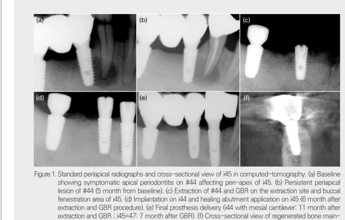
Figure 1. Standard periapical radiographs and cross-sectional view of i45 in computed-tomography. (a) Baseline showing symptomatic apical periodontitis on #44 affecting peri-apex of i45. (b) Persistent periapical lesion of #44 (5 month from baseline). (c) Extraction of #44 and GBR on the extraction site and buccal fenestration area of i45. (d) Implantation on i44 and healing abutment application on i45 (6 month after extraction and GBR procedure). (e) Final prosthesis delivery (i44 with mesial cantilever: 11 month after extraction and GBR ; i45=47: 7 month after GBR). (f) Cross-sectional view of regenerated bone main- tained on buccal fenestration area of i45 (14 month after GBR).
ing incision was performed to the periosteal region of the buccal flap, and the flaps were repositioned and sutured with 4-0 and 6-0 synthetic monofila- ment suture material (Monosyn®, B.Braun, Melsun- gen, Germany) to achieve primary closure (Fig. 1c &
Fig. 2a to 2e). Postoperative instructions were given, and medications were prescribed including 500 mg amoxicillin three times a day for 7 days. At 1 week postoperative follow-up appointment, healing on the surgical site was uneventful, and the sutures were stitched out another week later (Fig. 2f).
After six months post-operation, the flap was re- opened to evaluate the healing status of the opera- tion site and to perform implantation on the previ- ous extraction and guided bone regeneration (GBR) site of #44. The augmented buccal bone area of i45 was found to be clinically favorable as dense mature bone could be detected with firm, tactile quality us- ing periosteal elevator (Fig. 2g to 2h). Before implan- tation at the site #44, the trephine bur with an inner diameter of 3 mm was used under torque mode at depth of 6 mm to obtain the previously grafted bone tissue.
A 4 x 12 mm self-tapping SLA-surfaced implant (Superline III®, Dentium, Suwon, Republic of Korea) was placed with primary stability which was ob- tained along the residual lingual wall and a length of 2 mm at the apex of the implant and assessed by a final torque of 50 Ncm. The healing abutment was engaged to i44 and i45, and the stability of the i45 was assessed with AnyCheck® (Neobiotech, Seoul, Republic of Korea) with the measured value of 68
that closely resembles implant stability quotient val- ue of resonance frequency analysis (Fig. 1d & Fig. 2i to 2l). Standard postsurgical instructions and medi- cations were provided as aforementioned. After one month, a 3-unit prosthesis of i45=47 was loaded for allowing mastication with right molar area. Then, after five months from implantation of i44, a 2-unit prosthesis with a mesial cantilever was loaded on i44, taking a wide span of mesial-distal spatial re- lationship in the right posterior mandibular region into consideration (Fig. 2m to 2n).
III. Results
1. Post-operative clinical and radiographic findings The patient was recalled to the clinic after three months from final prosthesis delivery of i44 with mesial cantilever and seven months from prosthesis delivery of i45=47, equal to 14 months after regen- erative therapy performed on the extraction site of
#44 and buccal fenestration area of i45 (Fig. 1e). The patient did not feel any discomfort spontaneously or during functioning as speaking or mastication. On clinical observation regarding both implants, no sign of gingival redness or vestibular swelling were found, and the clinical probing depths were maintained normal on both the tooth and implant around 3 to 4 mm. On the cross-sectional view in computed-to- mography taken 14 months after GBR performed on the middle of i45, the buccal, crestal resorption to the second thread of the implant from the implant shoulder was observed (Fig. 1f). From both clinical
al Management of Retrograde Peri-Implantitis Associated with Adjacent Apical Periodontitis: a Case Report
Figure 2. Clinical photographs showing extraction of #44 and GBR on extraction site of #44 and buccal fenestration area of i45. (a) Pre-operative site showing buccal dehiscence defect on extraction site of #44 and extensive buccal fenestration defect of i45. (b) Application of de- proteinized porcine bone graft and collagen membrane. (c) Horizontal mattress suture over the membrane. (d) Lateral view and (e) occlusal view after primary closure. (f) Stitch-out after extraction and GBR. (g) Occlusal view of pre-implantation state. (h) Dense regener- ated bone around i45 and GBR site of #44. (i) Surgical stent application. (j) Implantation of i44. (k) Implant axis evaluation with guide pins. (l) Primary closure following implantation. (m) Occlusal view and (n) lateral view after final prosthesis delivery.
and radiographic observation, it could be consid- ered that the bone fill around the implant fixtures were well-maintained without any sign of inflam- matory reaction of adjacent soft tissue.
2. Histological preparation and findings
The biopsy sample obtained at the implantation site of #44, 6 months after GBR performed, was fixed in 4% paraformaldehyde solution for 2 days, decalcified, and embedded in paraffin before cut into serial sections using a microtome which were then stained using Hematoxylin and Eosin. Under observation of the histological slides under a light microscope (BX51®, Olympus, Tokyo, Japan), they were digitally scanned with a magnification rate of x200 for describing histological findings.
No marked inflammatory reaction from the graft materials was found in the histologic speci- men. The xenograft particles were clearly identi- fied, showing their typical structure surrounded by connective tissue with abundant occupation of cells and newly formed vessels. New bone tissue could be detected uniformly throughout the broad expanse of the biopsy sample, primarily along- side the grafted biomaterials. In particular within this living tissue, typical trabecular bone pattern with residence of osteocytes in the lacunae were observed, indicating active vitality of bone tissue while the typical osseous structure of the graft ma- terials stained with a pale eosinophilic color were shown to contain lacunae without the presence of osteocytes (Fig. 3).
IV. Discussion
In this case report, under correct diagnosis of ac- tive, type 2 retrograde peri-implantitis and surgi- cal intervention, the patient could save the implant placed two years ago by extracting the adjacent ailing tooth from symptomatic apical periodontitis and performing regenerative therapy on the buc- cal fenestration area of the implant after meticu- lous debridement.
The patient was diagnosed as such in that the clinical symtoms of pain and tenderness to pal- pation were accompanied in the form of ‘active lesion’ as disease progressed due to a spread of bacterial infection that initiated from the periapi- cal lesion of the adjacent tooth on account of deep crack line1,2). Sussman et al. described this type of periapical implant pathology as type 2, the tooth- to implant pathway where periapical lesion of a neighboring tooth due to caries involvement, ex- ternal root resporption, or poor endodontic seal can spread to cause periapical implant lesion9).
With regard to treatment modalities of retro- grade peri-implantitis, there is no current consen- sus owing to multifactorial etiology of this disease.
Waasdrop & Reynolds treated a patient only with amoxicillin and observed the lesion was resolved radiographically at 9-month follow-up after the treatment10). However, regarding that the size of the implant lesion in the described patient was relatively extensive and the disease progressed in time-dependent manner, surgical intervention was the first treatment of choice.
al Management of Retrograde Peri-Implantitis Associated with Adjacent Apical Periodontitis: a Case Report
Some authors suggested that explantation of the infected implant could preclude osteomyelitis and further bone loss11,12) while others proposed a con- servative surgical method as partial implant apical resection considering the size of infection and im- plant stability13). Trepanation and curettage without resection of the implant was considered effective as the third surgical treatment option that entails copious irrigation with saline and chlorhexidine solution14). Most commonly used chemical solution for decontamination of implant surface are saline, chlorhexidine, and tetracycline paste. However, treatment efficiency of any of theses agents, to
date, still remains questionable15-17).
Several authors reported on the successful clini- cal outcome of regenerative therapy where bone substitutes either with or without collagen mem- brane were applied on the implants with periapi- cal lesion. Bretz et al. also reported the successful treatment oucome of regenerative therapy using demineralized freeze-dried bone performed on the single implant affected by periapical implant pathology18). Quirynen et al. reported three out of four implants with periapial lesion that were treat- ed with deproteinized bovine bone mineral healed uneventfully while one implant experienced a fis- Figure 3. Photomicrograph of histological biopsy on extraction and GBR site of the right first premolar area immediately
before implant placement stained with Hematoxylin and Eosin. Scattered bone particles in the middle region of specimen and newly formed bone around and within the space between particles are shown on the left.
Boxed area refers to magnified view of the central region of specimen. XG: Residual xenograft material, NB:
Mineralized new bone, FT: Fibrous connective tissue
tula as biological complication19). Furthermore, it is stated that when the implants with single-tooth re- placement that are infected peri-apically, yet with its coronal part osseointegrated with intact bone, are treated with bone grafts, they can be success- fully loaded and function for many years. Lately, Lefever et al. demonstrated that 11 out of 15 im- plants with periapical lesion receving GBR proce- dure could function without clinical or radiological sign of inflammation5).
In our case study, the GBR procedure using de- proteinized porcine bone mineral was performed with a successful clinical result, yet with mild bone loss on the coronal portion of the implant. This may be attributable to application of insufficient amount of bone material and/or loss of osseoin- tegration potential of the contaminated implant surface itself. In addition, to better regenerate bone around implant surface, mechanical decon- tamination method could have been more care- fully selected. Cha et al. reported that glycine air abrasive was found to cause the least macroscopic alteration on implant surface topography, while demonstrating sufficient accessibility to its surface between the thread lines20).
Even though post-operatively taken CBCT dem- onstrates tight contact between the implant fixture and surrounding bone, it is hard to clearly conclude that regenerated bone promoted re-osseointegra- tion since it was impossible to obtain a block spec- imen for further analysis. However, with regard to qualitative evaluation of the regenerated bone around the implant, it can be interpreted with cau-
tion in a positive light that the bone tissue formed around the buccal fenestration of the implant is highly likely to be vital based on the histological assessment on the biopsy taken on the adjacent GBR site before implantation took place. A long- term clinical investigation, including retrospective cohort or case-control studies would be needed to evaluate the clinical efficacy of this surgical pro- cedure.
V. Conclusion
Despite its multifactorial etiological nature, ret- rograde peri-implantitis is well associated with apical infection of tooth at the site of or adjacent to an implant. If the disease occurs due to an apical lesion of the adjacent tooth, endodontic treatment or, if the disease persists regardless, extraction of the involved tooth should be performed. Surgical intervention is recommended in the state of active progression of the implant periapical infection, among which regenerative therapy using bone grafts and membrane can be suggested as one of the treatment options considering the extent of periapical lesion.
al Management of Retrograde Peri-Implantitis Associated with Adjacent Apical Periodontitis: a Case Report 1. Ayangco L, Sheridan PJ. Development and treatment of retrograde
peri-implantitis involving a site with a history of failed endodontic and apicoectomy procedures: a series of reports. Int J Oral Maxillofac Im- plants 2001;16(3):412-417
2. McAllister BS, Masters D, Meffert RM. Treatment of implants dem- onstrating periapical radiolucencies. Pract Periodontics Aesthet Dent 1992;4(9):37-41
3. Reiser GM, Nevins M. The implant periapical lesion: etiology, preven- tion, and treatment. Compend Contin Educ Dent 1995;16(8):768, 770, 772 passim
4. van Steenberghe D, Yoshida K, Papaioannou W, et al. Complete nose coverage to prevent airborne contamination via nostrils is un- necessary. Clin Oral Implants Res 1997;8(6):512-516
5. Lefever D, Van Assche N, Temmerman A, et al. Aetiology, microbiol- ogy and therapy of periapical lesions around oral implants: a retro- spective analysis. J Clin Periodontol 2013;40(3):296-302
6. Chang LC, Hsu CS, Lee YL. Successful medical treatment of an implant periapical lesion: a case report. Chang Gung Med J 2011;34(1):109-114
7. Dahlin C, Nikfarid H, Alsen B, Kashani H. Apical peri-implantitis: pos- sible predisposing factors, case reports, and surgical treatment sug- gestions. Clin Implant Dent Relat Res 2009;11(3):222-227 8. Sarmast ND, Wang HH, Sajadi AS, et al. Classification and Clinical
Management of Retrograde Peri-implantitis Associated with Apical Periodontitis: A Proposed Classification System and Case Report. J Endod 2017;43(11):1921-1924
9. Sussman HI. Endodontic pathology leading to implant failure--a case report. J Oral Implantol 1997;23(3):112-115; discussion 115-116 10. Waasdorp J, Reynolds M. Nonsurgical treatment of retrograde
peri-implantitis: a case report. Int J Oral Maxillofac Implants
2010;25(4):831-833
11. Silva GC, Oliveira DR, Vieira TC, et al. Unusual presentation of ac- tive implant periapical lesions: a report of two cases. J Oral Sci 2010;52(3):491-494
12. Sussman HI. Tooth devitalization via implant placement: a case re- port. Periodontal Clin Investig 1998;20(1):22-24
13. Oh TJ, Yoon J, Wang HL. Management of the implant periapical lesion: a case report. Implant Dent 2003;12(1):41-46
14. Zhou Y, Cheng Z, Wu M, et al. Trepanation and curettage treat- ment for acute implant periapical lesions. Int J Oral Maxillofac Surg 2012;41(2):171-175
15. Ataullah K, Chee LF, Peng LL, Lung HH. Management of ret- rograde peri-implantitis: a clinical case report. J Oral Implantol 2006;32(6):308-312
16. Chan HL, Wang HL, Bashutski JD, et al. Retrograde peri-implan- titis: a case report introducing an approach to its management. J Periodontol 2011;82(7):1080-1088
17. Penarrocha-Diago M, Boronat-Lopez A, Garcia-Mira B. Inflamma- tory implant periapical lesion: etiology, diagnosis, and treatment- -presentation of 7 cases. J Oral Maxillofac Surg 2009;67(1):168- 18. Bretz WA, Matuck AN, de Oliveira G, et al. Treatment of retrograde 173
peri-implantitis: clinical report. Implant Dent 1997;6(4):287-290 19. Quirynen M, Vogels R, Alsaadi G, et al. Predisposing conditions for
retrograde peri-implantitis, and treatment suggestions. Clin Oral Implants Res 2005;16(5):599-608
20. Cha JK, Paeng K, Jung UW, et al. The effect of five mechanical in- strumentation protocols on implant surface topography and rough- ness: A scanning electron microscope and confocal laser scanning microscope analysis. Clin Oral Implants Res 2019;30(6):578-587
References