Archives of Craniofacial Surgery
Copyright © 2012 The Korean Cleft Palate-Craniofacial Association
This is an Open Access article distributed under the terms of the Creative Commons Attribution Non-Commercial License (http://creativecommons.org/
licenses/by-nc/3.0/) which permits unrestricted non-commercial use, distribution, and reproduction in any medium, provided the original work is properly cited.
www.kcpca.or.kr ISSN 2287-1152 57
청소년기에 발견된 상구순 누공의 치험례
정한주·강석주·김진우·선 욱 인제대학교 부산백병원 성형외과학교실
Purpose: Congenital sinus of the upper lip is extremely rare and only 3 cases have been reported domestically. We report a case of congenital sinus of midline upper lip, which was found in an adolescent patient.
Methods: A 14-year-old girl presented with a small pit on midline of the upper lip, which was visible at birth. The patient had never been treated for the congenital sinus because it was asymptomatic. Surgical excision under local anesthesia was performed.
Results: The sinus had a tract extending into 5 mm posteroinferior and had not penetrated the oral cavity. Histological examination showed a fistulous tract lined by keratinized squamous epithelium. After complete excision, there was no recurrence and we obtained a satisfactory cosmetic result.
Conclusion: Congenital sinus of the midline upper lip is extremely rare. This is a special case that is reported because it did not cause symptoms for the patient until she reached adolescence.
Keywords: Congenital sinus, Upper lip
Congenital Upper Lip Sinus Found in Adolescent Patient: A Case Report
Han Ju Jung, Seok Joo Kang, Jin Woo Kim, Hook Sun
Department of Plastic and Reconstructive Surgery, Inje University Busan Paik Hospital, Inje University College of Medicine, Busan, Korea
Introduction
Congenital sinus occurring in the lips is so rare that its in- cidence rate is below 0.001%.1 Midline upper lip sinus is much rarer than that on the lower lip, and only one case has been reported in Korea by Lee et al.2 Most patients with congenital sinus of the lip are diagnosed during their infancy or early childhood and treated immediately through resection.
The authors found a very rare clinical case of an adolescent patient showing the characteristics of upper lip sinus that was treated successfully.
Case Report
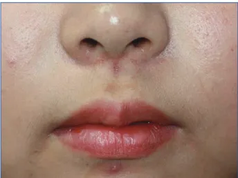
A 14-year-old girl visited our hospital with a deep hollow lesion in the middle of the philtrum. The lesion had been not- ed at birth, but was not treated because there were no symp- toms of pain, swelling, discharge, or inflammation. There was a small oval hole about 1 mm in diameter around 5 mm be- low the columella base on the midline of the philtrum. When a probe was inserted, the sinus was directed toward the poste- rior side, and its depth was around 6 mm (Fig. 1).
The authors thought that it might be the result of an injury during childhood that had not been treated properly and, as a result, the injury had left a spot-size lesion which had been ep- ithelialized, but the patient denied any history of facial injury or any other congenital deformity. The patient did not have family history of facial sinus. The purpose of visiting the hos- pital was only to remove the small spot for cosmetic reasons.
Arch Craniofac Surg Vol.13 No.1, 57-59 http://dx.doi.org/10.7181/acfs.2012.13.1.57
Correspondence: Seok Joo Kang
Department of Plastic and Reconstructive Surgery, Inje University Busan Paik Hospital, Inje University College of Medicine, 75 Bokji-ro, Busanjin-gu, Busan 614-735, Korea
Tel: +82-51-890-6236 / Fax: +82-51-894-7976 / E-mail: [email protected] Received October 20, 2011 / Revised January 17, 2012 / January 18, 2012 Accepted January 19, 2012
Case Report
Archives of Craniofacial Surgery Vol. 13, No. 1, 2012
www.kcpca.or.kr
58
The fistulous tract was removed under local anesthesia.
The lesion ended just beneath the mucosal surface of the up- per lip and did not communicate with the oral or nasal cavi- ties. The size of the specimen was 4 × 7 mm2 (Fig. 2). Micro- scopic examination revealed that it was lined by cornified squamous epithelium with sebaceous glands and hair follicles surrounding the end of the tract (Fig. 3). Ectopic tissue was not found. No postoperative recurrence was noted on one- year follow-up (Fig. 4).
Discussion
Congenital sinus in the upper lip is such a rare disease in which only 40 cases have been reported. The first reported midline sinuses of the upper lip were reported by Lan- nelongue and Menard in 1891 and Clavet in 1899. Among them, 26 occurred on the midline, 14 occurred laterally, and 3 occurred bilaterally.3
Congenital sinus that occurred on the midline was more frequent in women, and half of the cases were associated with congenital anormalies, such as cleft lip, lingual deformity, and Fig. 1. Preoperative view: 0.5 × 1 mm sized small pit is visible in mid-
line of the philtrum.
Fig. 2. Excised mass from the philtrum contains 4 × 5 × 7 mm sized mass, which contains a fistulous tract. The fistulous tract was observed in the center of this excised sample.
Fig. 3. Histologic finding showed fistulous tract lined by keratiniazed squamous epithelium. Sweat glands and hair follicles were around the tract, but heterotopic tissue was not found (H&E, × 40).
Fig. 4. Postoperative view 1 year after the complete excision was performed.
59
www.kcpca.or.kr
Han Ju Jung, et al. Congenital midline upper lip sinus
fistula of the nose.3 Around 20 cases of congenital midline si- nus have been reported in Bonn since the first case was re- ported by Tange in 1965. In Korea, 1 case of midline upper lip sinus reported by Lee et al.2 and 2 cases of congenital lateral sinus of upper lip have been by Lee et al.4
The size of opening has varied from between 1–2 mm and the depth between 5 to 30 mm, but there has been no case in which the opening was connected to the oral cavity.5 Accom- panying deformities include cleft lip, abnormalities of the max- illary labial frenulum, dental alveolus fissure, and bifid uvula, and some cases were found together with accompanying Pierre-Robin’s syndrome or idiopathic precocious puberty.6
Histologically, the sinus tract is covered with squamous epithelial cells, and in some reported cases, it was accompa- nied by sebaceous gland, salivary gland, hair follicles, hyaline cartilage, or mucous gland.
Differential diagnoses include tichofolliculoma, a carti- lage-shaped chondroid syringoma in the upper lip. Histologi- cally, tichofolliculoma is characterized by a number of other hair follicles formed radially centering on a hair follicle, and cartilage-shaped chondroid syringoma is characterized by the tubular structure of epithelial cells with 2 or more layers and homogenous basophilic cells filling the tubular lumen.
The pathogenesis of congenital sinus of the lip has not been explained clearly, but is explained by 3 theories. First, the fu- sion theory hypothesizes that sinus in the upper lip occurs due to failure in the complete fusion of the maxillary process- es in birth. The immersion theory assumes the insufficient in- flow of mesodermal cells into the ectodermal structure on the midline of the lip.1,3,5,7 These 2 theories are similar to the theo- ries on cleft face. Another hypothesis is the invagination the- ory in which upper lip sinus is formed by processes with in- vaginated epithelial cells as in the formation of nasal pit during the developmental stage.8 If upper lip sinus occurs due to failure in the inflow of mesodermal cells or in the fusion of the maxillary processes, there should have been cases of up- per lip congenital sinus connected to the oral cavity or the nasal cavity, but such a case has not been found. Thus, the in-
vagination theory seems to be more persuasive. However, the exact etiology and genetic course of congenital upper lip sinus remain obscure.
Most of upper lip midline sinus patients are usually detect- ed before their early childhood due to recurrent inflamma- tion. Thus, in general the disease is treated surgically for pre- venting infection when it is found. It is treated with surgical resection that includes the sinus and the sinus tract, and post- operative prognosis has been favorable and cosmetically sat- isfactory.
Our case showed the typical histological characteristics of very rare upper lip midline sinus without accompanying con- genital deformities. On the other hand it displayed an atypi- cal clinical pattern: a long-term asymptomatic state, which was detected at the oldest age among cases reported in Korea.
Therefore, we report the additional case of congenital upper lip sinus and hope this report will help further the under- standing of the upper lip sinus.
REFERENCE
1. Nakano Y, Somiya H, Shibui T, Uchiyama T, Takano N, Shibahara T, Hashimoto S: A case of congenital midline fistula of the upper lip. Bull Tokyo Dent Coll 51: 31, 2010
2. Lee BH, Park MC, Lee SH: Congenital midline sinus of the upper lip. J Korean Soc Plast Reconstr Surg 27: 565, 2000
3. Sen C, Agir H, Isken T, Alagoz S, Karadeniz E, Iscen D: Congenital mid- line upper lip sinus. J Craniofac Surg 17: 810, 2006
4. Lee TJ, Bin CW, Koh KS: Congenital lateral sinus of the upper lip. J Ko- rean Cleft Palate-Craniofac Assoc 4: 45, 2003
5. Asahina I, Sakakibara T, Miyashin M, Tachikawa N, Enomoto S: Con- genital midline sinus of the upper lip: case report and review of litera- ture. Cleft Palate Craniofac J 34: 83, 1997
6. Sumitomo S, Ogawa S, Hirai K, Takai Y: Congenital sinus of the upper lip with idiopathic precocious puberty. Oral Dis 8: 308, 2002
7. Charrier JB, Rouillon I, Roger G, Denoyelle F, Collon S, Garabedian EN:
Congenital isolated midline sinus of the upper lip: clinical and embryo- logical approaches. Cleft Palate Craniofac J 43: 488, 2006
8. Al-Qattan MM: Congenital midline sinus of the upper lip. Ann Plast Surg 44: 76, 2000