18β-Glycyrrhetinic acid가 lipopolysaccharide에 의한 생쥐 뇌조직의 염증성 사이토카인과 해마신경세포 자연사에 미치는 영향
이지승1#, 권만재1, 권수현1, 김지호1, 문지영1, 조윤정1, 신정원1, 이종수2, 손낙원1*
1 : 경희대학교 동서의학대학원 한의과학전공, 2 : 경희대학교 한의과대학 한방재활의학과
Effects of 18β-glycyrrhetinic acid on pro-inflammatory cytokines and neuronal apoptosis in the hippocampus of lipopolysaccharide-treated mice
Ji-Seung Lee
1#, Man-Jae Kwon
1, Su-Hyeon Kweon
1, Jeeho Kim
1, Ji-Young Moon
1, Yoon-Cheong Cho
1, Jung-Won Shin
1, Jong-Soo Lee
2, Nak-Won Sohn
1*1 : Department of Oriental Medical Science, Graduate School of East-West Medical Science, Kyung Hee University, 2 : Department of Rehabilitation Medicine, College of Korean Medicine, Kyung Hee University
ABSTRACT
Objectives : 18β -Glycyrrhetinic acid (18betaGA) is an metabolite of glycyrrhizin in
Glycyrrhiza(licorice). The present study investigated anti-inflammatory and anti-apoptosis effect of 18betaGA on the brain tissue of lipopolysaccharide (LPS)-treated C57BL/6 mice.
Methods : 18betaGA was administered orally with low (30 ㎎/㎏) and high (100 ㎎/㎏) doses for 3 days prior to LPS (3 ㎎/㎏) injection. Pro-inflammatory cytokines mRNA including tumor necrosis factor-α (TNF-α), interleukin (IL)-1β, IL-6, and inflammatory enzyme cyclooxygenase-2 (COX-2) mRNA were measured in the cerebral cortex, hippocampus, and hypothalamus tissue using real-time polymerase chain reaction at 24 h after the LPS injection.
Histological changes of Cornu ammonis area 1 (CA1) neurons, Bax, Bcl-2, and caspase-3 expression in the hippocampus was also evaluated by immunohistochemistry and Western blotting method.
Results : 18betaGA significantly attenuated the up-regulation of TNF-α , IL-1β , IL-6 mRNA, and COX-2 mRNA expression in the brain tissues induced by the LPS injection. 18betaGA also significantly attenuated the reductions of the thickness of CA1 and the number of CA1 neurons. The up-regulation of Bax protein expression in the hippocampal tissue by the LPS injection was significantly attenuated, while the ratio of Bcl-2/Bax expression was increased by 18betaGA treatment. 18betaGA also significantly attenuated the up-regulation of Bax and caspase-3 expression in the CA1 of the hippocampus.
Conclusion : This results indicate that 18betaGA has anti-inflammatory and anti-apoptosis effect under neuroinflammation induced by the LPS injection and suggest that 18betaGA may be a beneficial drug for various brain diseases accompanied with the brain tissue inflammation.
1)
Key words : 8β -Glycyrrhetinic acid, Pro-inflammatory cytokines, lipopolysaccharide, Hippocamus, Neuronal apoptosis
*Corresponding author : Nak-Won Sohn, Dept. of Oriental Medical Science, Graduate School of East-West Medical Science, Kyung Hee University.
·Tel:+82-31-202-2747 ·E-mail : [email protected]
#First author : Ji-Seung Lee, Dept. of Oriental Medical Science, Graduate School of East-West Medical Science, Kyung Hee [email protected]
·Tel:+82-31-202-2747 ·E-mail : [email protected]
·Received:10 October 2016 ·Revised:28 October 2016 ·Accepted:16 November 2016
Ⅰ. 서 론
甘草(
Glycyrrhiza,licorice)는 콩과에 속한 다년생 초본인 감초(
Glycyrrhiza glabraL.과
Glycyrrhiza uralensisFisch) 또는 동속식물의 뿌리를 건조한 약재로 다양한 염증성 질환에 응용되어 왔으며
1), 신경세포 손상 보호효능에 대해서도 많은 연구들이 이루어졌다
2,3). 또한 甘草의 신경세포손상 보호효능은 사포닌 성분 중 하나인 glycyrrhizin (GRZ)이 소화기계통 내 에서 가수분해된 대사체인 18β -glycyrrhetinic acid (18betaGA) 에 의한다는 보고가 있다
3,4). 18betaGA는 지질과산화를 억제 하는 항산화 작용
5), 사이토카인 분비를 억제하는 항염증 작용
6)과 미세아교세포 활성화를 억제하는 중추신경염증 억제작용
7)등을 통해 신경세포 손상을 보호하는 것으로 보고되고 있다.
Mizoguchi 등
8)의 연구에 의하면, 18betaGA는 혈액뇌관문 (blood-brain barrier)을 통과하여 뇌조직에 들어가며, 해마 에서 가장 높은 비율로 작용한다고 하였다. 한편으로 해마조직 신경세포 손상에 대해 GRZ의 작용은 많이 연구 보고되어 있
으나
9-12), 18betaGA의 작용과 기전에 대한
in vivo연구는
최근 Oztanir 등
5)의 뇌허혈에 대한 연구보고와 Zhou 등
7)의 자가면역성 뇌척수염에 대한 연구보고 이외에는 찾아보기 어 렵다.
뇌조직에서 염증성 사이토카인의 과도한 증가와 미세아교 세포의 활성화는 만성적인 뇌조직 염증의 가장 큰 특징 중 하 나로서
13), 수면과 식이 등 중추신경계 작용에 따른 생리적 행 동의 변화는 물론 질병행동(sickness behavior)을 유발하고 각종 뇌질환을 악화시키는 이차적인 요인이 된다
14,15). 그러므로 뇌조직 염증에 대한 한약성분의 효능과 작용기전 연구는 다양한 급만성 뇌질환은 물론 퇴행성뇌질환의 예방과 치료에 활용될 수 있는 한약물에 대한 유용한 기초적인 자료를 제공한다.
본 연구에서는 18betaGA가 중추신경염증 상태에서 염증성 사이토카인 발현에 미치는 영향을 관찰해보고자 하였다. 생쥐에 lipopolysaccharide (LPS)를 복강주사하는 방법으로 중추신 경염증을 유발하고, 대뇌피질과 해마 및 시상하부 뇌조직에서 염증성 사이토카인인 tumor necrosis factor-α (TNF-α ), interleukin (IL)-1β , IL-6의 mRNA, 염증매개 효소인 cyclooxygenase-2 (COX-2) mRNA 발현과 해마조직에서 Bax와 Bcl-2 발현의 변화를 통해 신경세포 자연사에 대한 영향을 관찰한바 유의한 결과를 얻었기에 이에 보고하는 바이다.
Ⅱ. 재료 및 방법
1. 실험동물
실험동물은 나라바이오텍(Nara Biotechnology, Korea)에서 구입한 25-28 g의 수컷 C57BL/6 생쥐를 사용하였다. 실험 동물의 사육과 처치는 00대학교 실험동물윤리위원회의 방침과 동물실험관련 규정을 따랐으며, 실험에 사용한 생쥐는 온도 (21-23
oC), 습도(40-60%)와 조명(12시간 명/암)이 자동적 으로 유지되는 사육실에서 무균음수와 사료를 자유롭게 공급 하며 사육되었고, 실험실 환경에 1주 이상 적응시킨 후 사용 하였다.
2. 약물 및 투여
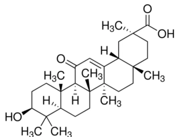
본 실험에 사용한 18β-glycyrrhetinic acid (18betaGA, Fig. 1)는 Sigma-Aldrich (St. Louis, MO, USA)에서 구입 하였으며, 1일 1회 3일간 각각 30 ㎎/㎏ 또는 100 ㎎/㎏을 생리식염수에 녹여 경구투여 하였다.
Fig. 1. Chemical structure of 18β-glycyrrhetinic acid.
3. 실험군의 구분
생쥐를 무작위로 4개 군으로 구분하였다. 정상군(Normal)은 약물처치 없이 통상적으로 사육하였으며, 대조군(LPS)은 LPS 3 ㎎/㎏을 복강주사 하였다. 18betaGA를 투여한 군은 이전의
in vivo연구들
5,7)에서 사용한 용량을 참고하여 저농도군 (18betaGA-L)과 고농도군(18betaGA-H)으로 구분하였으며, 18betaGA-L군은 30 ㎎/㎏, 18betaGA-H군은 100 ㎎/㎏의 18betaGA를 LPS의 복강주사 3일전부터 1일 1회 경구투여 하고, 3일째 경구투여 1시간 후에 LPS 3 ㎎/㎏을 복강주사 하였다. 실험동물은 각 군 당 12마리씩 총 48마리의 실험동 물이 사용되었으며, 무작위로 6마리는 면역조직화학염색용으로, 6마리는 real-time polymerase chain reaction (RT-PCR) 실험용 및 Western blotting 실험용으로 사용하였다.
4. 뇌조직의 처리
실험동물을 tiletamine과 zolazepam으로 마취한 상태에서 RT-PCR 실험용 생쥐는 단두로 희생시키고 뇌를 적출한 다음, 대뇌피질과 해마 및 시상하부의 뇌조직으로 분리하였다. 조직 염색용 생쥐는 개흉하고, 심장을 통하여 0.05 M phosphate buffered saline (PBS)과 4% paraformaldehyde로 충분히 관류하였다. 이후 뇌를 적출하여 24시간 정도 post-fixation 하고, sucrose 용액에 담궈 침전시킨 다음 뇌조직을 –40℃의 dry ice-isopentane 용액으로 동결시켰다. 동결조직은 cryocut (Leica, 2800N, Germany)으로 30 ㎛ 두께의 횡단절편으로 제작하여 조직염색에 사용하였다.
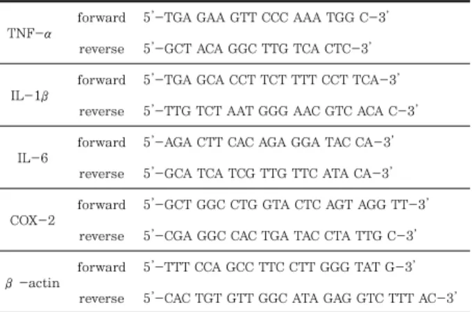
5. 염증성 사이토카인 mRNA의 RT-PCR 측정
대뇌피질과 해마 및 시상하부로 분리한 각각의 뇌조직으로
부터 Trizol (Qiagen, Germany)을 사용하여 total RNA을 추출하였다. 이후 1 ㎍의 total RNA와 Script cDNA synthesis Kit (Bio-Rad, USA)를 사용하여 DNA로 역전사 하였으며, 정량적 real-time PCR은 CFX 96 REAL-TIME PCR Detection System (Bio-Rad, USA)에서 iQ SYBR Green Supermix kit (Bio-Rad, USA)와 preoptimized primer/probe mixture를 사용하여 측정하였다. Housekeeping gene으로는 β-actin을 사용하였으며, 측정에 사용된 gene의 primer sequence는 아래 Table 1과 같다. 이외 측정방법은 제조사의 사용설명서에 따랐으며, 각 시료로부터 측정된 mRNA 발현량은 Normal군의 측정치를 1로 하여 그 배수 (fold change)를 자료로 사용하였다.
TNF-α
forward 5’-TGA GAA GTT CCC AAA TGG C-3’
reverse 5’-GCT ACA GGC TTG TCA CTC-3’
IL-1β
forward 5’-TGA GCA CCT TCT TTT CCT TCA-3’
reverse 5’-TTG TCT AAT GGG AAC GTC ACA C-3’
IL-6
forward 5’-AGA CTT CAC AGA GGA TAC CA-3’
reverse 5’-GCA TCA TCG TTG TTC ATA CA-3’
COX-2
forward 5’-GCT GGC CTG GTA CTC AGT AGG TT-3’
reverse 5’-CGA GGC CAC TGA TAC CTA TTG C-3’
β-actin
forward 5’-TTT CCA GCC TTC CTT GGG TAT G-3’
reverse 5’-CAC TGT GTT GGC ATA GAG GTC TTT AC-3’
Table 1. Primer sequences
6. Western blotting
분리된 뇌조직을 각각 lysis buffer (50 mM Tris–HCl, pH 8.0, 150 mM NaCl, 1% Triton X-100, 0.5% sodium deoxycholate, 0.1% sodium dodecyl sulfate (SDS), 1 mM EDTA, 1% protease inhibitor cocktail; Sigma, USA)에 넣어 균질화하고 원심분리하여 상층액을 분리하였다.
단백질 총량은 Bradford 방법을 사용하여 정량하였으며, 50 ㎍ 의 단백질을 포함한 표본을 SDS-10% polyacrylamide gel electrophoresis에 의해 세분하고, Western blotting을 시행 하였다. 1차항체는 anti-Bax (AB7977, Abcam, USA)와 anti-Bcl-2 (SC7382, Santa Cruz Biotechnology, USA)와 anti-β -actin antibody (MAB1501, Millipore, USA)를 사 용하였다.
7. 뇌조직의 염색
해마 신경세포들의 조직학적 소견을 관찰하기 위해 0.5%
cresyl violet 용액으로 염색하고 흐르는 물에서 수세하였으며, 이후 탈수하고 봉입하여 조직표본을 제작하였다. Bax와 caspase-3의 면역조직화학염색은 뇌조직 절편을 0.05 M PBS로 10분간 2회 씻어내고, 3% H
2O
2에서 30분간 반응시킨 다음 다시 2회 씻어낸 뒤 10% normal horse serum (Vectastain, USA)과 bovine serum albumin (Sigma-Aldrich, USA)을 PBS에 섞은 blocking solution에 한 시간 정도 반응시켰다.
이후 PBS로 2회 씻어 낸 후, rabbit anti-Bax antibody (1:200, ab7977, Abcam, UK)와 rabbit anti-caspase-3 antibody (1:300, BS1518, BioWorld, USA)를 1차항체로 사용하여 각각 PBS와 Triton X-100을 섞은 용액으로 희석한 후 4℃에서 반응시켰다. 이후 2차항체에 반응시키고, avidin–
biotin complex (Vector Laboratories, USA) 방법에 따라 0.05% DAB (Sigma-Aldrich, USA)로 발색한 다음 통상적 인 면역조직화학염색법에 따라 조직표본을 제작하였다.
8. 해마 신경세포의 관찰
각각 염색된 뇌조직을 CCD카메라(DP70, Olympus, Japan) 가 장착된 광학현미경(BX51, Olympus, Japan)을 사용하여 영상을 저장하고, ImageJ software (Ver. 1.44p, NIH, USA)를 사용하여 해마의 CA1 구역에서 신경세포의 수, 면역 염색 발현의 강도 등을 측정하였다. 각 실험동물 당 4장의 조 직표본, 각 조직표본 당 4개의 시야에서 측정하여 그 평균값을 각 실험동물의 측정치로 사용하였다.
9. 통계처리
본 논문에 제시된 자료들은 mean ± standard error로 표시되었다. 각 자료의 통계적 유의성 검증은 Student's t-test를 사용하여 p<0.05 이상의 유의수준으로 검정하였다.
Ⅲ. 결 과
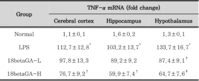
1. 뇌조직 TNF-α mRNA 발현의 변화
대뇌피질, 해마 및 시상하부 조직에서 TNF-α mRNA 발 현을 RT-PCR 방법으로 측정한 결과는 Table 2와 같다.
LPS군은 Normal군에 비해 모든 조직에서 매우 높은 유의성 (p<0.001)으로 TNF-α mRNA 발현이 증가하였으며, 18betaGA -L군은 LPS군에 비해 대뇌피질과 해마 조직에서는 유의한 변화가 없었고, 시상하부 조직에서만 TNF-α mRNA 발현 증 가가 유의하게(p<0.05) 억제되었다. 18betaGA-H군은 LPS 군에 비해 대뇌피질과 해마 조직에서는 p<0.05의 유의성, 시 상하부 조직에서는 p<0.01의 유의성 있게 TNF-α mRNA 발현 증가를 억제하였다(Table 2).
2. 뇌조직 IL-1β mRNA 발현의 변화
대뇌피질, 해마 및 시상하부 조직에서 IL-1β mRNA 발현을
RT-PCR 방법으로 측정한 결과는 Table 3과 같다. LPS군은
Normal군에 비해 모든 조직에서 매우 높은 유의성(p<0.001)
으로 IL-1β mRNA 발현이 증가하였다. 18betaGA-L군은
LPS군에 비해 대뇌피질과 해마, 시상하부 조직 모두에서 유
의한 변화가 없었고, 18betaGA-H군은 대뇌피질과 해마, 시
상하부 조직 모두에서 LPS군에 비해 IL-1β mRNA 발현 증
가가 유의하게(p<0.05) 억제되었다(Table 3).
Group
TNF-α mRNA (fold change)
Cerebral cortex Hippocampus Hypothalamus
Normal 1.1±0.1 1.6±0.2 1.3±0.1
LPS 112.7±12.8* 103.2±13.7* 133.7±16.7* 18betaGA-L 97.8±13.3 89.2±9.2 87.4±9.1†
18betaGA-H 76.7±9.2† 59.9±7.4† 64.7±7.6‡
Normal : group without the LPS injection and 18β-glycyrrhetinic acid treatment.
LPS : group of LPS injection without 18betaGA treatment.
18betaGA-L : group treated with 30 mg/kg of 18betaGA prior to the LPS injection.
18betaGA-H : group treated with 100 mg/kg of 18betaGA prior to the LPS injection.
Data are presented as mean ± standard error (n = 6 in each group; student t-test between the Normal and LPS groups (*, p<0.001) and between the LPS and 18betaGA groups (†, p<0.05; ‡, p<0.01; §, p<0.001).
Table 2. Changes of TNF-α mRNA Expression in the Brain Tissue of Mice Treated with Lipopolysccharide
Group
IL-1β mRNA (fold change)
Cerebral cortex Hippocampus Hypothalamus
Normal 1.2±0.2 0.9±0.1 1.3±0.2
LPS 114.3±12.7* 98.9±10.7* 141.3±20.7* 18betaGA-L 104.1±13.2 90.3±10.8 105.1±12.7 18betaGA-H 80.5±8.1† 67.2±7.2† 86.3±12.4†
Normal : group without the LPS injection and 18β-glycyrrhetinic acid treatment.
LPS : group of LPS injection without 18betaGA treatment.
18betaGA-L : group treated with 30 mg/kg of 18betaGA prior to the LPS injection.
18betaGA-H : group treated with 100 mg/kg of 18betaGA prior to the LPS injection.
Data are presented as mean ± standard error (n = 6 in each group; student t-test between the Normal and LPS groups (*, p<0.001) and between the LPS and 18betaGA groups (†, p<0.05; ‡, p<0.01; §, p<0.001).
Table 3. Changes of IL-1β mRNA Expression in the Brain Tissue of Mice Treated with Lipopolysccharide
3. 뇌조직 IL-6 mRNA 발현의 변화
대뇌피질, 해마 및 시상하부 조직에서 IL-6 mRNA 발현을 RT-PCR 방법으로 측정한 결과는 Table 4와 같다. LPS군은 Normal군에 비해 모든 조직에서 매우 높은 유의성(p<0.001) 으로 IL-6 mRNA 발현이 증가하였다. 18betaGA-L군은 LPS군에 비해 대뇌피질과 해마, 시상하부 조직 모두에서 유 의한 변화가 없었고, 18betaGA-H군 역시 대뇌피질과 해마 조직에서는 유의한 변화가 없었으며 시상하부 조직에서만 LPS군에 비해 IL-6 mRNA 발현 증가가 유의하게(p<0.05) 억제되었다(Table 4).
4. 뇌조직 COX-2 mRNA 발현의 변화
대뇌피질, 해마 및 시상하부 조직에서 COX-2 mRNA 발 현을 RT-PCR 방법으로 측정한 결과는 Table 5와 같다.
LPS군은 Normal군에 비해 모든 조직에서 매우 높은 유의성
(p<0.001)으로 COX-2 mRNA 발현이 증가하였다. 18betaGA- L군은 LPS군에 비해 대뇌피질과 해마, 시상하부 조직 모두에서 유의한 변화가 없었고, 18betaGA-H군은 대뇌피질과 시상하부 조직에서는 LPS군에 비해 유의한(p<0.05) COX-2 mRNA 발현 증가가 억제되었으나 해마 조직에서는 변화가 없었다 (Table 5).
Group
IL-6 mRNA (fold change)
Cerebral cortex Hippocampus Hypothalamus
Normal 1.1±0.1 1.1±0.2 0.9±0.1
LPS 11.5±2.1* 12.7±1.8* 13.6±1.9*
18betaGA-L 9.8±1.6 9.3±1.4 11.1±1.2
18betaGA-H 6.3±1.4 7.8±1.7 7.4±1.8†
Normal : group without the LPS injection and 18β-glycyrrhetinic acid treatment.
LPS : group of LPS injection without 18betaGA treatment.
18betaGA-L : group treated with 30 mg/kg of 18betaGA prior to the LPS injection.
18betaGA-H : group treated with 100 mg/kg of 18betaGA prior to the LPS injection.
Data are presented as mean ± standard error (n = 6 in each group; student t-test between the Normal and LPS groups (*, p<0.001) and between the LPS and 18betaGA groups (†, p<0.05; ‡, p<0.01; §, p<0.001).
Table 4. Changes of IL-6 mRNA Expression in the Brain Tissue of Mice Treated with Lipopolysccharide
Group
COX-2 mRNA (fold change)
Cerebral cortex Hippocampus Hypothalamus
Normal 1.0±0.1 0.9±0.1 0.9±0.1
LPS 7.8±0.9* 4.3±0.7* 8.2±1.1*
18betaGA-L 6.3±0.6 3.7±0.4 7.5±0.6
18betaGA-H 5.5±0.4† 2.5±0.4† 5.2±0.4†
Normal : group without the LPS injection and 18β-glycyrrhetinic acid treatment.
LPS : group of LPS injection without 18betaGA treatment.
18betaGA-L : group treated with 30 mg/kg of 18betaGA prior to the LPS injection.
18betaGA-H : group treated with 100 mg/kg of 18betaGA prior to the LPS injection.
Data are presented as mean ± standard error (n = 6 in each group; student t-test between the Normal and LPS groups (*, p<0.001) and between the LPS and 18betaGA groups (†, p<0.05; ‡, p<0.01; §, p<0.001).
Table 5. Changes of COX-2 mRNA Expression in the Brain Tissue of Mice Treated with Lipopolysccharide
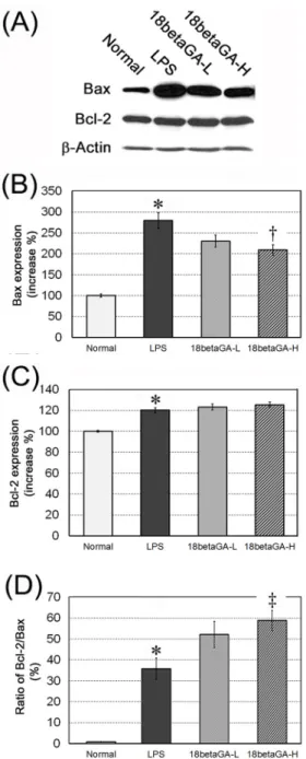
5. 해마 조직에서 Bax와 Bcl-2 발현의 변화
해마 조직에서 Western blotting 방법으로 Bax 발현을 측 정한 결과, Normal군의 100.0±4.2%에 비해 LPS군은 279.7
±19.0%로 Normal군에 비해 유의성(p<0.001) 있는 증가를
나타내었다. 이에 비해 18betaGA-L군은 230.6±14.2%로
LPS군에 비해 Bax 발현이 억제되지 않았으며, 18betaGA-H
군은 209.1±12.5%로 LPS군에 비해 Bax 발현이 유의하게
(p<0.05) 억제되었다(Fig. 2A, B). Bcl-2 발현은 Normal군
의 100.0±0.8%에 비해 LPS군은 120.3±2.3%로 Normal
군에 비해 유의성(p<0.001) 있는 증가를 나타내었다. 이에 비 해 18betaGA-L군은 123.2±2.9%로, 18betaGA-H군은 125.5±2.5%로 LPS군에 비해 Bcl-2 발현은 모두 유의한 변 화가 없었다(Fig. 2A, C). Bax 발현에 대한 Bcl-2 발현 비율 을 계산한 결과는 LPS군은 35.8±5.1%, 18betaGA-L군은 52.2±6.2%로 LPS군에 비해 유의한 차이가 없었으며, 18betaGA-H군은 58.9±4.8%로 LPS군에 비해 Bcl-2의 상대적 발현 비율이 유의성 있게(p<0.01) 높은 것으로 관찰되 었다(Fig. 2D).
Fig. 2. Effect of 18β-glycyrrhetinic acid (18betaGA) on Bax and Bcl-2 expression in the hippocampus. Representative western blots illustrating differences in the bands of Bax and Bcl-2 (A).
High-dose of 18betaGA significantly attenuated the Bax expression in the hippocampus. Bcl-2 expression was not different, but ratio of Bcl-2/Bax expression was significantly atennuated in the 18betaGA-H group. Data are represented by mean ± SEM (n=6 in each group). Statistical significances were compared between the Normal and LPS groups (*, p<0.001) and between the LPS and 18betaGA groups (†, p<0.05; ‡, p<0.01).
6. 해마 CA1 두께 및 신경세포 수의 변화
해마 CA1의 두께를 측정한 결과, Normal군은 54.0±2.1 ㎛, LPS군은 40.3±1.8 ㎛로 Normal군에 비해 유의성(p<0.001) 있는 감소를 나타내었다. 이에 비해 18betaGA-L군은 43.6
±2.2 ㎛로 LPS군에 비해 CA1 두께의 감소가 억제되지 못하 였고, 18betaGA-H군은 48.9±2.6 ㎛로 LPS군에 비해 CA1 두께의 감소가 유의하게(p<0.05) 억제되었다(Fig. 3A, B).
CA1의 500 ㎛ 길이에서 측정한 신경세포 수는 Normal군에서 188.3±2.3 개/500 ㎛, LPS군은 155.0±4.2 개/500 ㎛로 Normal군에 비해 유의성(p<0.001) 있는 감소를 나타내었다.
18betaGA-L군은 160.7±2.7 개/500 ㎛로 LPS군에 비해 신경세포 수의 감소가 억제되지 못하였고, 18betaGA-H군은 173.3±3.9 개/500 ㎛로 LPS군에 비해 신경세포 수의 감소 가 유의하게(p<0.05) 억제되었다(Fig. 3A, C).
Fig. 3. Effects of 18β-glycyrrhetinic acid (18betaGA) on the CA1 region in the hippocampus. Representative photographs of the cresyl violet-stained CA1 region (A). High-dose of 18betaGA significantly attenuated the reduction of the thickness of CA1 (B) and the number of CA1 neurons (C). Scale bar in the section A is 100 ㎛. Data are represented by mean ± SEM (n=6 in each group).
Statistical significances were compared between the Normal and LPS groups (*, p<0.001) and between the LPS and 18betaGA groups (†, p<0.05).
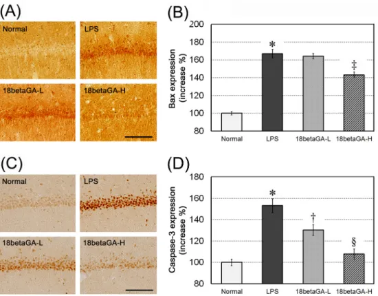
7. 해마 CA1 구역에서 Bax 및 caspase-3 발현의 변화
해마 CA1 신경세포에서 면역조직화학 반응에 의한 Bax 발현 강도의 증가율을 Normal군의 발현을 기준으로 측정한 결과, Normal군의 100.0±1.7%에 비해 LPS군은 167.1±4.8%로 Normal군에 비해 유의성(p<0.001) 있는 증가를 나타내었다.
이에 비해 18betaGA-L군은 164.3±2.9%로 LPS군에 비해 Bax 발현이 억제되지 못하였고, 18betaGA-H군은 143.2±
3.1%로 LPS군에 비해 Bax 발현이 유의하게(p<0.01) 억제되 었다(Fig. 4A, B). Caspase-3 발현 강도는 Normal군의 100.0±3.1%에 비해 LPS군은 153.2±6.4%로 Normal군에 비해 유의성(p<0.001) 있는 증가를 나타내었다. 이에 비해 18betaGA-L군은 130.3±5.1%로 LPS군에 비해 유의성 (p<0.05) 있는 caspase-3 발현의 억제를 나타내었으며, 18betaGA-H군은 107.9±4.8%로 역시 LPS군에 비해 caspase-3 발현이 유의하게(p<0.001) 억제되었다(Fig. 4C, D).
Fig. 4. Effects of 18β-glycyrrhetinic acid (18betaGA) on Bax and caspase-3 expressions in CA1 of the hippocampus. Representative photographs of the immuno-stained against Bax (A) and caspase-3 antibodies (C). High-dose of 18betaGA significantly attenuated the up-regulation of the Bax (B) and caspase-3 expression (D) in CA1 of the hippocampus. Scale bars in the section A and C are 100 ㎛.
Data are represented by mean ± SEM (n=6 in each group). Statistical significances were compared between the Normal and LPS groups (*, p<0.001) and between the LPS and 18betaGA groups (†, p<0.05; ‡, p<0.01; §, p<0.001).
Ⅳ. 고 찰
중추신경계에 대한 사이토카인의 작용은 전신적인 염증반 응에 의해 중추신경계 외부에서 분비된 사이토카인이 혈액뇌 관문을 통과하여 중추신경계 내로 들어와 작용하거나, 중추신 경계 내에서 생산 분비되어 작용한다
16). 어떠한 사이토카인이 든지 신경내분비계통에 영향을 미쳐 발열성(pyrogenic), 식 욕감퇴성(anorectic) 및 기면성(lethargic) 행동 양상의 변화 를 유발한다
17). 특히 TNF-α와 IL-1β 같은 염증성 사이토카 인은 알츠하이머병(Alzheimer’s disease), 다발성경화증, 뇌허혈, 뇌경색, 다양한 뇌병증(encephalopathies) 들에서 분비가 증가한다
14,18). 이러한 염증성 사이토카인의 과분비는 중추신경계에 신경독성으로 작용하고, 퇴행성 뇌질환을 악화 시키는 요인이 된다
19).
18betaGA의 항염증 및 사이토카인에 대한 연구들을 살펴 보면, 18betaGA가 mouse macrophages RAW264.7과
peritoneal macrophages에서 LPS 자극에 의한 TNF-α 분
비를 억제하고, 12-O-tetradecanoyl- phorbol-13-
acetate-(TPA-)에 의한 급성염증을 유의하게 억제하였으며
20),
macrophage에서 LPS에 의한 TNF-α , IL-6, IL-1β 분비
를 억제하고, inducible nitric oxide synthase (iNOS)와
COX-2의 단백질과 mRNA 발현 증가를 유의하게 억제하였
다
21). 또한 18betaGA가 혈관내피세포에서 NF-kB 발현을
줄이고 이에 따라 IL-8 발현도 감소되어, LPS에 의한 혈관
내피세포의 투과성 증가를 억제하는 항염증 작용을 발휘한다고
하였으며
22), dendritic cells에서 LPS 자극에 의한 surface
molecules cluster differentiation (CD)80, CD86 및 major
histocompatibility complex (MHC) class I과 MHC class
II 발현을 억제하는 항염증 작용을 나타내었다고 하였다
23).
이와 같은 18betaGA의 항염증 및 사이토카인에 대한 연구들은
주로 그 작용기전을 관찰하기 위해
in vitro세포실험을 통해
이루어졌으며, 18betaGA의 중추신경염증에 대한
in vivo연
구는 C57BL/J6 mouse의 뇌허혈 손상에 대해 지질과산화 억제 등의 항산화 작용을 발휘한다는 보고
5)와 C57BL/J6 mouse 의 자가면역성 뇌척수염 모형에서 미세아교세포 활성화를 억 제하여 염증을 줄인다는 보고가 있을 뿐이다
7). 그러므로 본 연구에서는 18betaGA가
in vivo동물실험에서 LPS에 의한 중추신경염증에서 대뇌피질, 해마 및 시상하부의 뇌조직 별로 TNF-α , IL-1β 및 IL-6의 mRNA 발현에 미치는 영향을 관 찰하였다. 그 결과 18betaGA는 30 ㎎/㎏의 저농도에서는 시 상하부 조직에서만 TNF-α mRNA 발현 증가를 유의하게 억 제하였다. 100 ㎎/㎏의 고농도에서는 대뇌피질과 해마 조직 에서 TNF-α와 IL-1β mRNA 발현 증가를 유의하게 억제하 였고, 시상하부 조직에서는 TNF-α , IL-1β와 IL-6 mRNA 발현 모두를 유의하게 억제하였다. 이러한 결과로 미루어보아 고농도의 18betaGA는 중추신경염증에서 전체적인 뇌조직의 염증성 사이토카인 발현 증가를 억제하는 작용이 있으며, 특 히 시상하부의 염증에 대해 효과적인 작용을 하는 것으로 추 측할 수 있다.
TNF-α는 뇌조직에서 모세혈관의 투과성을 증가시키고, 중성백혈구의 침윤을 증가시켜 염증반응을 유발하며
24), 신경 세포의 TNF-α receptor에 작용하여 TNF receptor- associated death domain을 작동시키므로 신경세포를 사망에 이르게 한다
25). 과도하게 발현된 IL-1β 역시 신경세포를 손 상시킨다
26). IL-6는 염증유발 및 염증억제의 이중적 작용을 가지고 있는 사이토카인이지만 과도한 IL-6의 발현은 허혈성 및 출혈성 뇌경색의 악화에 관여하고
27), 알츠하이머병에서 아 밀로이드플라크 주변의 염증반응을 악화시키는데 관여하며
28), 특히 시상하부에서 IL-6의 과발현은 체온조절 장애로 고열을 유발한다고 하였다
29). 그러므로 이러한 염증성 사이토카인의 작용과 이전
in vitro연구에서 보고된 18betaGA의 항염증 작용기전 및 본 연구의
in vivo결과들을 종합해보면 18betaGA는 중추신경계에 대한 항염증 작용으로 신경세포손 상에 대한 보호작용은 물론 대뇌피질과 해마 신경세포의 손상과 시상하부의 염증반응에 의한 질병행동의 개선 등에도 유의한 효능이 있을 것으로 추측된다.
나아가 18betaGA의 중추신경염증에 대한 항염증 작용을 확인하기 위해 뇌조직에서 COX-2 mRNA 발현을 관찰하였다.
COX-2는 급성 및 만성 염증에서 염증반응을 확산시키는데 중심적인 역할을 하는 효소로 염증성 사이토카인과 염증유발 매개물질들의 자극에 의해 빠른 시간 내에 발현이 촉진되어 prostaglandin을 생성하게 된다
30). 그러므로 COX-2 발현의 억제는 신경세포독성이나 퇴행성 뇌 병변에 대한 항염증 약물 개발의 유의한 목표가 된다
31). 본 실험의 결과, 저농도 18betaGA는 유의한 변화를 나타내지 못하였으나, 고농도 18betaGA는 대뇌피질과 시상하부 조직에서 COX-2 mRNA 발현 증가를 유의하게 억제하였다. 이러한 결과는 18betaGA가 중추신경염증에 대한 항염증 작용이 있다는 것을 보완하는 것 이다.
해마 신경세포 손상에 대한 18betaGA의 신경세포 보호효 능을 관찰한 본 연구의 결과, 18betaGA는 고농도에서 LPS에 의한 해마 CA1 두께의 감소와 CA1 신경세포 수의 감소를 유 의하게 억제하였다. 이는 18betaGA가 중추신경염증에 의한 해마 신경세포의 손상을 억제하는 작용이 있음을 추측하게 한다.
Bax와 Bcl-2는 세포자연사 기전을 조절하는 인자로서 Bax 는 세포자연사를 촉진하는 유발인자, Bcl-2는 세포자연사를 억제하는 인자로 알려져 있다
32). 그러므로 Bax 발현이 증가 하고 Bcl-2 발현이 감소되는 상태에서는 세포자연사가 유발 되고, Bax 발현이 억제되고 Bcl-2 발현이 증가하는 상태에 서는 세포자연사가 억제된다고 하였다
33). 18betaGA가 해마 조직에서 Bax와 Bcl-2 발현에 미치는 영향을 관찰한 결과, 18betaGA는 면역조직화학염색에서 해마 CA1 신경세포의 Bax 발현 강도 증가를 유의하게 억제하였으며, Western blotting으로 측정한 Bax 단백질 발현의 증가 또한 유의하게 억제하였다. Bcl-2 발현에 대해서는 유의한 변화가 없었으 나, Bax 발현에 대한 상대적 발현량을 관찰한 결과에서는 고 농도의 18betaGA가 Bcl-2/Bax의 발현비율을 유의하게 증 가시킨 것을 관찰할 수 있었다. 이러한 결과는 Bcl-2/Bax의 비율이 세포자연사 기전에서 세포의 사망 또는 생존을 결정하는 지표가 된다는 보고
34)를 감안하면 18betaGA가 신경세포손상 보호효능이 있다는 것을 보완하는 결과이다. Caspase-3은 세포자연사를 실행하는 주요 인자이다
35). 세포자연사 유발인 자인 Bax가 세포내의 mitochondria에 작용하여 막투과성을 증가시키고, cytochrome C를 생성하게 되며, 생성된 cytochrome C는 caspase-9로부터 caspase-3을 생성한다
36). Caspase-3 은 caspase-activated DNase를 생성하여 세포핵의 DNA를 조각내고, 세포핵에 작용하여 염색질의 응축 (chromatin condensation)을 일으키며, 세포골격을 분해하여 세포가 사 망하게 하는 자연사 기전을 유발한다
37). Caspase-3 발현세 포를 해마 조직에서 면역조직화학염색으로 관찰한 결과, 고농 도의 18betaGA는 LPS에 의한 해마 CA1 신경세포의 caspase-3 발현 강도 증가를 유의하게 억제하였다. 이러한 결과 역시 18betaGA가 신경세포손상 보호효능이 있다는 것 을 보완하는 결과이다.
본 연구의 결과들을 종합하면, 고농도(100 ㎎/㎏)의 18betaGA는 대뇌피질과 시상하부 조직에서 LPS에 의한 TNF-α , IL-1β, IL-6 mRNA 발현 증가를 유의하게 억제하 고, COX-2 mRNA 발현 증가를 유의하게 억제하였으며, 해 마 조직에서 LPS에 의한 CA1 신경세포 사망을 억제하고, Bax 발현과 caspase-3 발현 증가를 유의하게 억제하였다.
그러므로 18betaGA는 중추신경염증에 대해 항염증작용과 신 경세포손상에 대해 유의한 보호작용이 있는 것을 추측할 수 있으며, 중추신경염증을 수반하는 각종 뇌 병변을 예방 또는 치료하는데 유의한 약물이 될 수 있음을 추측하게 한다.
Ⅴ. 결 론
甘草의 소화기관 내 대사체인 18betaGA가 뇌조직의 염증
상태에서 염증성 사이토카인 발현과 해마 신경세포 자연사에
대한 영향을
in vivo동물실험에서 관찰하고자 하였다. 생쥐에
LPS를 복강주사하는 방법으로 염증을 유발하고 대뇌피질, 해마
및 시상하부 뇌조직에서 TNF-α와 IL-1β 및 IL-6의 mRNA
발현을 관찰하였으며, 염증매개 효소인 COX-2 발현을 관찰
하였다. 또한 해마 조직에서 신경세포 손상과 Bax, Bcl-2 및
caspase-3 발현을 관찰한바 아래와 같은 결과를 얻었다.
1. 18betaGA는 LPS에 의한 대뇌피질, 해마 및 시상하부 조직의 TNF-α mRNA 발현 증가를 유의하게 억제하였다.
2. 18betaGA는 LPS에 의한 대뇌피질, 해마 및 시상하부 조직의 IL-1β mRNA 발현 증가를 유의하게 억제하였다.
3. 18betaGA는 LPS에 의한 시상하부 조직의 IL-6 mRNA 발현 증가를 유의하게 억제하였다.
4. 18betaGA는 LPS에 의한 대뇌피질, 해마 및 시상하부 조직의 COX-2 mRNA 발현 증가를 유의하게 억제하였다.
5. 18betaGA는 LPS에 의한 해마 조직의 Bax 발현 증가를 유의하게 억제하였으며, Bax에 대한 Bcl-2의 상대적인 발현 비율을 유의하게 증가시켰다.
6. 18betaGA는 LPS에 의한 해마 CA1 두께의 감소를 유 의하게 억제하고, CA1 신경세포 수의 감소를 유의하게 억제하였다.
7. 18betaGA는 LPS에 의한 해마 CA1 신경세포의 Bax와 caspase-3 발현 강도 증가를 유의하게 억제하였다.
이상의 결과로 보아 고농도 18betaGA는 뇌조직의 염증성 사이토카인 발현 증가를 억제하고, 해마 신경세포의 자연사를 억제하는 작용이 있으므로 중추신경염증을 수반하는 다양한 만성적인 뇌질환에 유의한 약물이 될 수 있을 것으로 생각된다.
참고문헌