Original Articles 순환기:순환기:순환기:순환기:제제제 2 8 권제 권권 제권 제제제 1 호호호 1998호
Cisapride가 심근의 ATP-sensitive K 통로에 미치는 영향
전북대학교 의과대학 순환기내과학교실
문성기·인병현·김원호·고재기
=
=
=
= Abstract = = = =
Effect of Cisapride on ATP-sensitive K Channel of Ventricular Cell
Sung-Gi Moon, M.D., Byeong-Hyun In, M.D., Won-Ho Kim, M.D., Jae-Ki Ko, M.D.
Department of Cardiology, College of Medicine, Chonbuk National University, Chonju, Korea
Background:It has been generally accepted that Cisapride(Prepulsid
®or Propulsid
®), a widely used gastrointestinal prokinetic agent, is associated with Torsades de Points, a life-threatening arrhythmia.
Recently, cisapride-induced APD(action potential duration)-prolongation was inhibited by gliben- clamide, a K
ATPchannel blocker. But the direct effect of cisapride on K
ATPchannels has not been studied until now.
Therefore, we investigated cisapride’s effects on K
ATPchannels of isolated rat ventricular myocytes.
Method:After the isolation of rat ventricular myocytes, we analysed the single channel current with patch pipettes. The method of analysis was the student t-test.
Result:
1) Cisapride(10
-6M-10
-4M) inhibited K
ATPchannel opening without changing channel conductance K
iwas about 20μM, and Hill coefficient was 0.75.
2) Cisapride inhibited pinacidil-induced K
ATPchannel opening in the cell attached mode.
Conclusion:These results suggest that cisapride-induced APD prolongation and arrythmic effects may be partly related to K
ATPchannel inhibition.
KEY WORDS:Cisapride·K
ATPchannel·Ventricular cell.
서 론
2-methoxy-4-amino-5-chloro-substituted benzamide계 약물은 위, 식도역류, 위마비, 기능적 소 화불량과 변비 등의 위장계 운동질환의 치료에 사용
되는 약물이다1-3). Cisapride는 이중 대표약물의 하나 이며, 이 계통 약물은 5-HT4 수용체 작용물질로 알 려져 있고4,5), 콜린성 흥분 신경전달을 증가시키는 것 으로 보고되었다6). Cisapride는 돼지의 심방에서 5- HT4 수용체를 경유하여 빈맥을 발생시키고7) 이 작용 은 c-AMP 증가와 protein kinase A와 연관되어 있
음이 보고되었다8,9). 최근 분자생물학적인 연구에 의 해 심장에 5-HT4 수용체가 cloning되었고, 이 수용 체의 m-RNA를 발현시켰을 때 adenyl cyclase를 자 극함이 알려져 있다10). 이와 같은 cisapride의 심장에 서의 작용은 주로 심방에 대한 보고이며, long QT syndrome을 일으키고11), 심실부정맥을 일으킨다는 보고12), 퍼킨제 섬유에서 활동전위기간을 연장시키고, 조기탈분극을 일으킨다는 보고가 있다13).
최근에는 cisapride의 활동전위기간연장이 KATP 통 로차단제인 glibenclamide에 의해 약화됨이 보고되어14), cisapride가 KATP 통로의 활성 조절에 관여할 가능성 을 시사하였다. 따라서 저자는 본 실험에서 rat ven- tricular myocyte를 대상으로 cisapride가 KATP 통로 의 활성에 미치는 영향을 관찰하였다.
대상 및 방법
1. 단일심근세포의 분리
체중 250g 내외의 Sprague Dawley종 rat를 두부 타격으로 혼절시킨 후 가능한 빨리 Langendorff 장치 에 적출심장을 매달고 37℃의 Krebs-Henseleit 용액 (조성;118mM NaCl, 5.7mM KCl, 1.2mM MgSO4, 1.2mM KH2PO4, 10mM HEPES, 25mM NaHCO3, 10mM pyruvate, 11mM dextrose 및 1mM CaCl2) 을 분당 4ml의 속도로 관상동맥을 통해 5분간 관류시 켰다. 이 후 심장 박동이 멈출 때까지 Ca2+-free Krebs-Henseleit 용액으로 관류시킨 후 collagenase (Worthington) 22.5mg/30ml를 포함한 Ca2+-free Krebs-Henseleit 용액으로 관류시켜 소화시켰다. 소 화된 심장을 떼어내어 1% albumin을 포함한 Ca2+- free Krebs-Henseleit 영양액에서 가볍게 흔들어 단 일심근세포를 분리하고, 이와같이 분리된 세포들은 얼 음으로 냉각시킨 albumin 1%를 함유 한 Ca2+-free Krebs-Henseleit 용액에 보관하여 실험에 사용하였 다. 한편, 실험전에 세포를 도립현미경(inverted mi- croscope, Reichert-Jung, Biostar)의 대물대에 설 치한 bath에 도포한 후 막대기 모양의 무늬가 분명하 며 윤곽이 뚜렷한 세포들만 골라 실험에 사용하였다.
2. 미세전극 제작
단일통로전류기록에 사용한 미세전극(patch pipette)
은 내경 1.5mm의 borosilicate 유리관(Kimble, Ki- max-51, 1.5 -1.8×100mm, 미국)을 미세전극제작 기로(2-stage pipette puller, Narishige, PP-83, 일 본) 미세전극의 저항이 3~4MΩ 정도되게 뽑은 다음, 해부현미경을(stereozoom-microscope) 이용하여 가 능한 한 미세전극 말단까지 sylgard를 도포한 후 가 열된 니크롬선하에서 약 10초간 노출시켜 건조시켰다.
이렇게 실리콘이 도포된 미세전극의 말단을 현미경 시 야에서 열처리하여 단일통로기록에 사용하였는데 이 때 전극의 저항이 4MΩ 정도가 되도록 하였다.
3. 단일통로 전류의 기록
단일통로전류는 gigaohm-seal patch clamp 방법 중에서 cell atlach patch 및 excised inside-out patch 방법(Hamil 등 1981)으로 전기적인 신호를 patch clamp 증폭기(Axon Instruments, Axopatch- 1D, 미국)를 통하여 디지탈신호변환장치(digital pulse code modulator, Sony, PCM-501ES, 일본)를 거쳐 비디오(Gold Star, GHV-9000, 한국)에 녹화하였으 며, 모든 실험은 상온에서 시행하였다. 또한, 단일통로 전류를 분석할 때 cut-off frequency를 20K Hz로, 피지오그래프(Gould, 3400, 미국)에 기록할 때에는 300Hz로 여과하여 입력하였다. 한편, 단일통로의 기 록에 이용한 미세전극은 K-5 용액(조성;140mM KCl, 2mM MgCl2, 5mM EGTA 및 10mM HEPES, pH 7.2)으로 채웠으며, bath 용액 역시 K-5 용액을 사용하였다.
4. 단일통로전류의 자료분석
비디오 테이프에 저장된 단일통로기록들은 A/D converter(Digidata 1200, Axon Instruments, 미국) 를 이용하여 컴퓨터(Hyundae, 4038DX, 한국)에 저 장하여 분석하였다. 단일통로전류의 open probability (Po)는 Spruce등15)에 의해 유도된 공식을 이용하였다.
Po=(
∑
j=1 N
tj·j )/Td・N)
즉, 이 식에서 t j는(j=1, 2, 3, ...) N개의 통로가 열릴 때 각각의 전류단계에서의 통로가 열려 있는 시 간, j는 각 level에서 열려 있는 통로의 수, T d는 단 일통로전류를 기록한 시간, N은 control 상태에서
(ATP-free)에서 최대로 활성화되는 통로의 수를 가 리키며 본 실험에서 Po는 60초 이상의 연속된 단일통 로기록들을 사용하여 계산하였다. 한편, relative open probability는 컴퓨터와 pClamp를 이용하여 산출한 각각의 Po를 약물투여전의 단일통로 전류의 Po에 대 한 백분율로 표시하였다.
4. 통계학적 분석
통계학적 유의성 검정에는 Student’s t-test를 적 용하였다.
결 과
1. K
ATP통로에 대한 세포내의 ATP 및 gli- benclamide의 효과
본 실험에서 측정한 단일통로전류가 선택적인 KATP
통로를 경유한 전류인지, ATP 및 glibenclamide에 의해 얼마나 차단되는지 알아보기 위하여 excised inside-out patch 방법으로 단일통로 전류에 대한 여 러농도의 ATP 영향과(Fig. 1A) glibenclamide의 영 향을(Fig. 1B) 관찰하였다. ATP를 0.01, 0.03, 0.1, 0.3, 1mM로 증가시킴에 따라 단일통로 활성이 농도 에 비례하여 감소하였다. 이러한 ATP의 KATP 통로 억제에서, Hill 방정식, ralative open probability=
1/(1+([cisapride]/Ki)n)에서 50%의 억제효과를 나 타내는 ATP의 농도, Ki는 82.1μM이었으며, Hill coefficient, n은 1.96 이었다. 또한이 통로는 KATP
통로 차단제인 glibenclamide 0.03, 0.1, 0.3, 1, 3μM 에서 농도의존적인 활성의감소를 보였으며 이때 Ki는 3.71μM이었다. 이러한 ATP 및 선택적 KATP 통로 차단제 glibenclamide에 의한 통로의 억제효과를 통하 여 실험에 이용되는 통로가 KATP 통로임을 알 수 있었다.
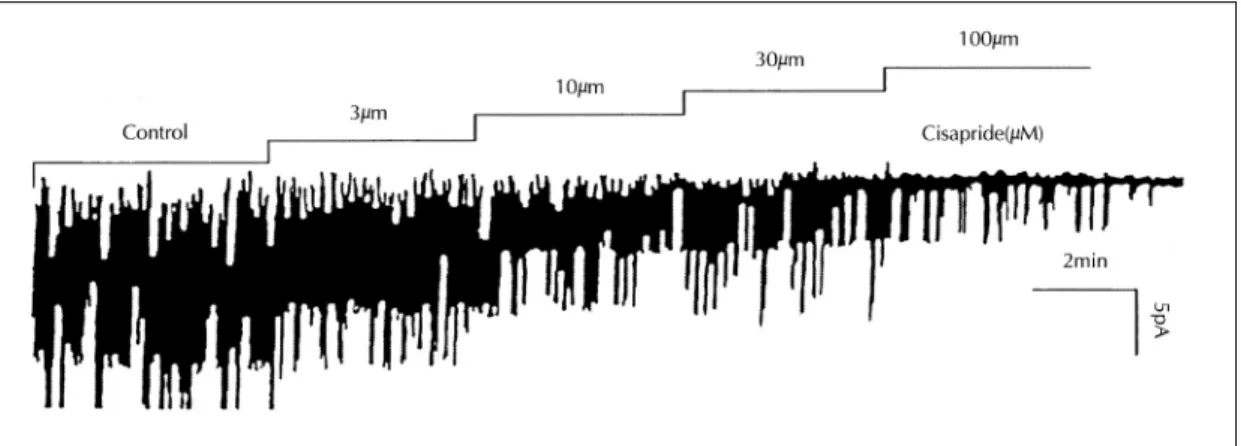
Fig. 2. Effects of intracellular cisapride on KATP channel activity cisapride inhibits KATP channel activity dose- dependently in excised inside-out membrane patches. Drug was treated in bath solution. Records were obtained as described in Fig. 2.
Fig. 1. Inhibitory effect of ATP (A) and glibenclamide (B) on KATP channel activity. obtained from isolated single rat ventricular myocyte. Records were made from excised inside-out membrane patches obtained from isolated single rat ventricular my- ocyte. In this and following figures, the membrane potentials was held at -60mV;dashed lines represent closed level for KATP channels. The current records were filtered at 300Hz.
2. K
ATP통로에 대한 세포내 cisapride의 영향
KATP 통로를 excised inside out patch 방법으로 기록하면서 세포내에 cisapride를 3, 10, 30, 100μM 로 증가시킴에 따라 용량 의존적으로 KATP 통로의 활
성을 억제하였다(Fig. 2). 세포내 cisapride에 의한 KATP 통로의 이러한 억제는 실험된 6예의 모든 patch 에서 관찰되었다. Cisapride에 의한 KATP 통로의 억 제효과를 용량-반응곡선으로 표시하기 위하여 대조군 에서 관찰되는 열림확률을 1로 할 때 cisapride 투여
Fig. 3. Dose-response relationship between relative ch- annel activity and concentration of cisapride.
Solid line is computer fit to Hill equation;y=
1/{1+([cisapride]/Ki)n}, where Ki is the concent- ration of cisapride causing half-maximal inhibition and n is the Hill coeffcient. Parameter values for the best fit are;Ki=1.793×10⁻⁵, n=0.75.
Fig. 4. Effects of intracellular cisapride on unitary con- ductance of KATP channels. Unitary currents were measured at various membrane potentials in the absence and presence of cisapride. Respective slope conductances of inward currents are 61.7, 62.5, 61.8 and 62.3pS in the presence of 0, 1, 3 and 10μM cisapride.
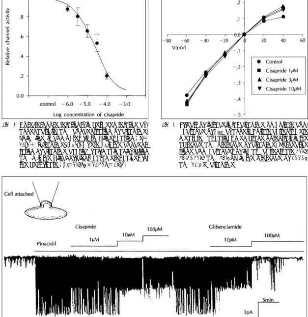
Fig. 5. Effects of extracellular cisapride on KATP channel activity. Cisapride inhibit pinacidil-induced KATP channel activity dose-dependently in cell-attached membrane patches. Drug was treated in bath solution. Records were obtained at membrane potentials, -60mV.
후 관찰되는 열림확률을 Hill 방정식으로 computer fitting하였다(Fig. 3). Hill 방정식, relative open probability=1/(1+([cisapride]/Ki)n)에서 50%의 억제효과를 나타내는 cisapride의 농도, Ki는 23.2μM 이었으며, Hill coefficient, n은 0.75였다.
3. K
ATP통로의 전도도에 미치는 세포내 ci- sapride의 영향
세포내의 cisapride가 K ATP 통로의 전도도에 미 치는 영향을 알아보기 위하여 cisapride 0, 1, 3, 10μM 존재하에서 전류-전압관계 그래프를 조사하였다. Ci- sapride 1, 3, 10μM을 축적적으로 투여한 후 각각의 전류-전압관계 그래프의 기울기로 얻어진 slope con- ductance는 각각 62.5, 61.8, 62.3pS으로 cisapride 투여 이전의 61.7pS과 유의한 차이가 없었다(Fig. 4).
한편, KAPT 통로의 전압-전류관계 그래프에서 관찰되 는 inward rectification이 +40mV에서 관찰되었다.
이러한 rectification은 cisapride 1, 3, 10μM 존재하 서도 대조군에서와 같은 정도로 관찰되었다.
4. Potassium channel opener, pinacidil의 K
ATP통로 개구에 미치는 cisapride의 영향
Cell attached mode patch에서 bath 내에 pota- ssium channel opener인 pinacidil 100μM을 투여하 여 KATP 통로 개구를 유도한 후 bath 내에 cisapride 1, 10, 100μM을 투여하였던 바 KATP 통로의 억제가 관찰되었다. 1, 10, 100μM cisapride에서 통로의 억 제 정도는 각각 77.8, 44.6, 35.6%였다. 한편, 이러한 억제는 세척에 의해 일부 회복되었다(Fig. 5).고 안
본 실험에서 excised inside-out patch mode에 서 세포내측에 투여된 cisapride는 dose-dependent 하게 KATP channel을 억제함을 관찰하였다. 이는 cisapride가 colliculi 신경세포에서 cyclic AMP- dependent protein kinase를 활성화시켜 voltage dependent K+ 통로를 억제함이 보고된 바16), 심장에 서도 cisapride의 K+ 통로 조절작용이 있음을 보여주 는 것이다.
Cell attached mode에서 pinacidil에 의해 개구된 KATP 통로가 bath내에 투여한 cisapride에 의해서 억
제된 것은 cisapride가 세포막을 투과한 후 세포막쪽 에서 KATP 통로를 차단할 수 있음을 보여주고 있고, inside-out configuration에서 실험한 결과가 유의함 을 보여주는 것이었다.
이러한 cisapride의 KATP 통로억제는 cisapride에 의한 심근 활동전위기간의 증가가 glibenclamide에 의하여 억압된다는 보고14)에 비추어 cisapride에 의 한 활동전위기간의 증가나 부정맥에 KATP 통로가 관 여할 수 있음을 시사하는 것이다.
Cisapride의 심근 KATP 통로억제효과는 curve fitting에서 1μM의 농도부터 대조군에 비하여 유의한 효과를 보였는데, 이 약량은 구강으로 치료목적으로 사용하였을 때 혈중 농도에 해당하는 약량이기 때문 에17) 실제 임상적으로도 중요할 것 같다. 특히, 심근 의 저산소증이나 허혈시 KATP 통로가 활동전위기간을 단축할 수 있다는 보고에서와 같이18,19), KATP 통로의 역할이 큰 상태에서 이러한 cisapride의 작용은 더욱 중요성을 더할 것으로 사료된다.
Cisapride의 K ATP 통로억제에서도 단일통로의 전도도는 변화하지 않는 것으로 보아 cisapride의 KATP 통로 억제는 전도도 이외의 다른 성질에 영향을 미치는 것으로 사료된다. 한편, 5-HT에 의한 혈관 수 축작용이 tyrosine kinase에 연관되어 있다는 보고20,21) 나, tyrosine kinase inhibitor인 genistein에 의해 cisapride의 APD 90 증가효과가 억제된다는 보고14) 등은 cisapride의 작용기전에 있어 tyrosine kinase 가 연관될 수 있음을 시사하고 있다. 따라서, cisa- pride에 의한 활동전위기간의 증가나 부정맥에 KATP
통로가 어떻게 관여할 수 있는지를 알기 위하여는 cisapride에 의한 KATP 통로의 억제기전에 명확한 규 명이 있어야 할 것이다. 이를 위하여는 cisapride에 의한 KATP 통로 특성의 변화 그리고 tyrosine kinase 를 비롯한 KATP 통로 조절인자와 cisapride의 관계에 대한 더 많은 연구가 필요할 것으로 사료된다.
요 약
연구배경:
Cisapride는 위식도역류, 기능적 소화불량과 변비 등의 위장계 운동질환의 치료에 널리 사용되는 약물 이다. 그러나 이와 같은 cisapride가 퍼킨제 섬유에서
활동전위기간을 연장시키고, 조기탈분극을 야기시켜 생명에 치명적인 long QT 증후군 및 심실부정맥을 일 으킨다는 보고가 있다. 최근에는 cisapride의 활동전 위기간연장이 KATP 통로차단제인 glibenclamide에 의 해 약화됨이 보고되어, cisapride가 KATP 통로의 활성 조절에 관여할 가능성을 시사하였다. 따라서 저자들은 rat ventricular myocyte를 대상으로 cisapride가 KATP 통로의 활성에 미치는 영향을 관찰하였다.
방 법:
체중 500g 내외의 쥐의 단일심근세포를 분리하여 미세전극에 의해서 단일통로 전류의 기록후 자료를 분 석하였다.
결 과:
1) 10-6-10-4 M의 cisapride에 의하여 KATP 통로 의 활성도가 억제되었고, Ki는 약 20μM이었다.
2) Cisapride에 의해 KATP 통로의 conductance는 변화가 없었다.
3) Cell attached mode에서 pinacidil에 의해 개구 된 KATP 통로는 cisapride에 의해 억제되었다.
결 론:
Cisapride에 의한 심장의 활동전위기간 증가효과와 부정맥의 일부가 KATP 통로 억제와 관계있음을 시사 하고 있다.
References
1) Reyntjens A, Verlinden M and Aerts T:Development and clinical use of the new gastrointestinal prokinetic drug cisapride(R 51 619). Drug Dev Res 8:251-265, 1986 2) M ller-Lissner SA:Treatment of chronic constipation with cisapride and placebo. Gut 28:1033-1038, 1987 3) Reynolds JC and Putnam PE:Prokinetic agents. Gast-
roenterol. Clin North Am 21:567-596, 1992
4) Bockaert J, Fozard JR, Dumuis A and Clarke DE:The 5-HT4 receptor:A place in the sun. Trends Pharmacol Sci 13:141-145, 1992
5) Ford APDW and Clarke DE:The 5-HT4 receptor. Med Res Rev 13:633-662, 1993
6) Taniyama K, Nakayama S, Takeda K, Matsuyama S, Shirakawa J, Sano I and Tanaka C:Cisapride stimulates motility of the intestine via the 5-hydroxytryptamine re- ceptors. J Pharmacol Exp Ther 258 :1098-1104, 1991 7) Kaumann AJ, Sanders L, Brown AM, Murray KJ and
Brown MJ:A 5-Hydroxytryptamine receptor in human atrium. Br J Pharmacol 100:879-885, 1990
8) Kaumann AJ, Sanders L, Brown AM, Murray KJ and Brown MJ:5-HT4-like receptor in human right atrium.
Naunyn-Schmied Arch Pharmacol 344:150-159, 1991a 9) Kaumann AJ, Brown AM and Raval P:Putative 5-HT4- like receptor in piglet left atrium. Br J Pharmacol 102: 98, 1991b
10) Gerald C, Adham N, Kao HT, Olsen MA, Laz TM, Schechter LE, Bard JA, Vaysse PJ, Hartig PR and Bra- nchek TA:The 5-HT4 receptor:Molecular cloning and pharmacological characterization of two splice variants.
EMBO J 14(12):2806-2815, 1995
11) Bran S, Murray WA, Hirsch AB and Palmer JP:Long QT syndrome during high-dose cisapride. Arch Intern Med 155:765-768, 1995
12) Ahmad SR and Wolfe SM:Cisapride and torsades de points. Lancet 345:508, 1995
13) Puisieux FL, Adamantidis MM, Dumotier BM and Dupuis BA:Cisapride-induced prolongation of cardiac action potential and early after depolarization in rabbit Purkinje fibers. Br J Pharmacol 117:1377-1379, 1996
14) 김성재:Effects of cisapride on action potential duration of guinea pig ventricular muscle. 전북대학교 대학원 의학과, 1997
15) Spruce AE, Standen NB and Standen PR:Voltage- dependent, ATP-sensitive potassium channels of skeletal muscle membrane. Nature London 316:736-738, 1985 16) Fagini L, Dumuis A, Sebben M and Bockaeret J:The 5-
HT4 receptor subtype inhibit K+ current in colliculi neu- rones via activation of a cyclic AMP-dependent protein kinase. Br J Pharmacol 105:973-979, 1992
17) Mccallum RW, Prakash C, Campoli-Richards DM and Goa KL:Cisapride. A preliminary review of its phar- macodynamic and pharmacokinetic properties and the- rapeutic use as a prokinetic agent in gastrointestinal motility disorder. Drugs 36:652-681, 1988
18) Cook DL and Hales CN:Intracellular ATP directly blocks K+ channels in pancreatic β-cells. Nature 311: 271-273, 1984
19) Castle NA and Haylett DG:Effect of channel blockers on potassium efflux from metabolically exahausted for frog skeletal muscle. J Physiol Lond 383:31-43, 1987 20) Watts SW, Yeum CH, Campbell G and Webb RC:
Serotonin stimulates protein tyrosyl phosphorylation and vascular contraction via tyrosine kinase. J Vasc Res 33 (4):288-298, 1996
21) Aiyar J, Grissmer S and Chandy KG:Full-length and truncated Kv1.3 K+ channels are modulated by 5-HT1C receptor activation and independently by PKC. Am J Physiol 265:C1571-1578, 1993