Thyroid Nodules with Macrocalcification:
Sonographic Findings Predictive of Malignancy
Yun Joo Park,
1,2Jeong-Ah Kim,
1Eun Ju Son,
1Ji Hyun Youk,
1Eun-Kyung Kim,
3Jin Young Kwak,
3and Cheong Soo Park
41Department of Radiology, Yonsei University College of Medicine, Gangnam Severance Hospital, Seoul;
2Department of Radiology, Soonchunhyang University Hospital, Soonchunhyang University College of Medicine, Seoul;
3Department of Radiology and Research Institute of Radiological Science, Yonsei University College of Medicine, Severance Hospital, Seoul;
4Department of Surgery, Yonsei University College of Medicine, Gangnam Severance Hospital, Seoul, Korea.
Received: May 23, 2013 Revised: July 9, 2013 Accepted: July 22, 2013
Corresponding author: Dr. Jeong-Ah Kim, Department of Radiology,
Yonsei University College of Medicine, Gangnam Severance Hospital, 211 Eonju-ro, Gangnam-gu, Seoul 135-720, Korea.
Tel: 82-2-2019-3510, Fax: 82-2-3462-5472 E-mail: [email protected]
∙ The authors have no financial conflicts of interest.
© Copyright:
Yonsei University College of Medicine 2014 This is an Open Access article distributed under the terms of the Creative Commons Attribution Non- Commercial License (http://creativecommons.org/
licenses/by-nc/3.0) which permits unrestricted non- commercial use, distribution, and reproduction in any medium, provided the original work is properly cited.
Purpose: To analyze which sonographic features of thyroid nodules with macrocal- cifications were predictable of thyroid malignancy. Materials and Methods: We reviewed sonographic findings of 854 macrocalcified thyroid nodules in patients who underwent fine needle aspiration biopsy between December 2009 and January 2011. There were 171 non-diagnostic aspirations, 34 nodules with category 3, 4, 5 based on Bethesda system, which were not confirmed by surgery, and these nodules were excluded from the analysis. Sonographic characteristics of the macrocalcifica- tions including its thickness, interruption, and existence of soft tissue rim outside the macrocalcification were analyzed. Other sonographic characteristics of nodules such as shape, margin, composition, echo pattern, vascularity, and underlying pa- renchymal echogenicity were also evaluated. The correlation of sonographic fea- tures with cytopathologic results and the diagnostic performance of sonographic features for the prediction of malignancy were analyzed. Results: Among 649 nod- ules, 179 (27.6%) nodules were malignant and 470 (72.4%) nodules were benign.
Among the features of the macrocalcification, interruption, irregular thickness, or the presence of soft tissue outside calcification rim were associated with malignan- cy (p<0.001). A high sensitivity and negative predictive values for the prediction of malignancy was found in sonographic characteristics of irregular thickness (92.2%
and 91.0%, respectively) and the presence of soft tissue (88.5% and 88.8%, respec- tively). Conclusion: Sonographic characteristics of macrocalcification such as in- terruption, irregular thickness and the presence of soft tissue rim were associated with malignancy in thyroid nodules with macrocalcifications.
Key Words: Sonography, papillary thyroid cancer, macrocalcification
INTRODUCTION
Evaluation of a thyroid nodule with sonography (US) is essential to determine whether it is likely to be benign or malignant and patients with malignant nodules can be properly diagnosed and treated. Several US characteristics that have been re-
system category 3 (atypia of undetermined significance), 4 (suspicious for a follicular neoplasm) or 5 (suspicious for malignancy) were excluded from the analysis due to the lack of definitive pathologic result. There were 171 (20.8%) non-diagnostic aspirations after FNAB that did not undergo subsequent surgery or without other cytologic results on re- peat FNAB, and these nodules were excluded from the analysis. A total of 649 nodules from 654 (535 women, 119 men, mean age 52 years±12.1) patients were included in the study for analysis.
Ultrasound imaging
All US examinations were performed with HDI 5000 (Phil- ips Advanced Technology Laboratories, Bothell, WA, USA), iU22 (Philips Medical Systems, Bothell, WA, USA) or Su- perSonic Imagine (Aix-en-Provence, France). Board-certi- fied radiologists specialized in thyroid imaging evaluated and recorded the following US findings of the lesion before FNAB and performed US-guided FNAB: size (maximal di- mension), shape (ovoid to round, taller-than-wide, or irregu- lar), margin (well-defined smooth, or irregular), composition (solid, cystic, mixed, or spongiform), echo pattern (hypere- choic, isoechoic, hypoechoic, or markedly hypoechoic), vascularity (peripheral, central, both, or absent) and under- lying parenchymal echogenicity (homogenous or heteroge- nous). The composition of a nodule was categorized ac- cording to the ratio of the cystic portion to the solid portion in the nodule. A solid nodule was defined when more than 90% of the nodule was solid, and a cystic nodule was defined when more than 90% of the nodule was cystic. A spongiform appearance was defined as the aggregation of multiple mi- crocystic components. A mixed nodule was defined when the nodule did not meet the criteria of solid, cystic, or spon- giform nodule. Echo pattern of solid portion was assessed with respect to the thyroid parenchyma and strap muscles and was classified as markedly hypoechoic (when a nodule showed a relatively hypoechoic pattern in regard to the ad- jacent strap muscle), hypoechoic, isoechoic, or hyperechoic (when a nodule showed a relatively hypoechoic, isoechoic, or hyperechoic pattern in regard to the normal thyroid pa- renchyma). Vascularity was assessed in respect to its loca- tion and was classified as peripheral, central, both, or ab- sence of vascularity. Underlying parenchymal echogenicity was categorized as homogeneous or heterogeneous.
Fine-needle aspiration cytology
US-guided FNAB was performed to localize the lesion.
ported as potential predictors of thyroid malignancy include irregular margins, hypoechogenicity, absence of a halo, a predominantly solid composition, or presence of calcifica- tion.1 It is well known that microcalcifications are associated with thyroid malignancy. However, there have been contro- versies about the interpretation of macrocalcifications.1,2
US-guided fine-needle aspiration biopsy (FNAB) has been proven accurate for the diagnosis of thyroid cancer and majority of FNAB is adequate for a cytological diagno- sis. However, 5-20% of FNAB results in inadequate sam- pling and the rate of non-diagnostic cytology are reported to be higher in nodules with macrocalcification.3 Thus, FNAB in nodules with macrocalcification is challenging for accurate diagnoses.4-6
Only a few reports about US findings of macrocalcified nodules are associated with malignancy.1,2 Moreover, insuf- ficient data was used to suggest which type of macrocalcifi- cations was suggestive of malignancy.
The purpose of our study was to investigate which US findings were associated with thyroid carcinoma in thyroid nodules with macrocalcification.
MATERIALS AND METHODS
This retrospective study was conducted with institutional review board approval and a waiver of patient informed consent.
Patients
Between December 2009 and January 2012, 2664 consecu- tive patients with 3012 thyroid nodules who had undergone FNAB at our institution were considered for the study. An retrospective review of our database about the US findings of the lesions in 2664 patients was performed to search nodules with macrocalcification. Macrocalcification was defined as echogenic foci of calcification larger than 1 mm at the longest diameter. These included nodules with com- plete or near complete peripheral calcification. When mi- crocalfications, which were defined as multiple punctate bright echoes of less than 1 mm with or without acoustic shadowing, were present, the lesions were excluded from the study. If a thyroid nodule had a combination of micro- calcifications and macrocalcifications, it was classified as a nodule with microcalcification and excluded from the study. Thus, 854 macrocalcified nodules from 845 patients were found, and 34 nodules with FNAB results of Bethesda
lection of significant variables was performed to determine independent US predictors for malignancy from the US characteristics that showed statistical significance (p<0.05).
For all analyses, results were considered statistically signifi- cant if the p value was 0.05 or less. Diagnostic performance of US characteristics for the prediction of malignancy was assessed using sensitivity, specificity, negative predictive value (NPV), positive predictive value (PPV), and accura- cy. Statistical analyses were performed using PASW Statis- tics, version 18.0.0 (SPSS Inc., Chicago, IL, USA).
RESULTS
Pathologic diagnosis
Out of 649 nodules, 197 nodules were pathologically con- firmed by surgical specimen, 18 nodules were proved be- nign, and 179 nodules were malignant.
Moreover, 452 nodules out of 649 nodules were diag- nosed by FNAB cytology, and category 2 benign according to two consecutive FNAB cytologies.
The diagnoses of malignancy at histologic examination included papillary carcinomas (n=177), follicular carcino- mas (n=1), and medullary carcinomas (n=1). Diagnosis of benign lesions included nodular hyperplasia (n=12), follic- ular adenoma (n=389), and chronic lymphocytic thyroiditis (n=69).
US findings
The size of nodules ranged from 2 mm to 70 mm (mean size, 12.2±8.8 mm). The size, shape, margin, composition, echo- pattern, vascularity, and underlying parenchymal echogenici- ty showed significant association with malignancy (p<0.05).
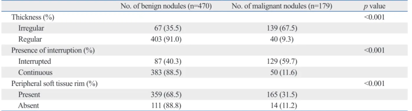
Among the US characteristics of macrocalcification, irregu- larity of thickening, interruption and soft tissue rim showed significant association with malignancy (Table 1).
A multivariate analysis with multiple logistic regression analysis was performed to determine independent US predic- tors for malignancy. Five US criteria including taller-than- wide or irregular shape, irregular margin of nodules, irregular thickness of calcification, interruption of calcification, or presence of soft tissue outside the calcification showed sig- nificant association with thyroid malignancy (p<0.05) (Table 2, Figs. 1 and 2).
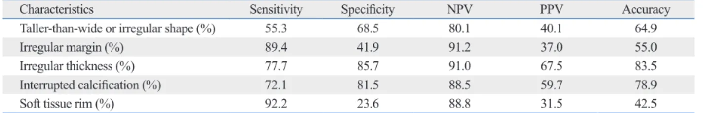
Sensitivity, specificity, NPV, PPV and accuracy of the US characteristics of macrocalcificationfor the prediction of malignancy are listed in Table 3.
Free hand FNAB was performed by radiologists special- ized in thyroid imaging with a 23- to 25-gauge needle and a 10-mL syringe. Ultrasonographic guidance was used to confirm the correct placement of the needle in the nodules.
At least two passes were made per nodule. Specimens were smeared on a slide, fixed in 95% ethanol immediately, and stained by the Papanicolaou method. Radiologists deter- mined the adequacy of the specimen and the number of passes.
Three pathologists with more than 6 years of experience in thyroid cytopathology interpreted the cytologic findings.
FNAB cytologic diagnosis was made based on the Bethes- da system; Nodules were classified as category 1 nondiag- nostic, category 2 benign, category 3 atypia of undeter- mined significance, category 4 suspicious for a follicular neoplasm/follicular neoplasm, category 5 suspicious for malignancy, or category 6 malignant.7
Image interpretation
Two radiologists (K.J.A and P.Y.J) blinded to the cytopa- thologic diagnosis retrospectively reviewed US images of macrocalcified nodules. The following findings of macro- calcifications were analyzed for each nodule: thickness of calcification (regular or irregular), presence of interruption (present or absent), and existence of soft tissue outside the calcification if the calcification has a shell appearance (pres- ent or absent). Regularity of the calcification thickness was assessed subjectively by consensus. Presence of interruption referred to loss of continuance of hyperechoic structures or a loss of approximation in the alignment of the macrocalcifi- cation. Existence of more than 1 mm thickness of a soft tis- sue rim outside the calcification was defined as presence of soft tissue echogenicity outside the macrocalcification. Di- agnostic impression regarding the US findings were classi- fied into three categories (probably benign, low suspicious for malignancy, or suspicious for malignancy) by consensus.
Statistical analysis
Each of the US characteristics was analyzed to determine its association with a cytopathologic diagnosis or surgical pa- thology. When FNAB cytology and the pathology of surgi- cal specimen were discordant, the pathology of surgical specimen was regarded as the standard histologic diagnosis.
Statistical comparisons were performed using the chi- square or Fisher’s exact tests for categoric data, and the Kruskal-Wallis test for continuous data. Multiple logistic regression analysis with a forward stepwise method for se-
7.5% to 26.8% and these nodules are frequently sampled in- adequately by the second FNAB.5,6,13,14 In this study, we ob- served a similar rate of 21% of inadequate FNAB cytology in nodules with macrocalcification. This might be explained by the procedural difficulty that a needle cannot break through the stiff calcification and approach the soft tissue component of the nodule, especially when there is no soft tissue rim located outside the calcification. Therefore, US- guided FNAB is a challenging diagnostic modality for mac- rocalcified thyroid nodules according to these limitations.
DISCUSSION
FNAB cytology has a sensitivity of 71-83% and a specifici- ty of 96%.6 However, regardless of the diagnostic perfor- mance of the FNAB cytology, 5-20% still remain insuffi- cient for diagnosis.4,8-12 Moreover, macrocalcification of thyroid nodules is frequently associated with inadequate sampling of FNAB.3 The portion of nondiagnostic aspirates are larger in nodules with macrocalcification ranging from
Table 1. Correlation of US Characteristics of Macrocalcification with Pathologic Diagnosis
No. of benign nodules (n=470) No. of malignant nodules (n=179) p value
Thickness (%) <0.001
Irregular 67 (35.5) 139 (67.5)
Regular 403 (91.0) 40 (9.3)
Presence of interruption (%) <0.001
Interrupted 87 (40.3) 129 (59.7)
Continuous 383 (88.5) 50 (11.6)
Peripheral soft tissue rim (%) <0.001
Present 359 (68.5) 165 (31.5)
Absent 111 (88.8) 14 (11.2)
p-value is by chi-square analysis.
Table 2. Multiple Logistic Regression Analysis of Malignancy Rate According to the US Features of Macrocalcified Thyroid Nodules
Characteristics Odds ratio 95% Confidence interval p value
Taller-than-wide or irregular shape 1.676 1.024, 2.742 0.040
Irregular margin 2.675 1.388, 5.155 0.003
Irregular thickness 7.139 4.285, 11.895 <0.001
Interrupted calcification 4.948 2.976, 8.226 <0.001
Presence of soft tissue rim 3.164 1.590, 6.293 <0.001
Fig. 1. US findings of malignant thyroid nodule with macrocalcification.
Transverse US image in a 42-year-old woman shows a nodule with inter- rupted macrocalcification (white arrows), irregular thickness and soft tis- sue rim outside the calcification (black arrow) and the nodule was diag- nosed as papillary thyroid carcinoma by fine needle aspiration cytology.
Fig. 2. US findings of benign thyroid nodule with macrocalcification.
Transverse US image of a 54-year-old woman shows a nodule with macro- calcification (arrows) which has regular margin and regular thickness without interruption. The soft tissue rim outside of the macrocalcification was not visible and the nodule was diagnosed as adenomatous hyperpla- sia by fine needle aspiration biopsy.
ruption or soft tissue outside the calcification rim showed significantly higher rate of malignancy than nodules with other features with odds ratios of 7.139, 4.948, and 3.164, respectively (Table 2). Irregular thickness of the macrocalci- fication showed high sensitivity, specificity, NPV, and diag- nostic accuracy for malignancy (77.7%, 85.7%, 91.0%, and 83.5%, respectively). Interrupted macrocalfication showed overall diagnostic accuracy that exceeded 75.0% with the specificity and NPV of 81.5% and 88.5%, respectively.
This result can be explained by the pathology that an inter- ruption of peripheral calcifications demonstrated tumor in- filtration through the broken calcification rim.6 The pres- ence of soft tissue outside calcification rim was a sensitive finding for malignancy with the sensitivity of 92.2% and showed high NPV of 88.8% (Table 3).
Other US findings of the thyroid nodules associated with macrocalcification were also evaluated for its value in pre- dicting malignancy. Previously reported US features predic- tive of malignancy included the presence of microcalcifica- tion, hypoechogenicity, irregular margins, and the absence of a halo.22 Park, et al.6 suggested that decreased internal echo- genicity of the nodule with peripheral macrocalcification was associated with malignancy. Kim, et al.1 reported that macrocalcified nodules with at least one of the following triple criteria (hypoechogenicity, irregular or microlobulated margins, or taller-than-wide shape) showed a significantly higher rate of malignancy than in cases without any triple criteria. In our study, nodules with taller-than-wide or irregu- lar shape and irregular margin showed a significantly higher rate of malignancy than nodules with other features with an odds ratio of 1.676 and 2.675, respectively. Among the pre- viously reported US features that are predictive of malignan- cy,22 only the finding of taller-than-wide or irregular shape and irregular margin were significantly associated with ma- lignancy in our study and the other findings including hy- poechogenicity and the absence of a halo were not associat- ed with malignancy in nodules with macrocalcification.
There are some limitations in our study. First, our study Calcifications can be present in both benign and malig-
nant thyroid nodules. It was reported that thyroid nodules with microcalcifications (1 mm or less in size) were associ- ated with thyroid malignancy.1,15-18 The results for thyroid nodules with macrocalcifications (larger than 1 mm at the longest diameter) are controversial. In macrocalcified thy- roid nodules, peripheral rim or eggshell calcification has been considered associated with multinodular goiters and has been generally considered as an indicator of a benign nodule.19-21 There are a few reports suggesting that a consid- erable portion of the macrocalcified nodules are malignant.
Taki, et al.2 reported that 43% (6/14) of nodules with pe- ripheral calcification were histopathologically proved to be papillary carcinoma. Kim, et al.1 evaluated 174 nodules with macrocalcification and 66% (116/174) proved to be malignant. In our study, 27.6% (179/649) nodules were di- agnosed as malignant. These results suggested that macro- calcification of the nodule was not infrequently associated with the thyroid malignancy.
Therefore, preoperative US features suggesting malignan- cy can provide useful information for preoperative prediction of pathology and can further lead to the proper management of the macrocalcified thyroid nodules. To our knowledge, only a few studies have reported the US findings of the thy- roid nodules associated with macrocalcification. Kim, et al.1 classified the macrocalcification into three subtypes (soli- tary, eggshell, and coarse not otherwise specified) and re- ported no significant difference in the prevalence of malig- nancy according to the types of calcification. Yoon, et al.5 divided the peripheral macrocalcification into stippled, smooth curvilinear, and irregular curvilinear calcification, but did not find any significant differences in the risk of malignancy.
We specifically classified the subtypes of macrocalcifica- tion in thyroid nodules by their regularity of thickness, pres- ence of interruption, and existence of soft tissue rim periph- eral to the calcification. We observed that thyroid nodules with macrocalcification showing irregular thickness, inter-
Table 3. Diagnostic Performance of US Characteristics for the Prediction of Malignancy in Thyroid Nodules with Macrocalci- fication
Characteristics Sensitivity Specificity NPV PPV Accuracy
Taller-than-wide or irregular shape (%) 55.3 68.5 80.1 40.1 64.9
Irregular margin (%) 89.4 41.9 91.2 37.0 55.0
Irregular thickness (%) 77.7 85.7 91.0 67.5 83.5
Interrupted calcification (%) 72.1 81.5 88.5 59.7 78.9
Soft tissue rim (%) 92.2 23.6 88.8 31.5 42.5
NPV, negative predictive value; PPV, positive predictive value.
Clin Ultrasound 2009;37:324-8.
7. Cibas ES, Ali SZ; NCI Thyroid FNA State of the Science Confer- ence. The Bethesda System For Reporting Thyroid Cytopatholo- gy. Am J Clin Pathol 2009;132:658-65.
8. Carmeci C, Jeffrey RB, McDougall IR, Nowels KW, Weigel RJ.
Ultrasound-guided fine-needle aspiration biopsy of thyroid mass- es. Thyroid 1998;8:283-9.
9. Danese D, Sciacchitano S, Farsetti A, Andreoli M, Pontecorvi A.
Diagnostic accuracy of conventional versus sonography-guided fine-needle aspiration biopsy of thyroid nodules. Thyroid 1998;8:
15-21.
10. Goellner JR, Gharib H, Grant CS, Johnson DA. Fine needle aspi- ration cytology of the thyroid, 1980 to 1986. Acta Cytol 1987;31:
587-90.
11. Rago T, Vitti P. Role of thyroid ultrasound in the diagnostic evalu- ation of thyroid nodules. Best Pract Res Clin Endocrinol Metab 2008;22:913-28.
12. Bastin S, Bolland MJ, Croxson MS. Role of ultrasound in the as- sessment of nodular thyroid disease. J Med Imaging Radiat Oncol 2009;53:177-87.
13. Seningen JL, Nassar A, Henry MR. Correlation of thyroid nodule fine-needle aspiration cytology with corresponding histology at Mayo Clinic, 2001-2007: an institutional experience of 1,945 cas- es. Diagn Cytopathol 2012;40 Suppl 1:E27-32.
14. Renshaw AA. Histologic follow-up of nondiagnostic thyroid fine needle aspirations: implications for adequacy criteria. Diagn Cyto- pathol 2012;40 Suppl 1:E13-5.
15. Berker D, Isik S, Ozuguz U, Tutuncu YA, Kucukler K, Akbaba G, et al. Prevalence of incidental thyroid cancer and its ultrasono- graphic features in subcentimeter thyroid nodules of patients with hyperthyroidism. Endocrine 2011;39:13-20.
16. Qian M, Wang J, Qiu Y. [The significance of calcification in the thyroid papillary carcinoma]. Lin Chung Er Bi Yan Hou Tou Jing Wai Ke Za Zhi 2011;25:673-5.
17. Petrone L, Mannucci E, De Feo ML, Parenti G, Biagini C, Panco- nesi R, et al. A simple ultrasound score for the identification of candidates to fine needle aspiration of thyroid nodules. J Endocri- nol Invest 2012;35:720-4.
18. Ozel A, Erturk SM, Ercan A, Yılmaz B, Basak T, Cantisani V, et al. The diagnostic efficiency of ultrasound in characterization for thyroid nodules: how many criteria are required to predict malig- nancy? Med Ultrason 2012;14:24-8.
19. Chen CY, Tseng HS, Lee CH, Chan WP. Primary squamous cell carcinoma of the thyroid gland with eggshell calcification: sono- graphic and computed tomographic findings. J Ultrasound Med 2010;29:1667-70.
20. Yaturu S, Rainer L. Thyroid nodule with eggshell calcification and oncocytic thyroid cancer. Med Sci Monit 2010;16:CS25-8.
21. Gooding GA. Ultrasonic appearance of a thyroid nodule invested in eggshell calcification. J Clin Ultrasound 1978;6:41-3.
22. Moon WJ, Jung SL, Lee JH, Na DG, Baek JH, Lee YH, et al. Be- nign and malignant thyroid nodules: US differentiation--multi- center retrospective study. Radiology 2008;247:762-70.
was retrospective and there could be selection bias. In our study, patients with benign findings at US usually did not undergo biopsy or surgery, which might have resulted in relatively fewer benign nodules in our study. This limitation could be overcome by a large-scale prospective study. Sec- ond, in clinical practice, FNAB is recommended in nodules with its diameter larger than 5 mm, but in our series, a nod- ule of 2 mm in diameter was included due to its posterior lo- cation; therefore, detailed evaluation was not available. In addition, we did not correlate the US findings and the patho- logic findings nodule by nodule. Further study with detailed correlation of US and pathologic findings could give sup- porting pathologic bases for the significant US findings.
In conclusion, US findings predictive of malignancy in thyroid nodules with macrocalcification were an interruption of macrocalcification, irregular thickness of macrocalcifica- tion, the presence of soft tissue outside the macrocalfication rim, taller-than-wide or irregular shape, and irregular margin of the thyroid nodules. Our study provides useful informa- tion about thyroid nodules with macrocalcification, and might be especially helpful in cases with inadequate FNAB cytology for predicting malignancy with US.
REFERENCES
1. Kim MJ, Kim EK, Kwak JY, Park CS, Chung WY, Nam KH, et al. Differentiation of thyroid nodules with macrocalcifications:
role of suspicious sonographic findings. J Ultrasound Med 2008;
27:1179-84.
2. Taki S, Terahata S, Yamashita R, Kinuya K, Nobata K, Kakuda K, et al. Thyroid calcifications: sonographic patterns and incidence of cancer. Clin Imaging 2004;28:368-71.
3. Choi SH, Han KH, Yoon JH, Moon HJ, Son EJ, Youk JH, et al.
Factors affecting inadequate sampling of ultrasound-guided fine- needle aspiration biopsy of thyroid nodules. Clin Endocrinol (Oxf) 2011;74:776-82.
4. Alexander EK, Heering JP, Benson CB, Frates MC, Doubilet PM, Cibas ES, et al. Assessment of nondiagnostic ultrasound-guided fine needle aspirations of thyroid nodules. J Clin Endocrinol Metab 2002;87:4924-7.
5. Yoon DY, Lee JW, Chang SK, Choi CS, Yun EJ, Seo YL, et al.
Peripheral calcification in thyroid nodules: ultrasonographic fea- tures and prediction of malignancy. J Ultrasound Med 2007;26:
1349-55.
6. Park M, Shin JH, Han BK, Ko EY, Hwang HS, Kang SS, et al.
Sonography of thyroid nodules with peripheral calcifications. J