INTRODUCTION
Patellar instability, seen frequently in adolescents and young adults, often is associated with previous knee injury.1,2 It is
caused by various factors, including genu valgum, trochlea dysplasia, femoral intorsion, and patella alta.1,2 Whereas initial episodes of patellar dislocation typically are managed conser- vatively by immobilization and physiotherapy, chronic/recur- rent dislocation and instability are treated primarily by sur- gery.3
The surgical treatment options include lateral release and medial plication,4,5 distal realignment procedures, such as tibial tubercle osteotomy,6,7 and procedures combining both proxi- mal and distal realignment techniques.8 Distal femoral oste- otomy (DFO), though most commonly advocated as a treat- ment option for lateral-compartment osteoarthritis,9 recently has been found to be effective for chronic patellar dislocation as well.10-13 DFO helps to correct excessive Q angle and genu
A Combined Closing Wedge Distal Femoral Osteotomy and Medial Reefing Procedure for Recurrent Patellar Dislocation with Genu Valgum
Chong Bum Chang1, Gautam M. Shetty2, Jong Seong Lee3, Young Chan Kim3, Jae Ho Kwon4, and Kyung Wook Nha3
1Department of Orthopedic Surgery, Seoul National University, Boramae Hospital, Seoul, Korea;
2Department of Orthopaedic Surgery, Breach Candy Hospital, Mumbai, India;
3Department of Orthopaedic Surgery, Inje University Ilsan Paik Hospital, Goyang, Korea;
4Department of Orthopedic Surgery, Barunsesang Hospital, Seongnam, Korea.
Purpose: Recurrent patellar dislocation is often associated with genu valgum. The purpose of this study was to analyze the short- term results of single-incision, closing-wedge distal femoral osteotomy (CWDFO) combined with medial reefing and lateral re- lease for recurrent patellar instability with genu valgum.
Materials and Methods: Combined CWDFO/medial reefing/lateral release was performed on 10 knees. Clinical evaluation was based on pre- and postoperative Knee Society Score (KSS) and Kujala patellofemoral score. Radiographic evaluation was per- formed with reference to the weight-bearing line (WBL), the femorotibial angle (FTA), and the mechanical lateral distal femoral angles in the knee-standing view.
Results: At a mean follow-up of 20±11.7 months (range, 12–42 months), KSS scores improved significantly, from 46.7±5.2 preoper- atively to 87±4.4 postoperatively (p<0.001), as did the Kujala score, from 44±8 preoperatively to 86.6±6.8 postoperatively (p<0.001).
The WBL decreased significantly, from 76±7% preoperatively to 41±11% postoperatively (p<0.001). The FTA was improved signifi- cantly, from 12.7±1.7° preoperatively to 4±4° postoperatively (p<0.001), as was the mLDFA, from 83±4° preoperatively to 91±1.3°
postoperatively (p<0.001).
Conclusion: Use of single-incision CWDFO combined with medial reefing and lateral release prevents patellar dislocation, cor- rects deformity, and improves clinical outcomes.
Key Words: Patellar instability, distal femoral osteotomy, genu valgum, medial reefing
pISSN: 0513-5796 · eISSN: 1976-2437
Received: September 1, 2016 Revised: February 21, 2017 Accepted: February 24, 2017
Corresponding author: Dr. Kyung Wook Nha, Department of Orthopaedic Surgery, Inje University Ilsan Paik Hospital, 170 Juhwa-ro, Ilsanseo-gu, Goyang 10380, Korea.
Tel: 82-31-910-7301, Fax: 82-31-910-7967, E-mail: [email protected]
•The authors have no financial conflicts of interest.
© Copyright: Yonsei University College of Medicine 2017
This is an Open Access article distributed under the terms of the Creative Com- mons Attribution Non-Commercial License (http://creativecommons.org/licenses/
by-nc/4.0) which permits unrestricted non-commercial use, distribution, and repro- duction in any medium, provided the original work is properly cited.
Yonsei Med J 2017 Jul;58(4):878-883 https://doi.org/10.3349/ymj.2017.58.4.878
valgum deformity, both of which are commonly associated with patellar instability.1,2 Furthermore, biplanar closing-wedge DFO (CWDFO) provides better stability and promotes bone healing, due to increased cancellous bone surface area at the osteotomy site.9,10,14,15 For these reasons, CWDFO could be a valuable procedure in a chronic patellar instability patient with excessive Q-angle stemming from significant genu valgum, Additional medial reefing with lateral release could be helpful when instability would be potentially intractable in the pa- tient.10-13,16
The purpose of this study was to analyze the short-term re- sults of single-incision CWDFO combined with medial reefing and lateral release for recurrent patellar instability with genu valgum. We hypothesized that the technique would provide satisfactory clinical outcomes in patient with chronic patellar instability and significant genu valgum at a minimum follow- up of 1 year.
MATERIALS AND METHODS
We reviewed the clinical and radiographic records of 10 knees in six patients (2 males, 4 females) undergoing CWDFO com- bined with medial reefing and arthroscopic lateral release for recurrent patellar instability between 2012 and 2015 with a min- imum follow-up of 1 year. The patients’ mean age at surgery was 26.5±6.4 years (range: 20–38 years). All of them had been operated on by one senior orthopaedic surgeon. The indication for surgical treatment was chronic patellar instability, either habitual or recurrent, combined with genu valgum deformity [criteria: exceeding 6–8° femorotibial angle (FTA) and 60%
Mikulicz line; less than 86° mechanical lateral distal femoral angle (mLDFA) on anteroposterior (AP) long-leg weight-bear- ing lower-extremity scanograph].17,18 The patients’ demograph- ic data are presented in Table 1.
All of the patients were assessed preoperatively and at final follow-up according to the Knee Society Score (KSS)19 and Ku- jala knee score.20 The radiographic evaluation included full- length, standing hip-to-ankle, knee AP, and lateral and patel-
lar merchant radiographs. The position of the weight-bearing line (WBL), as well as the FTA and mLDFA, were measured on full-length, standing hip-to-ankle radiographs both pre- and postoperatively (at final follow-up).21 This study was approved by the Institutional Review Board (IRB) of Inje University Ilsan Paik Hospital.
Surgical technique
Preoperative planning was performed using full-length, stand- ing hip-to-ankle radiographs. PACS View (PACS; Marotech Inc., Seoul, Korea) was used for preoperative determination of the extent of correction. Our target line traveled 45% across the width of the plateau medially to laterally.
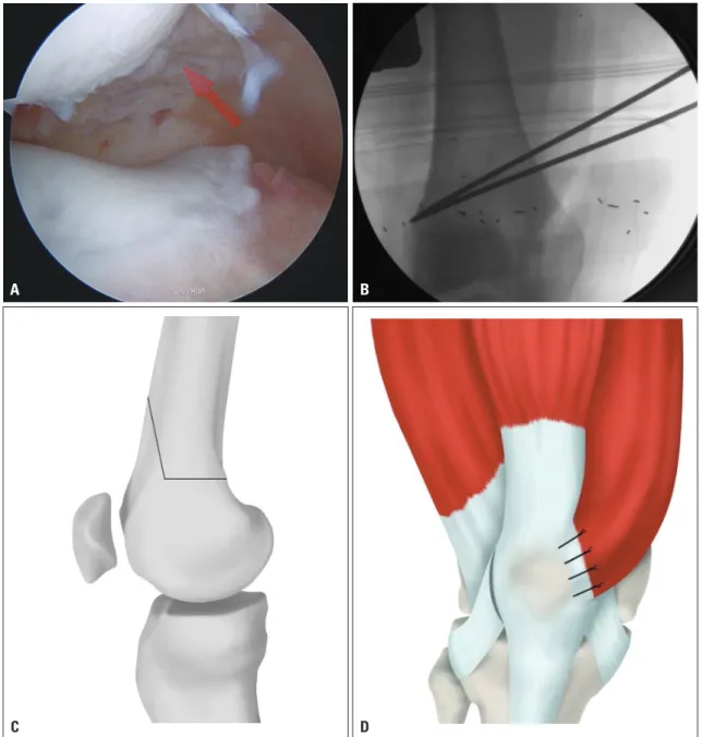
The patient was placed in a supine position on a radiolucent operating table, and a tourniquet was applied to the thigh. Ini- tially, knee arthroscopy was performed in order to visually confirm patellar subluxation or dislocation from the superolat- eral portal view (Fig. 1A). Then, lateral release was performed, with the knee in full extension, from the superior to the inferi- or pole of the patella using electrocauterization. For DFO, a longitudinal skin incision was made beginning 10 cm above the patella and extending distally to the patellar apex. By a sub- vastus approach, the anteromedial aspect of the supracondy- lar area of the femur was then exposed. After incising the muscle fascia, the vastus medialis was stripped from the intermuscular septum and retracted using a Hohmann retractor.
The DFO was planned as a medial biplanar CWDFO on a plane beginning approximately 2 cm superior to the adductor tubercle in the medial supracondylar area and ending in the lateral supracondylar area. The starting point was marked un- der fluoroscopy on the medial supracondylar area by electro- cauterization using a fixation plate as a reference. The target angle of correction was marked with two Kirschner wires un- der fluoroscopic guidance. The target point of the lateral cor- tex was above the lateral epicondyle (Fig. 1B). The axial part of the osteotomy involved the posterior two-thirds of the femur, and was performed up to 5 mm from the lateral femoral cortex in order to preserve it. Using a small oscillating saw, a medial- based wedge was created along the inner margins of the two Table 1. Demographic Data on Patients
No. Sex/Age Diagnosis Frequency (dislocation) Previous op (frequency)
1 M/27 (Rt)
(Lt) Congenital MED 3
3
None None
2 F/20 (Lt) Congenital MED 5 None
3 F/26 (Rt)
(Lt)
Genu valgum Genu valgum
7 3
Medial plication Medial plication
4 M/21 (Rt)
(Lt)
Genu valgum Genu valgum
5 5
None None
5 F/27 (Rt)
(Lt)
Congenital SED Congenital SED
Habitual Habitual
Medial reefing and Fulkerson osteotomy (7) Medial reefing and Fulkerson osteotomy (7)
6 F/38 (Lt) Genu valgum 4 Medial plication
Rt, right; Lt, left; MED, metaphyseal-epiphyseal dysplasia; SED, spondyloepiphyseal dysplasia; op, operation.
Kirschner wires previously applied.
The direction of the frontal plane was marked by electro- cauterization from the superior margin of the axial osteotomy to the anterior femoral cortex, and was angled approximately 110º from the axial osteotomy (Fig. 1C). The frontal-plane os- teotomy was performed with an oscillating saw from the me- dial cortex and by cutting completely through the lateral cor- tex. The medial bone wedge was then removed, and the medial osteotomy was carefully closed by application of consistent pressure. The alignment of the axis of the leg was evaluated and
confirmed by fluoroscopy. The osteotomy was stabilized using a TomoFix MDP plate (Synthes, West Chester, PA, USA). After fixation of the osteotomy, the medial retinaculum was careful- ly incised along the medial border of the patella, and approxi- mately 1 cm was reefed. The medial structure, including the vastus medialis obliquus, medial capsule and medial retinac- ulum, were overlapped and tightened on the medial border of the patella using No. 2 Ethibond sutures (Ethicon Inc., Johnson
& Johnson, Somerville, NJ, USA) with the knee at approxi- mately 30° flexion (Fig. 1D). A final evaluation was performed
Fig. 1. (A) Biplane distal femur osteotomy and medial reefing procedure arthroscopic superiorlateral view-subluxed patellar target point is above the lat- eral epicondyle of distal femur biplane osteotomy lateral release and medial reefing. (B) Biplane distal femur osteotomy and medial reefing procedure ar- throscopic superiorlateral view-subluxed patellar target point is above the lateral epicondyle of distal femur biplane osteotomy lateral release and medial reefing. (C) Biplane distal femur osteotomy and medial reefing procedure arthroscopic superiorlateral view-subluxed patellar target point is above the lateral epicondyle of distal femur biplane osteotomy lateral release and medial reefing. (D) Biplane distal femur osteotomy and medial reefing procedure arthroscopic superiorlateral view-subluxed patellar target point is above the lateral epicondyle of distal femur biplane osteotomy lateral release and me- dial reefing.
A
C
B
D
to confirm the patellofemoral alignment through full range of motion.
Rehabilitation
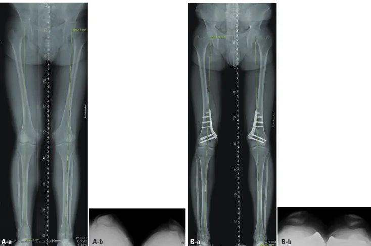
For a period of 4 weeks, the patients walked with partial weight bearing. Range-of-motion exercises were initiated on postop- erative day 3. Radiographs were obtained on postoperative day 7 to confirm the patellar femoral alignment and distal femur, and radiographs were reviewed after 4, and 8 weeks and 3, and 6 months in order to evaluate the healing of the osteotomy and the correction of the patellar instability (Fig. 2).
Statistical analysis
The assumption of the normal distribution of the variable was checked using the Kolmogorov-Smirnov test. The paired t-test was used to compare the preoperative and postoperative knee scores, WBL, FTA and mLDFA. SPSS software (version 18; SPSS Inc., Chicago, IL, USA) was used for the statistical analyses, with p<0.05 set as statistically significant.
RESULTS
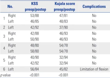
At a mean follow-up of 20±11.7 months (range: 12–42 months), KSS improved markedly, from 46.7±5.2 preoperatively to 87±4.4 at final follow-up (p<0.001), and Kujala score was significantly improved as well, from 44±8 preoperatively to 86.6±6.8 at final follow-up (p<0.001) (Table 2).
The WBL decreased significantly, from 76±7% preoperative- ly to 41±11% postoperatively (p<0.001). Similarly, the FTA was improved significantly, from 12.7±1.7° preoperatively to 4±4°
postoperatively (p<0.001), as was the mLDFA, from 83±4° pre- operatively to 91±1.3° postoperatively (p<0.001) (Table 3).
On postoperative patellar merchant views, none of the knees showed patellar subluxation or dislocation. At final follow-up, all of the knees showed complete union at the osteotomy site.
Two weeks postoperatively, one of the knees showed stiffness and limited flexion. This patient underwent additional manip- ulation under anesthesia. There was no infection, compart- ment syndrome, thrombosis, or non-union.
Fig. 2. (A-a) Preoperative standing, full length, hip-to-ankle radiograph of a 21-year-old male patient showing bilateral genu valgus deformity as shown by the weight bearing lines (green line) and subluxation of patellar both. (A-b) Postoperative standing, full length, hip-to-ankle radiograph of the same patient at 1-year follow-up showing significant change in the weight bearing lines after a combined medial closed-wedge distal femoral osteotomy and lateral release with medial reefing and normal alignment of patellofemoral joint. (B-a) Preoperative standing, full length, hip-to-ankle radiograph of a 21-year-old male patient showing bilateral genu valgus deformity as shown by the weight bearing lines (green line) and subluxation of patellar both. (B-b) Postopera- tive standing, full length, hip-to-ankle radiograph of the same patient at 1-year follow-up showing significant change in the weight bearing lines after a combined medial closed-wedge distal femoral osteotomy and lateral release with medial reefing and normal alignment of patellofemoral joint.
A-b
A-a B-a B-b
DISCUSSION
This paper describes the advantages of single-incision medial biplanar CWDFO combined with medial retinaculum reefing and lateral release for treatment of recurrent dislocation of the patella with genu valgus knee. Due to multiple complex con- tributing factors, recurrent dislocation of the patella is difficult to treat. The most common causes are reported to be disorders of the extensor mechanism such as soft-tissue restraints, pa- tella alta, genu valgum, and malrotation of the lower extremi- ty.4,5 The frequency of recurrent dislocation after acute patellar dislocation reportedly is 40–60% with conservative treatment and 10–30% with surgery.5
There are several techniques for treatment patellar disloca- tion, including proximal realignment, distal realignment, and combined proximal and distal realignment procedures; reports on CWDFO combined with proximal soft-tissue procedures for treatment of recurrent dislocation of the patella in valgus
knee remain rare. DFO can be performed using an opening- or closing-wedge technique, though the former has been impli- cated in patellofemoral contact pressure increment, bone graft use, collapse, and delayed rehabilitation, due to breakage of the far cortex. For these reasons, opening-wedge DFO (OWDFO) is unsuitable for the purposes described in this paper.
The clinical results of isolated lateral release for patellar in- stability, meanwhile, reportedly are poor, and include ongoing instability and poor patient satisfaction, among other issues.7 Medial reefing procedures for patellar instability frequently are performed (and typically in conjunction with lateral re- lease) to achieve proximal realignment. However, most of the relevant reports are not relevant to valgus knee. Medial patel- lofemoral ligament reconstruction, an effective surgical pro- cedure, requires a soft-tissue procedure (either autograft or al- lograft). Still, most reports, again, are not relevant to valgus knees.
This paper presents cases of patients with recurrent patellar dislocation in a valgus knee. Regardless of trauma, all of the pa- tients had valgus knee and more than one experience of patel- lar dislocation. It has been reported that proximal alignment should be performed when the tibial tubercle-trochlear groove (TTTG) distance is normal, whereas distal alignment combined with proximal alignment is preferable in cases where the TTTG distance is more than 20 mm.22 There have been several re- ports of the treatment of patellar dislocation with genu valgum.
Purushothaman, et al.13 described posttraumatic chronic pa- tellar dislocation using OWDFO and medial patellofemoral lig- ament reconstruction. Shen, et al.8 reported a procedure com- bining proximal soft-tissue, DFO, and distal tibial tuberosity realignment for treatment of genu valgum. Kwon, et al.12 report- ed a case of habitual patellar dislocation treated by CWDFO with medial reefing and lateral retinacular release. Kwak, et al.11 reported a case of habitual patellar dislocation treated by OWDFO with medial reefing in a patient with hypoplasia of the lateral femoral condyle. Hinterwimmer, et al.10 reported a Table 2. Preoperative and One-Year Follow-Up Clinical Outcomes for
Patients
No. KSS
preop/postop
Kujala score
preop/postop Complications 1 Right
Left
53/88 46/85
47/81 48/83
No No
2 Left 42/92 37/90 No
3 Right Left
42/88 50/90
46/93 46/93
No No 4 Right
Left
48/80 58/80
54/78 54/78
No No 5 Right
Left
40/90 42/92
32/94 32/94
No No
6 Left 56/84 45/82 Limitation of flexion
p value <0.001 <0.001
KSS, Knee Society Score; op, operation.
A p value<0.05 is significant.
Table 3. Preoperative and One-Year Follow-Up Radiologic Parameters
No. WBL preop/postop FTA preop/postop mLDFA preop/postop Patellofemoral joint
preop/postop 1 Right
Left
75/45 75/50
11/6 11/7
83/91 83/91
Subluxation/normal Subluxation/normal
2 Left 67/30 11/1 86/93 Subluxation/normal
3 Right Left
85/45 90/45
12/5 15/5
87/92 83/91
Subluxation/normal Subluxation/normal 4 Right
Left
75/50 75/50
14/6 13/6
86/89 86/89
Subluxation/normal Subluxation/normal 5 Right
Left
75/45 75/15
14/0 15/-5
77/92 76/89
Dislocation/normal Dislocation/normal
6 Left 65/35 11/0 85/91 Subluxation/normal
p value <0.001 <0.001 0.005
op, operation; WBL, weight-bearing line; FTA, femorotibial angle; mLDFA, mechanical lateral distal femur angle.
A p value<0.05 is significant.
method of biplanar lateral OWDFO for patellofemoral ma- lalignment.
There are several advantages to the new combined technique for the treatment of recurrent patellar dislocation with genu valgus knee. First, varus osteotomy can contribute to decreased risk of re-dislocation of the aligned patella, specifically by re- ducing the strong lateral vector applied to the patellar cause of the genu valgus.13 By correction of the valgus moment of the femur, the patellofemoral joint can be better stabilized. Second, medial CWDFO combined with medial retinaculum reefing can be performed via a single incision, which is more cosmetic than lateral OWDFO with medial reefing, which entails two incisions. Third, medial CWDFO offers an early-rehabilitation advantage over lateral OWDFO, due to its direct bone-to-bone contact. Additionally, biplanar CWDFO has been shown to im- prove the primary stability of the osteotomy and bone contact surface area, which can promote more rapid bone healing than is possible in the case of uniplanar CWDFO.11
The disadvantages of this technique, which include the risks of non-union and patellar re-dislocation, are of concern. No such complications, however, were observed in our study. In any event, the new technique requires further clinical study of primary cases with longer follow-up periods.
In conclusion, use of single-incision CWDFO combined with medial reefing and lateral release prevents patellar disloca- tion, corrects deformity, and improves clinical outcomes.
REFERENCES
1. Ries Z, Bollier M. Patellofemoral instability in active adolescents. J Knee Surg 2015;28:265-77.
2. Waterman BR, Belmont PJ Jr, Owens BD. Patellar dislocation in the United States: role of sex, age, race, and athletic participation. J Knee Surg 2012;25:51-7.
3. Smith TO, Song F, Donell ST, Hing CB. Operative versus non-oper- ative management of patellar dislocation. A meta-analysis. Knee Surg Sports Traumatol Arthrosc 2011;19:988-98.
4. Lee JJ, Lee SJ, Won YG, Choi CH. Lateral release and medial plica- tion for recurrent patella dislocation. Knee Surg Sports Traumatol Arthrosc 2012;20:2438-44.
5. Ricchetti ET, Mehta S, Sennett BJ, Huffman GR. Comparison of lateral release versus lateral release with medial soft-tissue realign- ment for the treatment of recurrent patellar instability: a system- atic review. Arthroscopy 2007;23:463-8.
6. Drexler M, Dwyer T, Dolkart O, Goldstein Y, Steinberg EL, Chakra- vertty R, et al. Tibial rotational osteotomy and distal tuberosity transfer for patella subluxation secondary to excessive external tibial torsion: surgical technique and clinical outcome. Knee Surg
Sports Traumatol Arthrosc 2014;22:2682-9.
7. Hall MJ, Mandalia VI. Tibial tubercle osteotomy for patello-femoral joint disorders. Knee Surg Sports Traumatol Arthrosc 2016;24:
855-61.
8. Shen HC, Chao KH, Huang GS, Pan RY, Lee CH. Combined proxi- mal and distal realignment procedures to treat the habitual dislo- cation of the patella in adults. Am J Sports Med 2007;35:2101-8.
9. Brinkman JM, Freiling D, Lobenhoffer P, Staubli AE, van Heer- waarden RJ. Supracondylar femur osteotomies around the knee:
patient selection, planning, operative techniques, stability of fixa- tion, and bone healing. Orthopade 2014;43 Suppl 1:S1-10.
10. Hinterwimmer S, Minzlaff P, Saier T, Niemeyer P, Imhoff AB, Feucht MJ. Biplanar supracondylar femoral derotation osteotomy for patellofemoral malalignment: the anterior closed-wedge tech- nique. Knee Surg Sports Traumatol Arthrosc 2014;22:2518-21.
11. Kwak JH, Sim JA, Kim NK, Lee BK. Surgical treatment of habitual patella dislocation with genu valgum. Knee Surg Relat Res 2011;
23:177-9.
12. Kwon JH, Kim JI, Seo DH, Kang KW, Nam JH, Nha KW. Patellar dislocation with genu valgum treated by DFO. Orthopedics 2013;
36:840-3.
13. Purushothaman B, Agarwal A, Dawson M. Posttraumatic chronic patellar dislocation treated by distal femoral osteotomy and medial patellofemoral ligament reconstruction. Orthopedics 2012;35:
e1668-72.
14. Brinkman JM, Hurschler C, Staubli AE, van Heerwaarden RJ. Axial and torsional stability of an improved single-plane and a new bi- plane osteotomy technique for supracondylar femur osteotomies.
Knee Surg Sports Traumatol Arthrosc 2011;19:1090-8.
15. van Heerwaarden R, Najfeld M, Brinkman M, Seil R, Madry H, Pape D. Wedge volume and osteotomy surface depend on surgical technique for distal femoral osteotomy. Knee Surg Sports Trau- matol Arthrosc 2013;21:206-12.
16. Iliadis AD, Jaiswal PK, Khan W, Johnstone D. The operative man- agement of patella malalignment. Open Orthop J 2012;6:327-39.
17. Forkel P, Achtnich A, Metzlaff S, Zantop T, Petersen W. Midterm results following medial closed wedge distal femoral osteotomy stabilized with a locking internal fixation device. Knee Surg Sports Traumatol Arthrosc 2015;23:2061-7.
18. Haviv B, Bronak S, Thein R, Thein R. The results of corrective oste- otomy for valgus arthritic knees. Knee Surg Sports Traumatol Ar- throsc 2013;21:49-56.
19. Insall JN, Dorr LD, Scott RD, Scott WN. Rationale of the Knee So- ciety clinical rating system. Clin Orthop Relat Res 1989;(248):13-4.
20. Kujala UM, Jaakkola LH, Koskinen SK, Taimela S, Hurme M, Neli- markka O. Scoring of patellofemoral disorders. Arthroscopy 1993;
9:159-63.
21. Eilert RE. Congenital dislocation of the patella. Clin Orthop Relat Res 2001;(389):22-9.
22. Sherman SL, Erickson BJ, Cvetanovich GL, Chalmers PN, Farr J 2nd, Bach BR Jr, et al. Tibial tuberosity osteotomy: indications, techniques, and outcomes. Am J Sports Med 2014;42:2006-17.