Mechanism Underlying a Proteasome Inhibitor,
Lactacystin-Induced Apoptosis on SCC25 Human Tongue Squamous Cell Carcinoma Cells
Chul-Jung Baek, D.D.S.,M.S.D.,Ph.D.
1, Gyoo-Cheon Kim, M.S.,Ph.D.
1, In-Ryoung Kim, M.S.,Ph.D.
1, Seung-Eun Lee, M.S.
1, Hyun-Ho Kwak, M.S.,Ph.D.
1,
Bong-Soo Park, D.D.S.,M.S.D.,Ph.D.
1, Il-Ho Tae, D.D.S.,M.S.D.
2, Myung-Yun Ko, D.D.S.,M.S.D.,Ph.D.
2, Yong-Woo Ahn, D.D.S.,M.S.D.,Ph.D.
2Dept. of Oral Anatomy, School of Dentistry, Pusan National University1 Dept. of Oral Medicine, School of Dentistry, Pusan National University2
Lactacystin, a microbial natural product synthesized by Streptomyces, has been commonly used as a selective proteasome inhibitor in many studies. Proteasome inhibitors is known to be preventing the proliferation of cancer cells in vivo as well as in vitro. Furthermore, proteasome inhibitors, as single or combined with other anticancer agents, are suggested as a new class of potential anticancer agents. This study was undertaken to examine in vitro effects of cytotoxicity and growth inhibition, and the molecular mechanism underlying induction of apoptosis in SCC25 human tongue sqaumous cell carcinoma cell line treated with lactacystin.
The viability of SCC25 cells, human normal keratinocytes (HaCaT cells) and human gingiva fibroblasts (HGF-1 cells), and the growth inhibition of SCC25 cells were assessed by MTT assay and clonogenic assay respectively. The hoechst staining, hemacolor staining and TUNEL staining were conducted to observe SCC25 cells undergoing apoptosis. SCC25 cells were treated with lactacystin, and Western blotting, immunocytochemistry, confocal microscopy, FAScan flow cytometry, MMP activity, and proteasome activity were performed.
Lactacystin treatment of SCC25 cells resulted in a time- and does-dependent decrease of cell viability and a does-dependent inhibition of cell growth, and induced apoptotic cell death. Interestingly, lactacytin remarkably revealed cytotoxicity in SCC25 cells but not normal cells. And tested SCC25 cells showed several lines of apoptotic manifestation such as nuclear condensation, DNA fragmentation, the reduction of MMP and proteasome activity, the decrease of DNA contents, the release of cytochrome c into cytosol, the translocation of AIF and DFF40 (CAD) onto nuclei, the up-regulation of Bax, and the activation of caspase-7, caspase-3, PARP, lamin A/C and DFF45 (ICAD). Flow cytometric analysis revealed that lactacystin resulted in G1 arrest in cell cycle progression which was associated with up-regulation in the protein expression of CDK inhibitors, p21WAF1/CIP1 and p27KIP1.
We presented data indicating that lactacystin induces G1 cell cycle arrest and apoptois via proteasome, mitochondria and caspase pathway in SCC25 cells. Therefore our data provide the possibility that lactacystin could be as a novel therapeutic strategy for human tongue squamous cell carcinoma.
Key words: Apoptosis, Proteasome inhibitor, Lactacystin, Human tongue squamous carcinoma
Corresponding author : Yong-Woo Ahn Dept. of Oral Medicine,
School of Dentistry Pusan National University, 1-10 Ami-Dong, Seo-Gu, Busan 602-739, Korea Tel: 82-51-240-7465
Fax: 82-51-247-0955
E-mail: [email protected] Received: 2009-06-26
Accepted: 2009-07-30
*This work was supported by for two years Pusan National University research grant.
I. INTRODUCTION
Apoptosis is implicated in a wide range of pathological conditions, including immunological diseases, allergy and cancer.
1,2)During apoptosis, cells undergo specific morphological and biochemical changes, including cell shrinkage, chromatin condensation, and internucleosomal cleavage of genomic DNA.
3)Multiple lines of evidence indicate that apoptosis can be triggered by the activation of caspase.
4)In addition, mitochondria are known as to central death regulators in response to several apoptotic stimuli.
5)In addition, the proteasome pathway is mostly known to work at the upstream of mitochondrial alterations and caspase activation.
6)The ubiquitin-proteasome system, which is a fundamental nonlysosomal machinery for degradation of short-lived proteins in the cells, has been known to be involved in apoptosis.
7-10)On the other hand, the ubiquitin-proteasome system has been empathized as an attractive target in proliferative disease therapy.
8,11-13)Recent studies have been demonstrated that the proteasome inhibitors had potential as chemotherapeutic agents for various cancer cells. Most of the proteasome inhibitors exert their chemotherapeutic effects through induction of cell cycle arrest and apoptotic cell death.
14-16)Therefore, the induction of apoptosis in cancer cells has become an indicator of the antitumor effect. Also, recent studies have focused on the development of target organ to specific apoptosis inducers as novel cancer preventive and therapeutic methods.
17,18)Although a variety of studies have been demonstrated that proteasome inhibitor induced apoptosis in various cancer cells, studies about a proteasome inhibitor-induced apoptosis on oral squamous cell carcinoma cells are little to date. Moreover, the mechanism of lactacystin-induced cell death in oral squamous cell carcinoma cells is not clearly defined yet.
Carcinoma of the oral cavity, especially oral squamous cell carcinoma (OSCC), is one of the most leading causes of cancer related to death and affects nearly 500,000 patients annually world-wide. And
OSCC is one of the most malignant carcinoma that remain incurable with current therapies.
19)In the present study, we investigated apoptosis of SCC25 human tongue squamous cell carcinoma cell through the treatment of a proteasome inhibitor, lactacystin which has been known to induce apoptosis in various cancer cells.
10)As will be shown, lactacystin induced apoptosis in SCC25 cells.
Importantly, lactacystin-induced apoptosis of SCC25 cell is mediated via proteasome, mitochondria and caspase. Furthermore, lactacystin leads to the inhibition of the cell cycle progression.
Ⅱ. MATERIALS AND METHODS 1. Reagents
The following reagents were obtained commercially: TUNEL reaction mixture was from Boehringer Mannheim (Mannheim, Germany); Suc- LLVY-AMC and lactacystin were from Calbiochem (EMD Biosciences, Germany); 5,5',6,6'-tetrachloro- 1,1',3,3-tetraethyl- benzimidazol carbocyanine iodide (JC-1) was from Molecular Probes (Eugene, OR, USA); Dulbecco's modified Eagle's medium (DMEM) and fetal bovine serum (FBS) were from Gibco (Gaithersburg, MD, USA); Dimethyl sulfoxide (DMSO), Hoechst 33342, RNase A, aprotinin, leupeptin, phenylmethylsulfonyl fluoride (PMSF), thiazolyl blue tetrazolium bromide, crystal violet, collagenase and propidium iodide (PI) were from Sigma (St. Louis, MO, USA); SuperSignal West Pico enhanced chemiluminescence Western blotting detection reagent was from Pierce (Rockford, IL, USA).
2. Antibodies
Mouse monoclonal anti-human p53 was from BD
biosciences (San Diego, CA, USA); rabbit polyclonal
anti-human AIF and cytochrome c antibodies were
from Upstate (NY, USA); mouse monoclonal
anti-human p27
KIP1, p21
WAF1/CIP1, caspase-3, caspase-
7, Bax, Bcl-xL, Lamin A/C, DFF45 (ICAD), poly
(ADP-ribose) polymerase (PARP) antibodies, and rabbit polyclonal anti-human β-actin antibody, and FITC-conjugated goat anti-mouse and anti-rabbit IgGs were from Santa Cruz Biotechnology (Santa Cruz, CA, USA); rabbit polyclonal anti-human DFF40 (CAD) antibody was from Stressgen (San Diego, USA); HRP-conjugated sheep anti-mouse and anti-rabbit IgGs were from Amershan GE Healthcare (Little Chalfont, UK);
3. Cell culture
SCC25 human tonuge squamous cell carcinoma cell line, HaCaT cells human keratinocyte and HGF-1 human gingival fibroblast were purchased from the ATCC (Rockville, MD, USA). These cells were maintained at 37℃ with 5% CO
2in air atmosphere in Dulbecco's modified Eagle's medium (DMEM) with 4 mM L-glutamine, 1.5 μg/L sodium bicarbonate, 4.5 g/L glucose and 1.0 mM sodium pyruvate supplemented with 10% fetal bovine serum (FBS).
4. In vitro treatment of lactacystin
24 h after SCC25 cells were subcultured, the original medium was removed. The cells were washed with phosphate-buffered saline (PBS) and then incubated in the same fresh medium.
Lactacystin from a stock solution was added to the medium to obtain 1, 2.5, 5, 7.5, 10, 12.5, 15 μM concentrations of the drug. Stock solutions of lactacytin (20 mM) made by dissolving the drug in DMSO were kept frozen at -20℃ until use. The concentrations of DMSO, 0.005-0.075%(vol/vol) used in this study, both as a vehicle for lactacystin and as a control, had no effect on SCC25 cells, HaCaT cells and HGF-1 cells proliferation in my preliminary studies.
5. Cell viability tests
The viability of the cultured cells was estimated by the MTT method. The cells were cultured in a
96-well plate and incubated for 24 h. The cells treated with various concentrations and time points of lactacystin. And then cells were treated with 500 μg/ml of MTT stock solution. After the cells were incubated at 37℃ with 5% CO
2for 4 h. The medium was aspirated and formed formazan crystals were dissolved in the mixture solution of DMSO and absolute ethanol (1 : 1). Cell viability was monitored on a ELISA reader (Sunrise Remote Control, Tecan, Austria) at 570 nm excitatory emission wavelength.
Since viability and proliferation assays demonstrated evident induction of SCC25 cell death at 10 μM lactacystin, this concentration was utilized for further assessment of apoptosis.
6. Clonogenic (Colony-forming) assay
Cells were seeded at 2.5 × 10
2per well (6-well culture plate) and incubated overnight. The cells were treated with lactacystin ranging from 1 to 3 μ M. After treatement, cells were washed and allowed to grow for 7 days. The colonies were then fixed 100% methanol and stained with a filtrated solution of 0.5% (w/v) crystal violet for 10 min. And after washing cells with tap water, they were dried at room temperature. The colonies, defined as groups of
≥ 50 cells, were scored manually and photographed under an IMT-2 inverted microscope (Olympus, Japan). Standard value was determined to value of control in 100 percentage. And then, the value of lactacystin treated cells were compared with control.
Three independent experiments were conducted.
7. Hemacolor staining
Cell suspensions were loaded into a cytospin chamber and centrifuged at 500 RPM for 2 min. The slides were allowed to air-dry for at least 5 min.
Cells were fixed by hemacolor fixative solution. Cells
were stained by dipping the slides for 10 sec in
hemacolor red reagent, then counter-staind by
dipping the slides for 10 sec in hemacolor blue
reagent. Slides were rinsed off excess dye and
allowed to air-dry. The samples were observed and
photographed under a light microscope (Carl Zeiss, German).
8. Hoechst staining
Hoechst staining method was employed for the identification of apoptosis nuclei. After cells were treated with lactacystin, they were harvested and cytocentrifuged onto a clean fat-free glass slide with a cytocentrifuge. Cells were stained in 4 μg/ml Hoechst 33342 solution for 10 min at 37℃ in the dark and washed twice in PBS. The slides were mounted with glycerol. The samples were observed and photographed under an epifluorescence microscope (Carl Zeiss, German). The number of cells that showed condensed or fragmented nuclei was determined by a blinded observer from a random sampling of 3 × 10
2cells per slide. Three independent experiments were done.
9. TUNEL technique
To
identify apoptotic cells by terminal deoxynucleotidyl transferase (TDT) - mediated dUTP nick and labelling (TUNEL), an In Situ Cell Death Detection Kit was used as recommended by the manufacturer. Cells were harvested after treatment of lactacystin on 60 mm dishes. The cell suspension was centrifuged onto a clean fat-free glass slide with a cytocentrifuge. After fixing with 4% paraformaldehyde for 1 h, the slides were washed with PBS and permeabilized with 0.1% Triton X-100 solution for 2 min on ice. Cells were added with reaction mixture for 1 h at 37℃. Total cell number, at least 300 cells from each group, was counted under DIC optics and the percentage of TUNEL positive cells were calculated and photographed under epifluorescence microscope (Carl Zeiss, German).
10. Proteasome activity assay
Cells were lysed in proteasome buffer [10 mM Tris-HCl, pH 7.5, 1 mM EDTA, 2 mM ATP, 20%
glycerol, and 4 mM dithiothreitol (DTT)], sonicated,
and then centrifuged at 13,000 g at 4℃ for 10 min.
The supernatant (20 μg of protein) was incubated with proteasome activity buffer [0.05 M Tris-HCl, pH 8.0, 0.5 mM EDTA, 50 μM Suc-LLVY-AMC] for 1 h 37℃. The fluorescenic intensity of each solution was measured by a modular fluorimetric system (Spex Edison, USA) at 380 nm excitatory and 460 nm emission wavelengths. All readings were standardized using the fluorescenic intensity of an equal volume of free AMC solution (50 μM).
11. Western blot analysis
Cells were plated at a density of 2 x 10
6cells in 100 mm culture dishes. Lactacystin treated cells were washed twice with ice-cold PBS and centrifuged at 2,000 rpm for 10 min. Total protein of cell was lysed with a RIPA buffer [300 mM NaCl, 50 mM Tris-HCl (pH 7.6), 0.5% TritonX-100, 2 mM PMSF, 2 μl/ml aprotinin and 2 μl/ml leupeptin] and incubated at 4°C for 1 h. The lysates were centrifuged at 14,000 rpm for 15 min at 4°C, and sodium dodecyl sulfate (SDS) and sodium deoxycholic acid (0.2% final concentration) were added. Protein concentrations of cell lysates were determined with Bradford protein assay (Bio-Rad, USA) and BSA was used as a protein standard. A sample of 25 μg protein in each well was separated and it were loaded onto 7.5-15%
SDS/PAGE. The gels were transferred to PVDF membrane (Amersham Pharmacia Biotech, UK) and reacted with each antibody. Immunostaining with antibodies was performed using SuperSignal West Pico enhanced chemiluminescence substrate and detected with Alpha Imager HP (Alpha Innotech, San Leandro, USA).
12. Assay of mitochondrial membrane potential (MMP)
Cells were plated in a standard 6-well plate at a
density of 5 × 10
5/ml. Lactacystin treated cells were
incubated for various time points. The cells were
harvested and then JC-1 was added directly (1 μM
final concentration) and incubated for 15 min at 37°C
in the dark. Flow cytometry to measure MMP (△Ψ m) was performed on a CYTOMICS FC500 flow cytometry system (Beckman Coulter, USA). The fluorescence emission were collected through a 530/30 band pass filter (FL-1) and through a 585/42 band pass filter (FL-2), both on a log scale. Data were acquired and analyzed using CXP software version 2.2. The analyzer threshold was adjusted on the FSC channel to exclude noise and most of the subcellular debris.
13. Immunofluorescent staining
Cells were placed on slides by cytocentrifuge and fixed for 10 min in 4% paraformaldehyde. After blocking nonspecific binding with 3% bovine serum albumin, the cells were incubated with a primary antibody at a dilution of 1 : 100 for 1 h. After the incubation, the cells were washed 3 times for 5 min, and then incubated with FITC-conjugated secondary antibody at a dilution of 1 : 100 for 1 h at room temperature. Fluorescent images were observed and analyzed under Zeiss LSM 510 laser-scanning confocal microscope (Göettingen, Germany).
14. Flow cytometry analysis
Cells were seeded into a 6-well plate at 1 × 10
6cells/ml and incubated overnight. Lactacystin treated cells were incubated for various time points. In each time, The harvested cells were washed with PBS containing 1% BSA and centrifuged at 2,000 rpm for 10 min. The cells were resuspended ice-cold 95%
ethanol with 0.5% Tween 20 to a final concentration of 70% ethanol. Fixed cells were pelleted, and washed in 1% BSA-PBS solution. They were resuspended in 1 ml PBS containing 20 μg/ml RNase A, incubated at 4℃ for 30 min, washed once with BSA-PBS, and resuspended in PI solution (10 μ g/ml). After cells were incubated at 4℃ for 5 min in the dark, DNA content were measured on a CYTOMICS FC500 flow cytometry system (Beckman Coulter, USA). The data were analyzed using the multicycle software which allowed a
simultaneous estimation of cell-cycle parameters and apoptosis.
Ⅲ. RESULTS
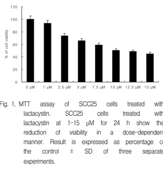
1. Lactacystin reduced the viability and inhibited the growth in SCC25 cells
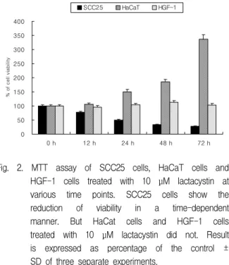
The cytotoxic effect of lactacystin was performed to measure the viability of SCC25 cells, HaCaT cells and HGF-1 cells by MTT assay. Lactacystin produced a significant does-dependent decrease in the viability of SCC25 cells over a period of 72 h (Fig.
1). The viability of SCC25 cells treated with lactacystin at 10 μM over a period of 72 h significantly reduced. But the viability of HaCaT cells treated with lactacystin at 10 μM over a period of 72 h remarkably increased. In addition, the viability of HGF-1 cells did not changed (Fig. 2). The half maximal inhibitory concentration (IC
50) of CGM was at the 10 μM for 24 h. Therefore this concentration was utilized for further assessment of apoptosis and alternation of the cell cycle.
To investigate whether lactacystin inhibited the growth of SCC25 cells, clonogenic assay was performed. After exposure to low level lactacystin
0 20 40 60 80 100 120
0 μM 1 μM 2.5 μM 5 μM 7.5 μM 10 μM 12.5 μM 15 μM
% of cell viability
Fig. 1. MTT assay of SCC25 cells treated with lactacystin. SCC25 cells treated with lactacystin at 1-15 μM for 24 h show the reduction of viability in a dose-dependent manner. Result is expressed as percentage of the control ± SD of three separate experiments.
0 50 100 150 200 250 300 350 400
0 h 12 h 24 h 48 h 72 h
% of cell viability
SCC25 HaCaT HGF-1
Fig. 2. MTT assay of SCC25 cells, HaCaT cells and HGF-1 cells treated with 10 μM lactacystin at various time points. SCC25 cells show the reduction of viability in a time-dependent manner. But HaCat cells and HGF-1 cells treated with 10 μM lactacystin did not. Result is expressed as percentage of the control ± SD of three separate experiments.
concentrations (0 to 3 μM) on SCC25 cells for 7 days, the inhibition of colony formation was determined and was shown in Fig. 3. The growth of lactacystin treated group was determined by percentage of control. The values on colony formation were 75.8%
(1 μg/ml lactacystin treated cells), 56.5% (1.5 μM
(A) (B)
0 20 40 60 80 100 120
0 μM 1 μM 1.5 μM 2 μM 2.5 μM 3 μM
colony number (%)
Fig. 3. Cell growth was examined by colony forming assay. Lactacystin treatment was performed as ranging from 0 to 3 μM on SCC25 cells. (A) Cells were allowed to grow for 7 days before staining with crystal violet. Colonies were decreased gradually according to medium-concentration as photograph. (B) Percentage of colony formation efficiency in different concentrations were displayed. Values are means
± SD of triplicates of each experiment.
lactacystin treated cells), 35.3% (2 μM lactacystin treated cells), 27.1% (2.5 μM lactacystin treated cells), 14.5% (3 μM lactacystin treated cells).
2. Morphological and biochemical changes in lactacystin treated SCC25 cells
SCC25 cells treated with lactacystin at 10 μM resulted in morphological and biochemical changes associated with apoptosis. Hoechst stain demonstrated that lactacystin induced a change in nuclear morphology. Compared with the typical round nuclei of the control cells, SCC25 cells treated with lactacystin at 10 μM for 24 h displayed condensed and fragmented nuclei (Fig. 4A and Fig.
4B). In addtion, to investigate whether lactacystin
was displayed morphological changes on SCC25 cells
and normal cells (HaCaT cells and HGF-1 cells) or
not, hemacolor staining was conducted. SCC25 cells
treated with lactacystin at 10 μM for 24 h displayed
condensed and dark stained nuclei whereas HaCaT
cells and HGF-1 cells showed morphology of normal
cells (Fig. 5). DNA fragmentation which is the
biochemical hallmark of apoptosis, was demonstrated
by TUNEL technique. The TUNEL positive SCC25
cells in the control cells did not show but the
(A) (B)
0 20 40 60 80 100
Con trol 24 h
% of nuclear condensation
Fig. 4. Hoechst staining. (A) SCC25 cells treated with lactacystin at 10 μM for 24 h show the nuclear condensation or fragmentation compared to the control group. Scale bar, 10 μm. (B) Quantification of the nuclear condensation determined by Hoechst staining. Result is expressed as percentage of the control ± SD of three separate experiments.
Fig. 5. Hemacolor staining of SCC25 cells, HaCaT cells and HGF-1 cells treated with lactacystin at 10 μM for 24 h.
numerous TUNEL positive SCC25 cells treated with lactacystin at 10 μM for 24 h were shown (Fig. 6).
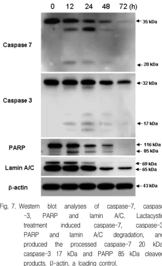
The western blotting study showed that lactacystin treatment at various time points induced degradations of caspase-7, caspase-3, PARP, lamin A/C and DFF45 (ICAD), and produced caspase-7 20 kDa, caspase-3 17 kDa, PARP 85 kDa, and DFF45 30 kDa cleaved products (Fig. 7, Fig. 8A). And confocal microscopy showed that lactacystin led to the translocation of DFF40 (CAD) from cytosol onto nuclei (Fig. 8B).
3. Proteasome activity in lactacystin treated SCC25 cells
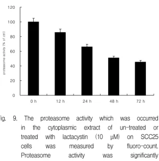
In order to investigate the inhibitory effect of proteasome activity on 10 μM lactacystin treated SCC25 cells, proteasome activity assay was conducted. Lactacystin significantly abolished proteasome activity in a time-dependent manner (Fig. 9).
4. Effect of mitochondrial events in lactacystin treated SCC25 cells
To dissect the molecular mechanism underlying
lactacystin-induced apoptosis in SCC25 cells,
alteration of the expression level of apoptotic factor,
Bax and antiapoptotic factor, Bcl-xL was assayed by
(A) (B)
0 20 40 60 80 100
Control 24 h
%TUNEL-positive cells
Fig. 6. Examination of apoptosis by TUNEL assay. SCC25 cells were treated with lactacystin at 10 μM for 24 h. (A) TUNEL positive cells in control group were not shown (left panel). Numerous TUNEL positive cells in experimental group were shown (right panel). Scale bar, 10 μm. (B) The values below micrographs are the mean ± SD of the means of TUNEL positive cells as determined by TUNEL method. Three independent assays were performed.
Fig. 7. Western blot analyses of caspase-7, caspase -3, PARP and lamin A/C. Lactacystin treatment induced caspase-7, caspase-3, PARP and lamin A/C degradation, and produced the processed caspase-7 20 kDa, caspase-3 17 kDa and PARP 85 kDa cleaved products. β-actin, a loading control.
western blotting. Whereas the expression level of Bax increased, Bcl-xL did not decreased after lactacystin treatment in a time-dependent manner (Fig. 10A). And confocal microscopy showed that lactacystin led to the release of cytochrome c from mitochondria into the cytosol (Fig. 10B).
Western blot assay and confocal microscopy were conducted to examine whether another mitochondrial apoptotic factor, AIF, is involved or not. Expression level of this protein increased till 24 h after treatment of lactacystin. AIF was shown to release from mitochondria, and translocation onto nuclei was evident in SCC25 cells (Fig. 11).
Loss of mitochondrial membrane potential (△ψm) is known to be common event in many pathways of apoptosis induction. Also in this study, mitochondrial membrane potential (△ψm) remarkably decreased after treatment of lactacystin in a time-dependent manner (Fig. 12).
These results suggests that lactacystin-induced
apoptosis was accompanied by modulation of
proapoptotic Bcl-2 family protein, Bax, resulting in
loss of MMP, AIF translocation, and cytochrome c
release.
(A) (B)
Fig. 8. Western blot analysis of DFF45 (ICAD) and confocal microscopy of DFF40 (CAD). (A) Lactacystin treatment induced DFF45 degradation, and produced the processed DFF45 30 kd cleaved product. β -actin, a loading control. (B) Confocal microscopy showing the translocation of DFF40 from cytosol onto the nuclei. Scale bar, 10 μm.
0 20 40 60 80 100 120
0 h 12 h 24 h 48 h 72 h
proteasome activity (% of ctrl)
Fig. 9. The proteasome activity which was occurred in the cytoplasmic extract of un-treated or treated with lactacystin (10 μM) on SCC25 cells was measured by fluoro-count.
Proteasome activity was significantly decreased in a time-dependent manner.
5. Effect of the cell cycle in lactacystin treated SCC25 cells
The evaluation of cell cycle and apoptotic cell percentages were confirmed with flow cytometry analysis. Time-course analysis of cell-cycle distribution after lactacystin treatment revealed an increase in the percentage of G1 phase cells, while
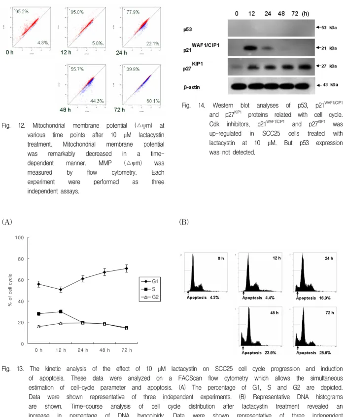
producing a concomitant fall in the percentage of S phase cells. The increase in the G1 phase cell percentage and concomitant fall in the percentage of S phase cells paralleled an increase in apoptotic cell portions (Fig. 13A & 13B). Furthermore, to investigate the expression of important cell-cycle proteins, western blot assay was conducted. In lactacystin treated SCC25 cells, the accumulation of p53 protein was not detected over a period of 72 h.
But the protein level of CDK inhibitor, p27
KIP1was remarkably up-regulated in a time-dependent manner. Furthermore, the remarkable up-regulation of another CDK inhibitor, p21
WAF1/CIP1was shown at 12 h and was not detected after 48 h (Fig. 14).
Ⅳ. DISCUSSION
Proteasome inhibitor was able to increase the
anti-proliferative effect of certain drugs and to
prevent the development of resistance to teniposide
induced by brefeldin A.
12)In addition, a number of
studies have shown that the proteasome inhibitor can
induce cell death. Furthermore, it has been shown
that proteasome inhibitors prevented the proliferation
of cancer cells in vivo as well as in vitro.
8,20,21)Lactacystin, a microbial natural product synthesized
(A) (B)
Fig. 10. Bax and cytochrome c involve apoptosis in SCC25 cells treated with lactacystin at 10 μM. (A) Western blotting analyses of Bax and Bcl-xL. Expression level of Bax increased compared to the control in a time-dependent manner. But expression level of Bcl-xL did not decreased. β-actin, a loading control.
(B) Confocal microscopy showing the release of cytochrome c from mitochondria into the cytosol. Scale bar, 10 μm.
(A) (B)
Fig. 11. AIF involves apoptosis in SCC25 cells treated with lactacystin at 10 μM. (A) Western blotting of AIF.
Expression level of AIF increased compared to the control over a period of 24 h. β-actin, a loading control. (B) Confocal microscopy showing that AIF was released from mitochondria, and that translocation onto nuclei was evident in SCC25 cells. Scale bar, 10 μm.
by Streptomyces, has been commonly used as a selective proteasome inhibitor in many studies.
In MTT assay, lactacystin showed a dose- and time-dependent decrease in the viability of SCC25 cells but not in human keratinocytes (HaCaT cells) and human gingival fibroblast (HGF-1 cells).
Moreover, the colony forming assay confirmed that lactacystin ranging from 1 to 3 μM remarkably inhibited the growth of SCC25 cells. Interestingly, lactacytin remarkably revealed cytotoxicity in SCC25 cells but not normal cells. These data indicate that lactacystin exerts a specific cytotoxic effect on SCC25 cells. Based on these data, lactacystin could have an anti-cancer effect for human oral squamous
cell carcinoma.
Apoptosis and necrosis are conceptually distinct forms of cell death and can be distinguished by their specific morphological and biological changes.
During apoptosis, the cells undergo specific
morphological and biochemical changes such as cell
blebbing, reduction of cell size, cell shrinkage,
chromatin condensation and DNA fragmentation.
3,22)In the results of Hoechst stain and TUNEL assay,
SCC25 cells treated with lactacystin showed
apoptotic hallmarks such as the formation of
apoptotic bodies and the increase of TUNEL-positive
cells. These results indicate that lactacystin induced
SCC25 cell death via activating the apoptosis.
Fig. 12. Mitochondrial membrane potential (△ψm) at various time points after 10 μM lactacystin treatment. Mitochondrial membrane potential was remarkably decreased in a time- dependent manner. MMP (△ψm) was measured by flow cytometry. Each experiment were performed as three independent assays.
(A) (B)
0 20 40 60 80 100
0 h 12 h 24 h 48 h 72 h
% of cell cycle
G1 S G2
Fig. 13. The kinetic analysis of the effect of 10 μM lactacystin on SCC25 cell cycle progression and induction of apoptosis. These data were analyzed on a FACScan flow cytometry which allows the simultaneous estimation of cell-cycle parameter and apoptosis. (A) The percentage of G1, S and G2 are depicted.
Data were shown representative of three independent experiments. (B) Representative DNA histograms are shown. Time-course analysis of cell cycle distribution after lactacystin treatment revealed an increase in percentage of DNA hypoploidy. Data were shown representative of three independent experiments.
Fig. 14. Western blot analyses of p53, p21WAF1/CIP1 and p27KIP1 proteins related with cell cycle.
Cdk inhibitors, p21WAF1/CIP1 and p27KIP1 was up-regulated in SCC25 cells treated with lactacystin at 10 μM. But p53 expression was not detected.
Apoptotic stimuli may induce apoptosis by inhibiting the proteasome activity of the target cells.
9)However, other studies have reported that a proteasome inhibitor itself can induce apoptosis in certain cells.
10)Generally, the proteasome-mediated step(s) in apoptosis are located at the upstream of mitochondrial changes and caspase activation, and can involve different systems, including various cyclins, p53, NF-kB, Bax and Bcl-2.
6,23,24)Thus, the possibility existed that lactacystin may have affected proteasome activity in SCC25 cells and induced the mitochondrial pathway of apoptosis. In this study, lactacystin treated SCC25 cells showed a remarkable reduction in a time-dependent manner.
Mitochondria are known to be central death regulators in response to several apoptotic stimuli.
5)The induction of increased mitochondrial permeability plays a definite role in the regulation of apoptosis.
5,25-27)The mitochondrial pathway can also be triggered by various intracellular and extracellular stress signals, which result in activation of pro-apoptotic proteins, including Bax and Bak, or inactivation of anti-apoptotic Bcl-2 family members, such as Bcl-2 and Bcl-xL.
27)As a result of activation/inactivation of Bcl-2 family proteins, changes in mitochondrial membrane lead to the dissipation of inner membrane potential and permeabilization of the outer mitochondrial membrane (OMM). These in turn induce the release of various proapoptotic proteins, such as cytochrome c, Smac/Diablo, endonuclease G and AIF.
28-30)It has been reported that the pro-apoptotic Bcl-2 family in isolated mitochondria induces cytochrome c release, the loss of mitochondrial membrane potential and results in AIF release.
31,32)Cytochrome c release and disruption of mitochondrial membrane potential (MMP) are known to contribute to apoptosis triggered by proteasome inhibition.
33,34)Generally, cytochrome c is released into the cytosol during apoptosis, where it binds to Apaf-1. This cytochrome c/Apaf-1 complex (the apoptosome) promotes the autoactivation of procaspase-9 to caspase-9.
Caspase-9 then acts on procaspase-3 to initiate a caspase activation cascade.
35,36)Released AIF
through proapoptotic Bcl-2 family activation induces its translocation to the nucleus, resulting in chromatin condensation and large-scale DNA fragmentation.
37)In the present study, lactacystin treatment also induced Bax up-regulation, translocation of AIF into nuclei, cytochrome c release from mitochondria into the cytosol and a significant loss of MMP These data here elucidate that the lactacystin-induced apoptosis of SCC25 cells involves the representative mitochondrial events as mentioned above.
Multiple lines of evidence indicate that apoptosis
can be triggered by the activation of caspase.
4)Commonly, the final event of apoptosis is nuclear
condensation, which is controlled by the known
caspase substrates, DFF and PARP.
38)Caspases,
which are aspartate-specific intracellular cysteine
proteases, play an essential role during apoptotic cell
death.
39)Once activated, the effector caspases
(caspase-3, caspase-6 or caspase-7) are responsible
for the proteolytic cleavage of a broad spectrum of
cellular targets, ultimately leading to cell death. The
known cellular substrates include structural
components (such as actin and nuclear lamin),
inhibitors of deoxyribonuclease (such as DFF45
(ICAD)) and DNA repair proteins (such as
PARP).
40,41)In apoptotic cells, activation of DFF40
(CAD), also known as a substrate of caspase-3,
occurs with the cleavage of DFF45 (ICAD). Once
DFF40 (CAD) is activated and released from the
complex of DFF45 (ICAD) and DFF40 (CAD), it can
translocate to the nucleus where it degrades
chromosomal DNA and produces DNA
fragmentation.
42)In this study, cleavage of
caspase-7, DFF45 (ICAD), and PARP and
degradation of caspase-3, -7, DFF45 (ICAD), and
PARP were shown in lactacystin-treated SCC25
cells. Furthermore, confocal microscopy showed that
lactacystin led to the translocation of DFF40 (CAD)
from cytosol into nuclei in SCC25 cells. Therefore,
these data demonstrate that lactacystin-induced
apoptosis in SCC25 cells is associated with
caspase-3 and caspase-7 activation, and that
activated caspase-3 and caspase-7 leads to the
cleavage of PARP and DFF45 (ICAD), and translocation of DFF40 (CAD) from cytosol into nucleus.
Various studies on the molecular analyses of cancers have revealed that cell cycle regulators are frequently mutated in most common malignancies.
Therefore, control of cell cycle progression in tumor cells is considered to be a potentially effective strategy for the control of tumor growth.
43)It has been reported that lactacystin is able to inhibit differentiation of a neuroblastoma cell line as well as cell cycle progression.
44)The present study also demonstrated that lactacystin inhibited SCC25 cells growth by G1 cell cycle arrest and apoptosis. Our flow cytometry data indicated that the increase in the G1 phase cell percentage and concomitant fall in the percentage of S phase cells paralleled an increase in apoptotic cell portion.
The p53 tumor suppressor is normally degraded by the proteasome.
6)Cells treated with proteasome inhibitors were noted to undergo apoptosis and, in parallel, to accumulate p53 in cells.
45)Also, proteasome inhibition is deficient to induce apoptosis in HL60 cells, which are p53-null,
7)and U937 cells undergo apoptosis without detectable accumulation of p53.
46)Generally, p53 protein induced the Cdk inhibitor p21
WAF1/CIP1transactivation which, in combination with cyclin D and Cdk4, will result in G1/S arrest.
6)Likewise, another Cdk inhibitor p27
KIP1, is present in maximal amounts during the G0 and G1 phases of the cell cycle progression, decreasing when the cell is induced to enter into proliferation.
47)Recent studies have demonstrated that p27
KIP1is degraded by the ubiquitin-mediated pathway. Also it has been associated with cytotoxicity caused by proteasome inhibition which lead to the conclusion that p27
KIP1casuse apoptosis.
48)In present study, we investigated the expression level of these proteins after the treatment of lactacystin. Our results showed that the accumulation of p53 protein was not detected in SCC25 which were p53-null cell, and p21
WAF1/CIP1showed the remarkable up-regulation at 12 h.
Besides, the expression level of p27
KIP1showed a significant increase in a time-dependent manner.
These data show that significant up-regulation of p21
WAF1/CIP1and p27
KIP1plays a crucial role in lactacystin-induced G1 cell cycle arrest. In addition, we suggest that up-regulation of p27
KIP1regarded as an essential protein of G1 cell cycle arrest is associated with proteasome inhibition in lactacystin treated SCC25 cells. Although the up-regulated level of p21
WAF1/CIP1and p27
KIP1may represent their role in this G1 cell cycle arrest, further investigation is needed.
In conclusion, lactacystin induces apoptosis via proteasome, mitochondrial and caspase pathways in SCC25 cells. Our experimental data contribute to new insights to the role of lactacystin and support the view that the clinical effect of lactacystin may depend on its pharmacological efficacy in regulating human oral squamous cell carcinoma cells.
Furthermore we suggest that lactacystin treatment can be considered as a novel therapeutic strategy for human tongue squamous cell carcinoma from its cell cycle arrest and apoptosis-inducing activity.
V. CONCLUSION
Lactacystin, a microbial natural product synthesized by Streptomyces, has been commonly used as a selective proteasome inhibitor in many studies. Proteasome inhibitors is known to be preventing the proliferation of cancer cells in vivo as well as in vitro. Furthermore, proteasome inhibitors, as single or combined with other anticancer agents, are suggested as a new class of potential anticancer agents. This study was undertaken to examine in
vitro effects of cytotoxicity and growth inhibition,and the molecular mechanism underlying induction of apoptosis in SCC25 human tongue sqaumous cell carcinoma cell line treated with lactacystin.
The viability of SCC25 cells, human normal
keratinocytes (HaCaT cells) and human gingiva
fibroblasts (HGF-1 cells), and the growth inhibition
of SCC25 cells were assessed by MTT assay and
clonogenic assay respectively. The hoechst staining,
hemacolor staining and TUNEL staining were
conducted to observe SCC25 cells undergoing
apoptosis. SCC25 cells were treated with lactacystin, and Western blotting, immunocytochemistry, confocal microscopy, FAScan flow cytometry, MMP activity, and proteasome activity were performed.
Lactacystin treatment of SCC25 cells resulted in a time- and does-dependent decrease of cell viability and a does-dependent inhibition of cell growth, and induced apoptotic cell death. Interestingly, lactacytin remarkably revealed cytotoxicity in SCC25 cells but not normal cells. And tested SCC25 cells showed several lines of apoptotic manifestation such as nuclear condensation, DNA fragmentation, the reduction of MMP and proteasome activity, the decrease of DNA contents, the release of cytochrome c into cytosol, the translocation of AIF and DFF40 (CAD) onto nuclei, the up-regulation of Bax, and the activation of caspase-7, caspase-3, PARP, lamin A/C and DFF45 (ICAD). Flow cytometric analysis revealed that lactacystin resulted in G1 arrest in cell cycle progression which was associated with up-regulation in the protein expression of CDK inhibitors, p21
WAF1/CIP1and p27
KIP1.
We presented data indicating that lactacystin induces G1 cell cycle arrest and apoptois via proteasome, mitochondria and caspase pathway in SCC25 cells. Therefore our data provide the possibility that lactacystin could be as a novel therapeutic strategy for human tongue squamous cell carcinoma.
REFERENCES
1. Carson DA, Ribeiro JM. Apoptosis and disease. Lancet 1993;341:1251-1254.
2. Ohta K, Yamashita N. Apoptosis of eosinophils and lymphocytes in allergic inflammation. J Allergy Clin Immunol 1999;104:14-21.
3. Williams GT. Programmed cell death: apoptosis and oncogenesis. Cell 1991;65:1097-1098.
4. Thornberry NA, Rosen A, Nicholson DW. Control of apoptosis by proteases. Adv In Pharmacol 1997;41:
155-177.
5. Green DR and Reed JC. Mitochondria and apoptosis.
Science 1998;281:1309-1312.
6. Orlowski RZ. The role of the ubiquitin-proteasome pathway in apoptosis. Cell Death Differ 1999;6:303-313.
7. Drexler HC. Activation of the cell death program by inhibition of proteasome function. Proc Natl Acad Sci USA 1997;94:855-860.
8. Orlowski RZ, Eswara JR, Lafond-Walker A, Grever MR, Orlowski M, Dang CV. Tumor growth inhibition induced in a murine model of human Burkitt's lymphoma by a proteasome inhibitor. Cancer Res 1998;58:4342-4348.
9. Meng L, Kwok BH, Sin N, Crews CM. Eponemycin exerts its antitumor effect through the inhibition of proteasome function. Cancer Res 1999;59:2798-2801.
10. Drexler HC, Risau W, Konerding MA. Inhibition of proteasome function induces programmed cell death in proliferating endothelial cells. FASEB J 2000;14:65-77.
11. Delic J, Masdehors P, Omura S et al. The proteasome inhibitor lactacystin induces apoptosis and sensitizes chemo- and radio-resistant human chronic lymphocytic leukemia lymphocytes to TNF-alpha- initiated apoptosis. Br J Cancer 1998;77:1103-1107.
12. Lin ZP, Boller YC, Amer SM et al. Prevention of brefeldin A- induced resistance to teniposide by the proteasome inhibitor MG-132: involvement of NF-kappaB activation in drug resistance. Cancer Res 1998;58:3059-3069.
13. Adams J. Development of the proteasome inhibitor PS-341. Oncologist 2002;7:9-16.
14. An WG, Hwang SG, Trepel JB, Blagosklonny MV.
Protease inhibitor-induced apoptosis: accumulation of wt p53, p21WAF1/CIP1, and induction of apoptosis are independent markers of proteasome inhibition.
Leukemia 2000;14:1276-1283.
15. Pasquini LA, Besio Moreno M, Adamo AM, Pasquini JM, Soto EF. Lactacystin, a specific inhibitor of the proteasome, induces apoptosis and activates caspase-3 in cultured cerebellar granule cells. J Neurosci Res.
Mar 2000;59:601-611.
16. Inoue T, Shiraki K, Fuke H et al. Proteasome inhibition sensitizes hepatocellular carcinoma cells to TRAIL by suppressing caspase inhibitors and AKT pathway.
Anticancer Drugs. 2006;17:261-268.
17. Kudo Y, Takata T, Ogawa I et al. p27Kip1 accumulation by inhibition of proteasome function induces apoptosis in oral squamous cell carcinoma cells. Clin Cancer Res 2000;6:916-923.
18. Takezawa J, Ishimi Y, Yamada K. Proteasome inhibitors remarkably prevent translesion replication in cancer cells but not normal cells. Cancer Sci 2008;99:
863-871.
19. Shen J, Huang C, Jiang L et al. Enhancement of
cisplatin induced apoptosis by suberoylanilide hydroxamic acid in human oral squamous cell carcinoma cell lines. Biochem Pharmacol 2007;73:
1901-1909.
20. Adams J, Palombella VJ, Sausville EA et al.
Proteasome inhibitors: a novel class of potent and effective antitumor agents. Cancer Res 1999;59:
2615-2622.
21. Kim JH, Bae HR, Park BS et al. Early mitochondrial hyperpolarization and intracellular alkalinization in lactacystin-induced apoptosis of retinal pigment epithelial cells. J Pharmacol Exp Ther 2003;305:
474-481.
22. Wyllie AH, Kerr JF, Currie AR. Cell death: the significance of apoptosis. Int Rev Cytol 1980;68:
251-306.
23. Grimm LM, Goldberg AL, Poirier GG, Schwartz LM, Osborne BA. Proteasome play an essential role in thymocyte apoptosis. EMBO J 1996;15:3845-3852.
24. Li B, Dou QP. Bax degradation by the ubiquitin/
proteasome-dependent pathway: Involvement in tumor survival and progression. Pro Natl Acad Sci USA 2000;97:3850-3855.
25. Kroemer G, Zamzami N, Susin SA. Mitochondrial control of apoptosis. Immunol Today 1997;18:44-51.
26. Susin SA, Lorenzo HK, Zamzami N et al. Molecular characterization of mitochondrial apoptosis-inducing factor. Nature 1999;397:441-446.
27. Orrenius S. Mitochondrial regulation of apoptotic cell death. Toxicol Lett 2004;149:19-23.
28. Barczyk K, Kreuter M, Pryjma J et al. Serum cytochrome c indicates in vivo-apoptosis and it can serve as a prognostic marker during cancer therapy. In.
J Cancer 2005;114:167–173.
29. Brouckaert G, Kalai M, Saelens X, Vandenabeele P.
Apoptotic Pathways and Their Regulation. In: Los, M., Gibson, S.B. (Eds.), Apoptotic Pathways as Target for Novel Therapies in Cancer and Other Diseases. New York. 2005, Springer Academic Press
30. Hengartner MO. The biochemistry of apoptosis. Nature 2000;407:770-776.
31. Hunter JJ, Parslow TG. A peptide sequence from Bax that converts Bcl-2 into an activator of apoptosis. J Biol Chem 1996;271:8521-8524.
32. Narita M, Shimizu S, Ito T et al. Bax interacts with the permeability transition pore to induce permeability transition and cytochrome c release in isolated mitochondria.Proc Natl Acad Sci U S A. 1998;95:
14681-14686.
33. Wagenknecht B, Hermission M, Groscurth P, Liston P, Krammer PH, Weller M. Proteasome inhibitor-induced apoptosis of glioma cells involves the processing of multiple caspases and cytochrome c release. J Neurochem 2000;75:2288-2297.
34. Marshansky V, Wang X, Bertrand R et al. Proteasomes modulate balance among proapoptotic and antiapoptotic Bcl-2 family members and compromise functioning of the electron transport chain in leukemic cells. J Immunol 2001;166:3130-3142.
35. Li P, Nijhawan D, Budihardjo I et al. Cytochrome c and dATP-dependent formation of Apaf-1/caspase-9 complex initiates an apoptotic protease cascade. Cell 1997;91:479-489.
36. Zou H, Li Y, Liu X, Wang, X. An APAF-1, cytochrome c multimeric complex is a functional apoptosome that activates procaspase-9. J Biol Chem 1999;274:11549–
11556.
37. Dauglas E, Susin SA, Zamzami N et al.
Mitochondrio-nuclear translocation of AIF in apoptosis and necrosis. FASEB J 2000;14:729-739.
38. Thornberry NA, Lazebnik Y. Caspase: Enemies within.
Science 1998;28:1312-1316.
39. Acehan D, Jiang X, Morgan DG, Heuser JE, Wang X, Akey CW. Three-dimensional structure of the apoptosome: Implications for assembly, procaspase-9 binding, and activation. Mol Cell 2002;9:423-432.
40. Gross A, McDonnell JM, Korsmeyer SJ. BCL-2 family members and the mitochondria in apoptosis. Genes Dev 1999;13:1899-1911.
41. Porter AG. Protein translocation in apoptosis. Trends Cell Biol 1999;9:394-401.
42. Cheng AC, Jian CB, Huang YT, Lai CS, Hsu PC, Pan MH. Induction of apoptosis by Uncaria tomentosa through reactive oxygen species production, cytochrome c release, and caspases activation in human leukemia cells. Food Chem Toxicol 2007;45:2206-2218.
43. Pavletich NP. Mechanisms of cyclin-dependent kinase regulation: structures of Cdks, their cyclin activators, and Cip and INK4 inhibitors. J Mol Biol. 1999;287:
821-828.
44. Fenteany G, Standaert RF, Lane WS, Choi S, Corey EJ, Schreiber SL. Inhibition of proteasome activities and subunit specific-amino terminal threonine modification by lactocystin. Science 1995;268:726–731.
45. Shinohara K, Tomioka M, Nakano H, Toné S, Ito H, Kawashima S. Apoptosis induction resulting from proteasome inhibition. Biochem J 1996;317:385-388.
46. Meriin AB, Gabai VL, Yaglom J, Shifrin VI, Sherman MY. Proteasome inhibitors activate stress kinases and induce Hsp72. Diverse effects on apoptosis. J Biol Chem 1998;273:6373-6379.
47. Elledge S, Harper W. Cdk inhibitors: on the threshold of checkpoints and development. Curr Op Cell Biol 1994;6:847–852.
국문초록
사람혀편평상피세포암종세포에서 proteasome 억제제인 lactacystin에 의해 유도된 세포자멸사의 기전에 대한 연구
부산대학교 치의학전문대학원 구강해부학교실1
부산대학교 치의학전문대학원 구강내과학교실2
백철중
1․김규천
1․김인령
1․이승은
1․곽현호
1․박봉수
1․ 태일호
2․고명연
2․안용우
2Sreptomyces라는 세균에서 추출한 lactacystin은 선택적인 proteasome 억제제로서 많은 연구에서 사용되어져 왔다.
Proteasome 억제제는 최근의 많은 연구를 통해서 암세포증식의 억제에 대한 효과가 증명되었으며, 특히 다른 항암제와 병용처 리 시, 상호작용에 의한 상승효과가 있다고 알려져 있다. 현재 proteasome 억제제는 새로운 강력한 항암제로서 분류되어 있다.
본 연구는 사람혀편평세포암종세포(SCC25 cells)에서 lactacystin의 세포독성과 성장억제 효과, 그리고 세포자멸사의 유도에 대한 분자생물학적 기전을 밝히기 위해 실험을 시행하였다.
SCC25 세포, 사람정상각화세포 (HaCaT cells) 그리고 사람치은섬유모세포(HGF-1 cells)의 생존율 측정은 MTT법을 시행 하였고, SCC25 세포의 성장억제를 확인하기 위해서는 clonogenic assay를 사용하였다. lactcystin이 SCC25 세포에서 세포자멸 사가 유도되는지를 확인하기 위해서 hoechst 염색법, hemacolor 염색법 그리고 TUNEL법을 시행하였다. 그리고 SCC25 세포 에 lactacystin을 적용한 후, Western blot 분석, 세포면역화학염색, 공초점레이저주사현미경 검경, FACScan flow cytometry, 사립체막 전위변화, proteasome 활성도 측정 등을 시행하였다.
Lactacystin으로 처리된 SCC25 세포는 시간 및 용량 의존적인 세포생존율의 감소, 용량의존적인 세포성장억제 그리고 세포 자멸사에 의한 세포죽음을 보였다. 흥미롭게도 lactacytin은 정상세포인 HaCat 세포와 HGF-1 세포에서는 세포독성을 전혀 보이지 않았다. 그리고 lactacystin이 적용된 SCC25세포에서 핵 응축, DNA의 조각남, 사립체막전위와 proteasome 활성도의 감소, DNA 양의 감소, cytochrome c의 사립체에서의 세포질로의 유리, AIF와 DFF40 (CAD)의 핵으로의 이동, Bax의 증가, caspase-7, caspase-3, PARP, lamin A/C 그리고 DFF45 (ICAD)의 활성화 혹은 파괴와 같은 아주 다양한 세포자멸사 증거를 보였다. Flow cytometry 분석에서는 CDK 억제제인 p21WAF1/CIP1와 p27KIP1의 발현 증가와 관계있는 것으로 추정되어 지는 G1 세포주기 정지를 보였다.
이러한 결과는 lactacytin이 SCC25 세포에서 G1 세포주기정지와 proteasome, 사립체 및 caspase 경로의 연속반응을 통한 세포자멸사를 유도함을 명확하게 증명하고 있다. 이와 같은 세포주기 정지와 세포자멸사 유도능은 lactacytin이 사람혀편평상 피세포암종의 새로운 치료전략으로서의 가능성을 제공한다고 생각한다.
주제어: 세포자멸사, Proteasome inhibitor, Lactacystin, 사람혀편평상피암종
48. Pagano M, Tam SW, Theodoras AM et al. Role of the ubiquitin-proteasome pathway in regulating abundance of the cyclin-dependent kinase inhibitor p27. Science 1995;269:682–685.