• 검색 결과가 없습니다.

Tannic acid-induced apoptosis in FaDu hypopharyngeal squamous cell carcinoma

N/A
N/A
Protected

Academic year: 2021

Share "Tannic acid-induced apoptosis in FaDu hypopharyngeal squamous cell carcinoma"

Copied!
7
0
0

로드 중.... (전체 텍스트 보기)

전체 글

(1)

Introduction

Head and neck squamous cell carcinoma (HNSCC) is a group of cancer mostly arising in head and the region comprising epithelial layer of oral cavity, nasal cavity, larynx and pharynx [1,2]. It is not clear what causes the mutations in squamous cells leading to HNSCC but alcohol use and tobacco smoking have been identified as risk factors. Statistical research in the UK showed that more than 75% of mouth and oropharyngeal cancers are attributed to the consumption of alcohol and to- bacco [3,4]. In addition these factors have synergistic effects that people who use both alcohol and tobacco are at higher

risk of oral cancer because alcohol has dehydrating effect on cell membrane inducing specific carcinogens in tobacco to be absorbed into oral cells [5]. Other risk factors which contribute to the incidence of HNSCC in community include human papil- loma virus infection, chronic immunodeficiency and exces- sive sunlight exposure [3]. Despite the extensive efforts for early diagnosis and development in therapeutics, over half of patients with HNSCC were diagnosed at advanced stage and high proportion of them developed cancer [6]. Conventional treatment was not successful in improving five-year survival rate of HNSCC patients which remains in the ranges of 40% to 50%. Surgery or radiotherapy was applied for the treatment of Int J Oral Biol 44:43-49, 2019

pISSN: 1226-7155 • eISSN: 2287-6618 https://doi.org/10.11620/IJOB.2019.44.2.43

Tannic acid-induced apoptosis in FaDu hypopharyngeal squamous cell carcinoma

Loan Thi Ta 1 , Trang Thi Kieu Nguyen 2 , and Hoon Yoo 2 *

1 Institute of Biotechnology, Vietnam Academy of Science and Technology, Hanoi 1000, Vietnam

2 Department of Pharmacology and Dental Therapeutics, College of Dentistry, Chosun University, Gwangju 61452, Republic of Korea

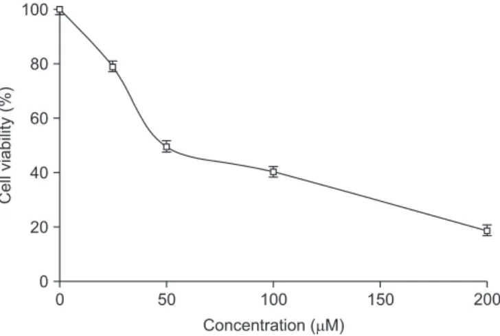
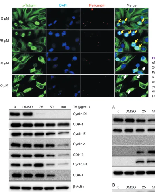
Tannic acid (TA) is a water-soluble polyphenol compound found in various herbal plants. We investigated the chemopreventive effects of TA on FaDu hypopharyngeal squamous carcinoma cells. In an 3-(4,5-dimethylthiazol-2-yl)- 2,5-diphenyltetrazolium bromide (MTT) assay, TA showed dose-dependent cytotoxicity with a half maximal inhibitory concentration (IC50) of 50 µM. Cell cycle analysis and immunofluorescence imaging demonstrated that under low- dose (25 µM) treatment, FaDu cells were arrested in G2/M phase, and as the dose of TA was increased, apoptosis was induced with the increase of cell population at sub-G1 phase. The expressions of various cyclins, including cyclin D1 and cyclin-dependent kinases (CDK-1 and CDK-2), were down-regulated at low doses of TA, whereas apoptotic effectors such as cleaved caspase 3, cleaved caspase 7, and poly (ADP-ribose) polymerase (PARP) were expressed in a dose-dependent manner in Western blotting. In addition, TA-induced apoptosis of FaDu cells might be mediated by the extracellular signal-regulated kinase (ERK)/mitogen-activated protein kinase pathway, with the upregulation of p-AKT/p-PKB (phosphorylated protein kinase B) and p-ERK. Overall, our data support the hypothesis that TA is a potential candidate agent for the treatment of hypopharyngeal cancer.

Keywords: Apoptosis, Hypopharyngeal cancer, FaDu cells, Tannic acid

Received May 7, 2019; Revised June 5, 2019; Accepted June 6, 2019

*Correspondence to: Hoon Yoo, E-mail: hoon_yoo@chosun.ac.kr https://orcid.org/0000-0002-9249-1446 Copyright © The Korean Academy of Oral Biology

CC

This is an open-access article distributed under the terms of the Creative Commons Attribution Non-Commercial License (http://creativecommons.org/licenses/by- nc/4.0/), which permits unrestricted non-commercial use, distribution, and reproduction in any medium, provided the original work is properly cited.

Original Article IJOB

(2)

the early stage while multimodal treatment such as combina- tion of radiotherapy with chemotherapy and/or molecular-tar- geted therapies using monoclonal antibodies were applied for the advanced HNSCCs [7]. Especially hypopharyngeal cancer which shows the worst prognosis and the lowest 5-year sur- vival rate (25% to 40%) [8,9] is in urgent need for the efficient therapeutics.

Tannic acid (TA), also called gallotannin, is a water-soluble polyphenols with multi-gallic acid moieties in chemical struc- ture which is found in various plants. Especially grape and green tea contain high level of TA, and in the production of commercial wine and tea TA plays a critical role for the genera- tion of color and flavor. TA is also used as a flavoring agent in food industry or as additives in ink manufacturing [10]. Previ- ous reports showed that TA has various therapeutic effects of anti-inflammation [11], antioxidation [12,13], and anti-tumor [14,15].

For anti-tumor effects, TA suppressed cell proliferation on cancer cell lines such as prostate cancer [16], breast cancer [17,18], and malignant glioma [19]. Interestingly, TA effects on tumor cell lines were cell-type dependent. In breast cancer cells, TA induced G1-arrest and apoptosis through regulating both canonical and non-canonical signal transducer and acti- vator of transcription (STAT) pathway [18] whereas TA inhib- ited cell cycle arrest at G2/M phase and stimulated apoptosis through p53 induction in T98G glioma cell lines [20]. In case of gingival cancer cells, TA exhibited apoptosis by cell cycle arrest at G1 phase by inhibiting Jak2/STAT3 pathway [21]. Thus, our intent in this study was to evaluate whether TA has any spe- cific cellular effects on hypopharyngeal squamous cancer cells and if so what would be the underlying mechanism to eluci- date. Here, we report that TA suppresses the proliferation of FaDu hypopharyngeal squamous cells by arresting cell cycle at G2/M phase and induces apoptosis possibly through the sig- naling pathway of extracellular signal-regulated kinase (ERK)/

mitogen-activated protein kinase (MAPK) and AKT.

Materials and Methods

1. Materials and reagents

Dimethyl sulfoxide (DMSO), 3-(4,5-dimethylthiazol- 2-yl)-2,5-diphenyltetrazolium bromide (MTT), TA was pur- chased from Sigma Aldrich (St Louis, MO, USA). Ten percent fetal bovine serum (FBS), 0.05% trypsin-EDTA (ethylene diamine tetraacetic acid) and antibiotic-antimycotic solution

were obtained from WELGENE (Gyeongsan, Korea). Primary antibodies of cyclins, cyclin-dependent kinases (CDKs), pro- caspase 3 and caspase 9 were from Santa Cruz Biotechnology Inc. (Santa Cruz, CA, USA) and cleaved caspase 3, cleaved caspase 7 and phosphorylated-ERK (p-ERK) were from Cell Signaling Technology Inc. (Danver, CA, USA). Anti-tubulin, anti-pericentrin and cleaved-PARP (poly [ADP-ribose] poly- merase) antibodies were purchased from Abcam (Cambridge, MA, USA). Secondary antibodies were purchased from Novus Biologicals (Centennial, CO, USA).

2. Cell culture

FaDu hypopharyngeal squamous cell carcinoma was ob- tained from Korean Cell Line Bank (Seoul, Korea) and cultured in minimun essential media (MEM) media containing 10% FBS and 1% amoxicillin and streptomycin. The cells were seeded at the density of 2 × 10 5 cells/mL and incubated in 37℃, 5% CO 2 for 24 hours.

3. MTT assay

Two × 10 4 cells/well were seeded in 96-well plate and incu- bated for 24 hours. After treating with various concentrations of TA for 24 hours, the media were replaced by 200 µL of MTT solution (0.5 mg/mL). The cells were further incubated for 4 hours at 37℃ until formazan crystals were formed. The old media were aspirated and 200 µL of DMSO was added to dis- solve formazan. The absorbance was measured at 540 nm by using Multiskan TM FC Microplate Photometer (Thermo Fisher Scientific Inc., Waltham, MA, USA).

4. Cell cycle analysis

FaDu cells were seeded in 100 mm culture dish for 24 hours

and then followed by TA treatment at a concentration range

from 0 to 100 µM. After 24 hours, cells were trypsinized,

washed and fixed in 70% ethanol. The fixed cells were washed

by phosphate buffered saline (PBS) to remove ethanol trace,

treated with ribonuclease (200 µg/mL) and stained with prop-

idium iodide (50 µg/mL). Flow cytometer was used to identify

DNA content of 10,000 cells per sample and the data was

analyzed by Cellquest software (BD Biosciences, San Jose,

CA, USA).

(3)

5. Western blotting

After TA treatment on FaDu cells, cells were collected to carry out protein extraction. The total proteins were separated in sodium dodecyl sulfate –polyacrylamide gel electrophoresis (SDS-PAGE) and transferred to polyvinylidene difluoride (PVDF) membranes. The membranes were blocked with skim milk 5%

in PBS with Tween 20 for 1 hour, incubated overnight at 4℃

with primary antibody of a target protein. After washing (×3) with tris buffered saline (TBS) 0.1% Tween-20 for 15 minutes, the membrane was incubated with corresponding horserad- ish peroxidase (HRP)-conjugated secondary antibodies for 2 hours at room temperature, and then followed by additional washing (×3). Proteins were exposed with chemiluminescent HRP substrate (Millipore Corporation, Billerica, MA, USA) and detected by imaging system (Kodak image station; Bruker Bio- Spin, Billerica, MA, USA).

6. Immunofluorescence imaging

FaDu cells (1 × 10 5 /well) were seeded in coverslip contain- ing 24-well plate and incubated for 24 hours prior to TA treat- ment. After treating with TA, cells were washed with PBS and fixed with 4% paraformaldehyde for 30 minutes at room tem- perature. The chamber slides were incubated with antibodies of tubulin or pericentrin overnight at 4℃. Prior to the treatment with secondary antibodies, cells were completely washed with TBS plus 0.1% Tween to eliminate residual unconjugated anti- bodies. For nuclei staining DAPI (4’,6-diamino-2-phenylindole)

was added and incubated for 20 minutes at room tempera- ture. After washing with TBS, cells on coverslips were finally observed by a fluorescence microscopy (Zeiss International, Oberkochen, Germany).

Results

1. The cytotoxicity of tannic acid on FaDu cell

The cytotoxicity of TA on FaDu cell growth was examined by an MTT assay. TA significantly inhibited cell growth dose- dependently in the range of 0 to 200 µM with 50 µM of IC50.

Treating cells with 200 µM of TA for 24 hours caused 80% of cell death (Fig. 1).

2. Tannic acid induced cell cycle arrest and apoptosis

Flow cytometry was used to examine the cell cycle distribu- tion under the TA treatment. The analysis on cell cycle revealed that under the low concentration of TA (25 µM) cells were sig- nificantly inhibited in cell cycle progression with 30% of cells arrested at G2/M phase. Meanwhile, cell population at sub-G1 phase was dose-dependently increased indicating that high dose of TA induced apoptosis on FaDu cells (Fig. 2). To further confirm the changes in cell division and cellular nucleus, the effect of TA on cell cycle progression was examined by immu- nofluorescence imaging. Under the treatment of 25 µM TA for 24 hours, the number of cells in division process was increased with doubled centrosomes and bipolar mitotic spindles while at higher doses cells were in apoptosis with nuclei condensed

Fig. 1. Tannic acid (TA) inhibited the growth of FaDu cells. Cells were treated with various concentrations of TA and incubated for 24 hours. MTT (3-(4,5-dimethylthiazol-2-yl)-2,5-diphenyltetrazolium bromide) assay was carried out to determine the cell viability. Vertical bars indicate means and standard errors (n = 3).

G0/G1 100

80

60

40

20

Cell (%)

Cell cycle 0

S G2/M Sub G1

0 25 50 100


  M

M M M

Fig. 2. Tannic acid (TA) regulated cell cycle distribution. FaDu cells were

treated with TA (0, 25, 50, and 100 µM) and incubated for 24 hours. TA

treated cells were stained with propidium iodide and analyzed by flow cy-

tometry.

(4)

and fragmented (Fig. 3).

3. Tannic acid suppressed cell cycle regulatory proteins

The effects of TA were further investigated by Western blot- ting to elucidate the expression of regulatory proteins in cell cycle. As shown in Fig. 4, TA inhibited the expression of cyclins and CDKs. Noticeably cyclin D1 was dramatically suppressed at low dose (25 µM) condition while the expression of cyclin

D1 dependent kinase CDK-4 was slightly at 25 µM and gradu- ally decreased at higher doses condition. Other cyclins (cyclin A, B1, and E) and CDKs (CDK-1 and CDK-2) were also down- 0 M 

25 M 

50 M 

100 M 

-Tubulin DAPI Pericentrin Merge

Fig. 3. Immunofluorescence staining images of FaDu cells. After tannic acid treatment for 24 hours, cells were fixed with paraformalde- hyde and stained for the following detection:

α-tubulin for microtubules, DAPI for nuclei and pericentrin antibody for centrosomes. The yellow arrows indicate the cell at G2/M phase and the white arrows the cells in apoptosis (×400).

0 DMSO 25 50 100 TA ( g/mL) 

Cyclin D1

CDK-4 Cyclin E Cyclin A

CDK-2 Cyclin B1

CDK-1

-Actin

Fig. 4. Tannic acid (TA) suppressed the expression of cell cycle regulatory proteins. The cells were treated with 25, 50, 100 µM of TA for 24 hours and Western blotting was carried out as described in Materials and Methods.

DMSO, dimethyl sulfoxide; CDK, cyclin-dependent kinase.

0 DMSO 25 50 100 TA ( g/mL) 

Procaspase 9 Procaspase 3

Cleaved caspase 3 (17 kDa)

Cleaved caspase 7 (20 kDa) Cleaved PARP (89 kDa)

-Actin

p-AKT p-ERK

A

B 0 DMSO 25 50 100 TA ( g/mL) 

-Actin

Fig 5. Tannic acid (TA) activated caspase-dependent apoptosis (A) and phosphorylated both AKT and ERK/MAPK (B). FaDu cells were treated with various concentrations of TA for 24 hours and various proteins were detected by Western blotting.

DMSO, dimethyl sulfoxide; PARP, poly (ADP-ribose)polymerase; ERK,

extracellular signal-regulated kinase; MAPK, mitogen-activated protein ki-

nase.

(5)

regulated dose-dependently.

4. Tannic acid-induced apoptosis was through caspase-dependent pathway

TA-induced apoptosis was further investigated to elucidate the underlying molecular mechanism. As shown in Fig. 5A, the apoptosis was mediated by caspase-dependent pathway. Un- der TA treating condition, the expression of procaspase 9 and procaspase 3 was dose-dependently decreased in Western blotting while the active forms of apoptotic effectors, cleaved caspase-3 and caspase-7 as well as cleaved PARP were in- creased at higher doses of TA. Under the same condition both AKT and ERK were highly phosphorylated indicating that TA- induced apoptosis was mediated through the signaling path- way of AKT and ERK/MAPK (Fig. 5B).

Discussion

In this study we investigated the cellular effect of TA on FaDu hypopharyngeal squamous cell carcinoma to elucidate TA- induced apoptosis and its underlying mechanism. TA treatment for 24 hours reduced the viability of FaDu cells by arresting cell cycle at G2/M phase at low dose (25 µM) and inducing apopto- sis at higher dose (50 to 100 µM). Immunofluorescence imag- ing and cell cycle analysis revealed that at low dose condition cells were at G2/M phase in cell division but at higher doses undergone into apoptosis with nuclei condensed and frag- mented. The expression of various cell CDK and cyclins were reduced under TA treatment. Especially the suppression of cyclin D1 and overexpression of cyclin B1 at 25 µM of TA were dramatic. Considering that cyclin D1 overexpression has been associated with cancer onset and tumor progression [22], the strong suppression of cyclin D1 might be the main contributor against cell division, consequently arresting cell cycle at G2/

M phase. Other cyclins (cyclin A and B1) and CDKs (CDK-1 and CDK-2) also contributed to cell cycle arrest at G2/M phase since cyclin A/CDK-2 complex which drives cells from S phase to G2/M phase, and mitotic cyclin/CDK-1 complex (A/CDK- 1 and cyclin B1/CDK-1) which promotes G2/M phase entry, were not affected at the condition of 25 µM of TA (Fig. 4). The decreased expression of cyclin E and CDK-2 might be associ- ated with the poor activity of cyclin E/CDK-2 complex which is responsible for G2/M phase transition. Contrarily, at higher dose (50 to 100 µM) condition programmed cell death was overtaken with rapidly increasing cell population at sub-G1 phase (Fig. 2).

Sequential activation of caspase 9, caspase 7 and caspase 3 were confirmed by Western blotting, and the activated forms of these proteolytic caspases catalyzed the cleavage of PARP generating cleaved PARP (89 kDa), which has the catalytic function on DNA fragmentation in nuclear and protein degrada- tion in cytosol thus leading to programmed cell death.

Interestingly TA-induced apoptosis was through the up- regulation of ERK and AKT pathways with dose-dependent increase of p-ERK and p-AKT. It is unclear how TA-induced apoptosis was mediated through the signaling pathway of AKT and ERK/MAPK. ERK/MAPK was known to be associ- ated with various functions of cell proliferation, differentiation, migration, senescence and apoptosis [23,24]. AKT activation in response to extracellular stimuli generally promoted cell survival, metabolism and proliferation [25,26]. However, there have been other reports demonstrating that AKT activation and its relocalization into nucleus induce apoptosis when cells were treated with antitumor drugs, methotrexate and docetaxel [27].

Reactive oxygen species also promoted apoptosis through the activation of AKT in prostate cancer cells [28].

TA effects on hypopharyngeal cancer cells have been rarely reported. The most recent report by Schmidt et al. [29] shows TA effects on FaDu cells by using the extract of Osmunda regalis root, which contains tannin and other phytochemicals.

The root extract inhibited the cell proliferation, motility and in- vasion which were accompanied by various gene modulations involved in adhesion and metastasis. However it was unclear whether these inhibition effects were attributed by tannin itself. From this context, our results provide the convincing evidence of anti-tumor effect by TA action on hypopharyngeal cancer cells. Whether or not TA has other effects on cell sig- naling pathway including cell motility remains to be answered in future study. Overall our data support that TA may be a potential candidate as an alternative for the treatment of hypo- pharyngeal cancer.

Acknowledgements

This study was supported by the research funds from Edu- cation and Cultural Foundation of College of Dentistry (2016), Chosun University.

Conflicts of Interest

No potential conflict of interest relevant to this article was

reported.

(6)

References

1. Sanderson RJ, Ironside JA. Squamous cell carcinomas of the head and neck. BMJ 2002;325:822-7. doi: 10.1136/

bmj.325.7368.822.

2. Torre LA, Bray F, Siegel RL, Ferlay J, Lortet-Tieulent J, Jemal A. Global cancer statistics, 2012. CA Cancer J Clin 2015;65:87-108. doi: 10.3322/caac.21262.

3. Majchrzak E, Szybiak B, Wegner A, Pienkowski P, Pazdrowski J, Luczewski L, Sowka M, Golusinski P, Malicki J, Golusinski W. Oral cavity and oropharyngeal squamous cell carcinoma in young adults: a review of the literature. Radiol Oncol 2014;48:1-10. doi: 10.2478/raon-2013-0057.

4. Negri E, La Vecchia C, Franceschi S, Tavani A. Attributable risk for oral cancer in northern Italy. Cancer Epidemiol Biomarkers Prev 1993;2:189-93.

5. IARC Working Group on the Evaluation of Carcinogenic Risks to Humans. Tobacco smoke and involuntary smoking. IARC Monogr Eval Carcinog Risks Hum 2004;83:1-1438.

6. Sacco AG, Cohen EE. Current treatment options for recurrent or metastatic head and neck squamous cell carcinoma. J Clin Oncol 2015;33:3305-13. doi: 10.1200/JCO.2015.62.0963.

7. Pignon JP, le Ma ître A, Maillard E, Bourhis J. Meta-analysis of chemotherapy in head and neck cancer (MACH-NC): an up- date on 93 randomised trials and 17,346 patients. Radiother Oncol 2009;92:4-14. doi: 10.1016/j.radonc.2009.04.014.

8. Suh Y, Amelio I, Guerrero Urbano T, Tavassoli M. Clinical up- date on cancer: molecular oncology of head and neck cancer.

Cell Death Dis 2014;5:e1018. doi: 10.1038/cddis.2013.548.

9. Wycliffe ND, Grover RS, Kim PD, Simental A Jr. Hypopharyn- geal cancer. Top Magn Reson Imaging 2007;18:243-58. doi:

10.1097/RMR.0b013e3181570c3f.

10. Frutos P, Herv ás G, Giráldez FJ, Mantecón AR. Review. Tan- nins and ruminant nutrition. Span J Agric Res 2004;2:191- 202. doi: 10.5424/sjar/2004022-73 .

11. Erd èlyi K, Kiss A, Bakondi E, Bai P, Szabó C, Gergely P, Erdödi F, Virag L. Gallotannin inhibits the expression of chemokines and inflammatory cytokines in A549 cells. Mol Pharmacol 2005;68:895-904. doi: 10.1124/mol.105.012518.

12. Khan NS, Ahmad A, Hadi SM. Anti-oxidant, pro-oxidant prop- erties of tannic acid and its binding to DNA. Chem Biol Inter- act 2000;125:177-89. doi: 10.1016/S0009-2797(00)00143- 5.

13. Hamiza OO, Rehman MU, Tahir M, Khan R, Khan AQ, Lateef A, Ali F, Sultana S. Amelioration of 1,2 Dimethylhydrazine (DMH) induced colon oxidative stress, inflammation and

tumor promotion response by tannic acid in Wistar rats.

Asian Pac J Cancer Prev 2012;13:4393-402. doi: 10.7314/

apjcp.2012.13.9.4393.

14. Hayatsu H, Arimoto S, Negishi T. Dietary inhibitors of muta- genesis and carcinogenesis. Mutat Res 1988;202:429-46.

doi: 10.1016/0027-5107(88)90204-7.

15. Savelyev N, Baykuzina P, Dokudovskaya S, Lavrik O, Rubtsova M, Dontsova O. Comprehensive analysis of telomerase in- hibition by gallotannin. Oncotarget 2018;9:18712-9. doi:

10.18632/oncotarget.24642.

16. Nagesh PKB, Hatami E, Chowdhury P, Kashyap VK, Khan S, Hafeez BB, Chauhan SC, Jaggi M, Yallapu MM. Tannic acid induces endoplasmic reticulum stress-mediated apoptosis in prostate cancer. Cancers (Basel) 2018;10:68. doi: 10.3390/

cancers10030068.

17. Jordan LG, Booth BW. HER2+ breast cancer cells undergo apoptosis upon exposure to tannic acid released from re- modeled cross-linked collagen type I. J Biomed Mater Res A 2018;106:26-32. doi: 10.1002/jbm.a.36205.

18. Darvin P, Joung YH, Kang DY, Sp N, Byun HJ, Hwang TS, Sasidharakurup H, Lee CH, Cho KH, Park KD, Lee HK, Yang YM. Tannic acid inhibits EGFR/STAT1/3 and enhances p38/

STAT1 signalling axis in breast cancer cells. J Cell Mol Med 2017;21:720-34. doi: 10.1111/jcmm.13015.

19. Zhang J, Chen D, Han DM, Cheng YH, Dai C, Wu XJ, Che FY, Heng XY. Tannic acid mediated induction of apoptosis in hu- man glioma Hs 683 cells. Oncol Lett 2018;15:6845-50. doi:

10.3892/ol.2018.8197.

20. Zieli ńska-Przyjemska M, Kaczmarek M, Krajka-Kuźniak V, Łuczak M, Baer-Dubowska W. The effect of resveratrol, its naturally occurring derivatives and tannic acid on the induction of cell cycle arrest and apoptosis in rat C6 and human T98G glioma cell lines. Toxicol In Vitro 2017;43:69-75. doi: 10.1016/

j.tiv.2017.06.004.

21. Darvin P, Baeg SJ, Joung YH, Sp N, Kang DY, Byun HJ, Park JU, Yang YM. Tannic acid inhibits the Jak2/STAT3 pathway and induces G1/S arrest and mitochondrial apoptosis in YD- 38 gingival cancer cells. Int J Oncol 2015;47:1111-20. doi:

10.3892/ijo.2015.3098.

22. Diehl JA. Cycling to cancer with cyclin D1. Cancer Biol Ther 2002;1:226-31. doi: 10.4161/cbt.72.

23. Mordret G. MAP kinase kinase: a node connecting multiple

pathways. Biol Cell 1993;79:193-207. doi: 10.1016/0248-

4900(93)90138-5.

(7)

24. Sun Y, Liu WZ, Liu T, Feng X, Yang N, Zhou HF. Signaling pathway of MAPK/ERK in cell proliferation, differentiation, mi- gration, senescence and apoptosis. J Recept Signal Transduct Res 2015;35:600-4. doi: 10.3109/10799893.2015.1030412.

25. Franke TF. PI3K/Akt: getting it right matters. Oncogene 2008;27:6473-88. doi: 10.1038/onc.2008.313.

26. Bhaskar PT, Hay N. The two TORCs and Akt. Dev Cell 2007;12:487-502. doi: 10.1016/j.devcel.2007.03.020.

27. Maddika S, Ande SR, Wiechec E, Hansen LL, Wesselborg S, Los M. Akt-mediated phosphorylation of CDK2 regulates its dual role in cell cycle progression and apoptosis. J Cell Sci

2008;121(Pt 7):979-88. doi: 10.1242/jcs.009530.

28. Chetram MA, Bethea DA, Odero-Marah VA, Don-Salu-Hew- age AS, Jones KJ, Hinton CV. ROS-mediated activation of AKT induces apoptosis via pVHL in prostate cancer cells. Mol Cell Biochem 2013;376:63-71. doi: 10.1007/s11010-012- 1549-7.

29. Schmidt M, Skaf J, Gavril G, Polednik C, Roller J, Kessler M, Holzgrabe U. The influence of Osmunda regalis root extract on head and neck cancer cell proliferation, invasion and gene expression. BMC Complement Altern Med 2017;17:518. doi:

10.1186/s12906-017-2009-4.

수치

Fig. 2. Tannic acid  (TA) regulated cell cycle distribution. FaDu cells were  treated with TA (0, 25, 50, and 100 µM) and incubated for 24 hours
Fig. 3.  Immunofluorescence staining images  of FaDu cells. After tannic acid treatment for  24 hours, cells were fixed with  paraformalde-hyde and stained for the following detection:

참조

관련 문서

These results suggest that the bilobalide inhibits the cell proliferation and induces the apoptotic cell death in FaDu human pharyngeal squamous cell carcinoma via both

Taken together, these results suggested that latex containing the ficin inhibited the cell growth and induced apoptosis by caspase and Bcl-2 family signaling pathway in

It also suggest that cell proliferation inhibits by a novel signal transduction for adenosine effect in human FaDu hypopharynx squamous cell carcinoma cells....

Differential Expression of Desmoglein1, Desmoglein3, Epithelial Membrane Antigen, Ber-EP4 and CD10 in Basal Cell Carcinoma and Squamous Cell Carcinoma

XRD analysis, surface roughness test, FE-SEM imaging, and biaxial flexural strength test were performed... Results: In the result of XRD analysis, an phase change occurred

In vitro cell migration in APE or JAG1 siRNA-treated M059K cell line Fig,11 Down-regulation of JAG1 induces S phase arrest in

Objective: The purpose of this study was to analyze recent trend in incidence of basal cell carcinoma and squamous cell carcinoma in patients from the Gwangju City

Glutamate excitotoxicity induced by excessive activation of NMDA receptor causes various damage to cells, which leads to cell death.. In previous studies, increased ROS