I. 서론
치주질환은 세균이 중요한 원인인자로 작용하여 치아주위의 조직에 염증을 일으켜 비가역적인 소실 및 그 형태에 변형을 유발하여, 그 결과 치은의 출혈, 부종, 궤양, 치아동요 등의 증상을 보이게 되며, 결국 에는 35세 이상의 성인에 있어서 치아상실의 가장 큰 원인이 되고 있다.
단근치와는 달리 치근이개부를 갖는 상악제1소구 치, 상악제1, 2대구치, 하악제1, 2대구치와 같은 다근 치에서는 치조골 소실로 말미암아 치근이개부병변 이 흔히 나타난다. 치근이개부 병변이 생기는 가장 일반적인 원인은 치주질환이 점진적으로 진행되어 치근이개부까지 치조골 및 치주인대의 소실이 생기 기 때문인데, 이러한 염증의 진행정도 뿐만 아니라 치근의 해부학적인 형태가 치주질환의 진행 양상이 나 치료의 결과에 많은 영향을 미친다. 영향을 미치 는 해부학적 형태에는 치경부법랑돌기(cervical enamel projection)1), 법랑진주(enamel pearl)2), 구치 치근함요(molar root concavity)3), 이개부융선(bifur- cation ridge)4, 5), 치 근 이 개 부 입 구 (furcation entrance)의 구조6-8), 치근의 융합(root fusion)9, 10)과 더불어 치근이개부의 위치(location of furcation)11)등
을 들 수 있다. 이러한 형태적 특성들은 환자 스스로 의 치태조절을 어렵게 할 뿐 아니라, 치료시에도 기 구의 접근을 어렵게 하여 결과적으로 치료의 성공률 을 낮추게 된다. Wasserman 등은 치주치료 후 유지 기 기간중 전체 치아의 상실율보다 치근이개부병변 을 갖고 있던 치아의 상실율이 더 높다고 보고한 바 있다12).
Mandelaris등13)은 134개의 발치한 하악대구치를 조사한 결과, 치근본체(root trunk)의 길이는 2.23- 2.93mm이며, 일반적으로 설측에서 협측보다 치근 본체의 길이가 길다고 하였다. Roussa14)는 60개의 발치한 대구치를 대상으로 조사한 결과 치근본체의 길이가 가장 짧은 것은 하악제1대구치의 협측이며, 가장 긴 것은 상악제2대구치의 원심측이라고 하였 다. 중국인에 있어서 89개의 상악대구치와 93개의 하악대구치를 조사한 결과를 살펴보면, 치근본체의 평균길이는 상악제1대구치의 협측, 근심측, 원심측 에서 3.42mm, 3.55mm, 3.69mm이고, 상악제2대구 치는 3.01mm, 4.04mm, 3.00mm이며, 하악제1대구 치의 협측과 설측에서 1.90mm, 2.90mm이며 하악제 2대구치에서는 2.82mm, 3.46mm이라고 보고하였다
15). Hou 등16)은 중국인을 대상으로 한 조사에서, 상 악대구치에서는 협측이 짧고 근심측이 길며, 하악대 대한치주과학회지 : Vol. 28, No. 4, 1998
한국인의 대구치 치근이개부의 위치
김승남·구 영·손성희*·최상묵
서울대학교 치과대학 치주과학교실
*성균관대학교 의과대학 치과학교실
이 논문은 서울대학교병원 지정연구비(02-1996-2410-0)의 지원에 의한 것임
구치에서는 협측이 짧고 설측이 긴 것으로 보고하였 다. 또 제2대구치가 제1대구치보다 치근본체의 길이 가 일반적으로 길며, 치근본체의 길이가 긴 경우는 치근의 길이가 짧다고 보고하였다. Michele 등17)은 203개의 상악대구치와 207개의 하악대구치를 대상 으로 조사하였는데, 치근본체의 길이를 상악제1대구 치는 3.8mm, 상악제2대구치 4.4mm, 하악제1대구치 는 3.3mm, 하악제2대구치는 4.5mm라고 보고하였 다.
구치부의 경우 위치에 따른 기구의 접근도가 나쁘 기 때문에 초기의 치근이개부 병변을 발견하지 못하 는 경우가 있다. 방사선사진도 진단시 많은 도움이 되는 보조도구로 유용하게 사용될 수 있지만, X선의 조사각도나 방향에 따라서 치근이개부병변이 나타 나지 않을 수도 있다. 따라서 치근이개부병변을 초 기에 진단하지 못하여 치아의 예후를 나쁘게 하거나, 수술시 판막모양이나 수술방법의 선택에 있어서 과 오를 범할 수가 있다. 따라서 평균적인 치근이개부 의 위치를 미리 잘 이해함으로써 치주질환의 진단 및 치료계획의 수립, 예후 평가에 있어 많은 도움을 얻을 수 있을 것이며, 따라서 보다 예측 가능한 치료 방법을 제공함으로써 보다 나은 치료결과를 얻을 수 있을 것이다. 이 연구는 발거된 구치를 대상으로 상, 하악 구치부 치근이개부의 수직적 위치와 수평적 위 치에 대한 조사가 그 목적이다.
II. 연구 대상 및 방법
1. 연구대상서울대학교병원 치과진료부 치주과에 내원한 환 자 중 방사선 사진 및 임상검사를 통하여 치료불능 으로 판단되어 발거된 치아를 대상으로 하였다. 발 거된 치아 중 보철물이나 수복물이 있는 경우나 백 악법랑경계부나 치근이개부에 치아우식증이 있는 치아는 처음부터 제외시켰다. 조사에 사용된 치아는 모두 252개 였으며, 이들 중 상악대구치는 132개였 고 하악대구치는 120개였다. 상악대구치 중 치근이 융합된 것은 47개였고, 하악대구치는 34개였으며 이 들은 치근이개부의 수직, 수평 위치에 대한 조사에서 는 제외시켰다.
2. 측정항목 및 방법
3% 차아염소산나트륨에 3시간동안 보관한 뒤 큐 렛과 초음파 치석제거기를 이용하여 백악법랑경계 부와 치근이개부부위의 연조직과 치근침착물 등을 완전히 제거하였다.
1) 치근이개부의 수직적 위치 : 백악법랑경계부에 서 치근이개부까지의 거리를 divider로 측정하 여 Digimatic Micrometer(Mitutoyo, Japan)로 수 치화하여 평균과 표준편차를 계산하였다. 단 법 랑돌기가 존재하는 경우는 인접 법랑백악경계
Maxillary Maxillary Mandibular Mandibular
first molars second molars first molars second molars
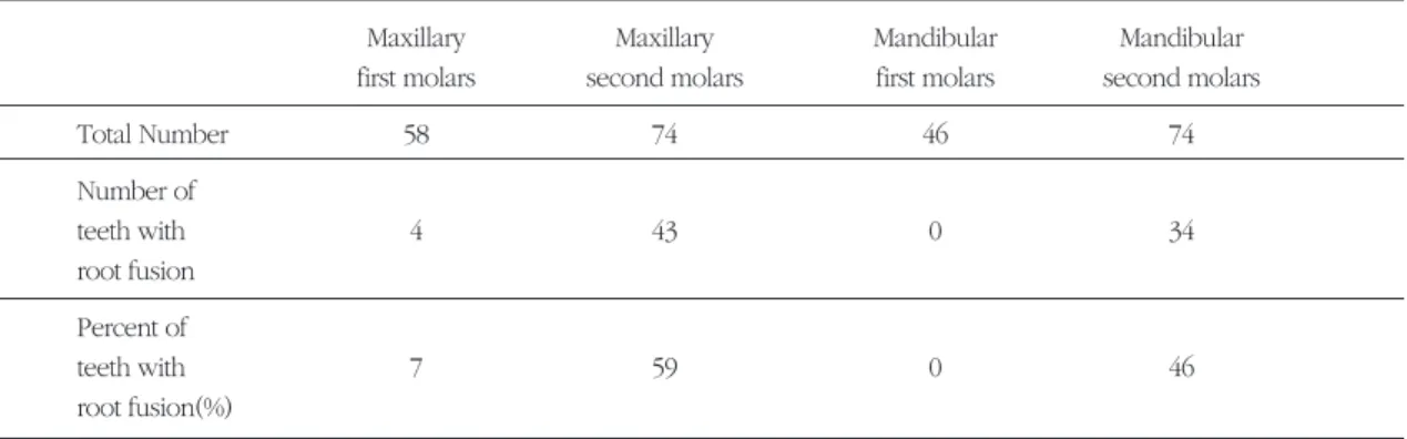
Total Number 58 74 46 74
Number of
teeth with 4 43 0 34
root fusion Percent of
teeth with 7 59 0 46
root fusion(%)
Table 1. Total tooth number and percent of teeth with root fusion
부에 가상선을 그어 측정하였다.
2) 치근이개부의 수평적 위치 : 치근이개부에서, 백악법랑경계부에 평행하게 가상선을 그어 치 근의 측면과 치근이개부까지의 거리를 수직적 위치와 같은 방법으로 측정하였다.
III. 연구결과
1. 치근융합의 빈도연구에 사용된 치아의 총 개수는 252개이고 상악 대구치중 치근이 융합된 것은 47개이고 하악대구치 중 치근이 융합된 것은 34개이었다(Table 1).
2. 치근이개부의 수직적 위치
상악제1대구치의 경우 법랑백악경계부와 치근이 개 부 의 거 리 가 원 심 측 은 5.06mm, 근 심 측 은 4.52mm, 협측은 4.01mm로 측정되었다. 상악제2대 구 치 의 경 우 는 원 심 측 이 4.04mm, 근 심 측 이 4.02mm, 협측이 3.87mm로 측정되었다(Table 2).
하악제1대구치의 경우 법랑백악경계부와 치근이 개부사이의 거리가 협측은 2.81mm, 설측은 3.69mm 로 측정되었고, 하악제2대구치의 경우는 협측이 3.61mm, 설측이 3.87mm인 것으로 측정되었다 (Table 3).
3. 치근이개부의 수평적 위치
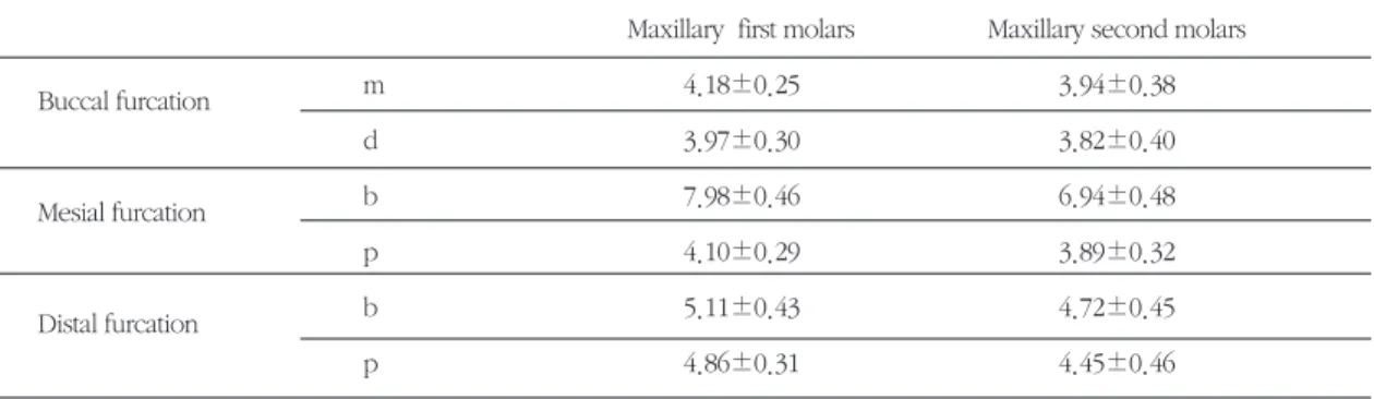
상악대구치의 경우 협측과 원심측치근이개부는 중앙에 위치하는 반면, 근심측치근이개부는 구개측 2/3에 위치하였으며(Table 4), 하악대구치는 협측과
Maxillary first molars Maxillary second molars
B 4.01±0.41 3.87±0.60
M 4.52±0.45 4.02±0.38
D 5.06±0.47 4.04±0.35
B : Length between CEJ and buccal furcation(mm) M : Length between CEJ and mesial furcation(mm) D : Length between CEJ and distal furcation(mm)
Table 2. Vertical location of maximally molar furcations
B : Length between CEJ and buccal furcation(mm) L : Length between CEJ and lingual furcation(mm)
Mandibular first molars Mandibular second molars
B 2.81±0.51 3.61±0.64
L 3.69±0.53 3.87±0.63
Table 3 : Vertical location of mandibular molar furcations
m: Thickness of root of mesial side(mm) d: Thickness of root of distal side(mm) b: Thickness of root of buccal side(mm) p: Thickness of root of palatal side(mm)
Maxillary first molars Maxillary second molars
Buccal furcation m 4.18±0.25 3.94±0.38
d 3.97±0.30 3.82±0.40
Mesial furcation b 7.98±0.46 6.94±0.48
p 4.10±0.29 3.89±0.32
Distal furcation b 5.11±0.43 4.72±0.45
p 4.86±0.31 4.45±0.46
Table 4. Horizontal location of maxillary molar furcations
설측치근이개부 모두 중앙에 위치하였다(Table 5).
IV. 총괄 및 고안
치주질환을 일으키는 원인 중 가장 중요하게 여겨 지고 있는 것은 세균이지만, 외상성교합, 치근의 근 접도, 악궁내에서의 치아의 위치 및 치아형태학적 특 성과 같은 국소적인 인자들도 이러한 치주질환을 발 생시키기거나 진행을 가속화시키는 역할을 한다. 이 중 치아형태학적인 면에서 살펴보면 다근치의 경우 치경부법랑돌기(cervical enamel projection)1), 법랑 진주(enamel pearl)2), 구치치근함요(molar root con- cavity)3), 이개부융선(bifurcation ridge)4, 5), 치근이개 부 입구(furcation entrance)의 구조6-8), 치근의 융합 (root fusion)9, 10)등의 형태적 이상이나 특징이 영향 을 줄 뿐 아니라,. 치근이개부의 위치(location of fur- cation)11) 또한 질환의 진행에 영향을 미친다.
치근이 융합된 경우는 일반적으로 치근의 길이가 짧아져서 치관-치근의 비율이 불리하게 되어, 치근 이 분리되어있는 경우보다 예후가 좋지 못하게 된 다. Ross등10)은 170명의 환자에서 1,340개의 상, 하 악대구치를 대상으로 치근융합의 분포와 발생빈도 를 조사하였는데, 상악구치의 경우는 35%로 하악의 24%보다 높게 나타났다고 하였다. 상악의 경우는 제 2대구치가 52.9%, 제1대구치가 7.7%이고 하악은 제 2대구치 3.2%, 제 1대구치는 3.3%로 상악제2대구치 에서 그 발생빈도가 가장 높게 나타난다고 하였다.
Hou 와 Tsai9)는 중국인에서 발치된 상악구치 158개 와 하악구치 151개에서 치근융합을 조사한 결과 상
악(39.7%)이 하악(28.1%)보다 높게 나타났다고 하였 으며, 제2대구치가 제1대구치보다 치근융합의 비율 이 높게 나타났다고 보고하였다. 우리들의 조사에서 는 조사대상 총 252개의 치아 중 치근융합의 발생 빈 도가 상악제1대구치에서는 7%, 상악제2대구치에서 는 59%로 조사된 반면, 하악제1대구치에서는 치근 융합이 전혀 나타나지 않았으며, 하악제2대구치의 경우에서는 46%로 나타났다(Table 1). 치근융합의 발생빈도에 있어 제2대구치의 비율이 높게 나타난 것은 다른 연구결과들과 일치하였지만, 그 발생 비율 은 다른 보고에서 보다 매우 높게 나타났다. Ross 등 은 비교치아형태학적 연구에서 치아의 해부학적 특 성의 양상은 인종에 따라 매우 다양하다고 보고하였 지만10), 우리들의 결과는 치주질환에 의해 발거된 치 아를 대상으로 조사를 하였기 때문에 상대적으로 높 은 비율을 보인 것으로 생각된다. 치근융합이 있는 경우 일반적으로 이개부 하방의 치근의 길이가 짧고, 불완전한 융합으로 인한 치근면의 발육구나 함몰부 등이 빈발하며, 기형적인 치근이 많이 존재하기 때문 에 치주질환에 쉽게 이환된다고 생각된다.
상하악대구치의 치근이개부의 위치에 대한 조사 에서 수직적인 위치의 경우, 상악 제1대구치에서는 원심측에서 치근이개부와 백악법랑경계부사이의 길 이가 가장 큰 것으로 나타났으며, 근심측, 협측의 순 이었고 이들 세 부위에서 그 길이는 평균 5mm 정도 였다. 상악제2대구치의 경우는 근심측과 원심측의 길이가 협측보다 큰 것으로 나타났으며(Table 2), 하 악 제1대구치에서는 설측이 협측보다 크게 나타났 Table 5. Horizontal location of mandibular molar furcations
Mandibular first molars Mandibular second molars
Buccal furcation m 4.13±0.52 3.79±0.4
d 4.30±0.53 4.04±0.37
Lingual furcation m 3.65±0.44 3.89±0.33
d 3.78±0.40 4.04±0.24
m : Thickness of mesial root(mm) d : Thickness of distal root(mm)
다. 하악 제2대구치의 경우도 비슷한 양상을 보였으 나 설측과 협측에서의 차이는 그다지 크지 않았다 (Table 3).
치근이개부의 수평적 위치에 대한 조사에서, 상악 대구치의 경우는 협측과 원심측치근이개부, 하악대 구치의 경우는 협측과 설측치근이개부 모두에서 대 체적으로 치아의 중앙에 위치하였다. 그러나 상악대 구치의 근심측 치근이개부의 경우엔 이개부의 위치 가 구개측 2/3에 존재하는 것으로 나타났다(Table 4, 5). Rosenberg18) 와 Ross19)는 상악대구치에 있어서 원심측이 근심측보다 치근이개부병변의 발생빈도가 높다고 하였는데, 이는 이개부병변 발생에 있어 이개 부의 수평적인 위치의 중요성을 말해주는 것이다.
그들은 근심측의 경우와는 달리 원심측의 치근이개 부는 협설중앙에 위치하기 때문에 인접치사이의 접 촉점 하방에 가깝게 위치하게 되므로, 치주질환에 이 환되면 접촉점 하방에 생긴 골분화구(osseous crater)의 영향을 더 쉽게 받기 때문이라고 하였다.
Ochsenbein20)는 다근치에 있어서 치근본체를 그 길이에 따라 세가지로 나누었는데, 상악에 있어서는 치근 본체의 길이가 3mm, 4mm, 5mm이상, 하악의 경우는 2mm, 3mm, 4mm이상인 경우에 각각 short, average, long에 속한다고 정의하였으며, Larato21)는 188개의 치근이개부병변을 보이는 치아중 75%가 short에 속했다고 하였다. 한편, Rosenberg 등은 치 근본체가 긴 경우는 상대적으로 치근이개부병변의 발생은 적지만, 일단 이개부병변이 발생되면 오히려 치료의 결과는 좋지않았으며, 반대로 짧은 치근본체 를 갖는 경우는 치근이개부 병변의 발생은 쉽지만, 보다 다양한 치료가 가능하며 예후도 양호하다고 하 였다18). Dunlap22)은 하악제1대구치에서 치근본체의 길이는 약 4mm 정도되며 6mm 보다 더 긴 것은 없 었다고 보고하였으며, Gher23)은 상악 제1대구치에 서 근심측의 치근본체의 길이는 5.0mm이고 원심측 의 치근본체의 길이는 5.5mm로 조사되어, 따라서 이 부위의 부착상실이 6mm이상인 경우는 치근이개 부병변이 존재한다고 하였다.
이와 같이 다근치에서 치근이개부의 위치는 치아 의 진단, 치료계획의 수립, 치아의 예후를 판단하는
데 많은 영향을 미치며 치료에 앞서 대상 치아의 치 근이개부의 위치와 치근본체의 길이에 대한 충분한 이해는 치료의 성공에 큰 영향을 미치게 된다. 우리 들의 연구에서는 발거된 치아를 대상으로 치근융합 발생빈도, 치근이개부의 수평적 및 수직적 위치에 대 한 조사를 실시하였는데, 향 후 이들 해부학적 형태 의 특성과 임상 지수와의 관련성, 성별에 따른 차이 점 등에 대한 더 많은 연구가 필요하리라 생각된다.
V. 결론
총 발거된 치아 252개 중 치근융합된 81개의 치아 를 제외하고서 남은 상악대구치 85개와 하악대구치 86개를 조사하여 다음과 같은 결과를 얻었다.
1. 치근융합의 비율은 상악제2대구치에서 59%로 가장 높았고, 하악제2대구치(46%), 상악제1대 구치(7%)순이였으며 하악제1대구치의 경우에 는 발견되지 않았다.
2. 치근이개부의 수직적 위치에 대한 조사에서 상 악제1대구치에서는 원심측(5.06mm), 근심측 (4.52mm), 협측(4.01mm) 순이었으며, 상악제2 대 구 치 에 서 는 원 심 측 (4.04mm), 근 심 측 (4.02mm), 협측(3.87mm) 순이었으며, 하악제1 대구치에서는 설측(3.69mm), 협측(2.81mm) 순 이었고, 하악제2대구치에서는 설측(3.87mm), 협측(3.61mm) 순이었다.
3. 치근이개부의 수평적 위치에 대한 조사에서는 상악대구치의 협측과 원심측 치근이개부, 하악 대구치의 협측과 설측치근이개부의 위치는 대 체로 중앙에 존재하지만, 상악대구치의 근심측 치근이개부는 구개측 2/3에 존재한다.
VI. 참고문헌
1. Hou GL & Tsai CC :“Relationship between periodontal furcation involvement and molar cervical enamel projections”J Periodontol 58:715-721 1987
2. Risnes S. :“The prevalence, location, and size
of enamel pearls on human molars”J Dent Res 82:403-412 1974
3. Bower RC :“Furcation morphology relative to periodontal treatment:furcation root surface anatomy”J Periodontol 50:366-374 1979 4. Everett FG, Jump EB et al :"The intermediate
bifurcational ridge: a study of the morphology of the bifurcation of the lower first molars" J Dent Res 37:162-169 1958
5. Svardstrom G & Wennstrom JL :“Furcation topography of the maxillary and mandibular first molars”J Clin Periodontol 15:271-275 1988 6. Bower RC :“Furcation morphology relative to periodontal treatment: furcation entrance archi- tecture”J Periodontol 50:23-27 1979
7. Chiu BM, Zee KY et al :“Periodontal implica- tions of furcation entrance dimensions in Chinese 1st permanent molars”J Periodontol 62:308-311 1991
8. Hou GL & Tsai CC :“Root separation and tun- neling therapy in a molar with narrow furca- tion entrance diameter: periodontal & prosthet- ic therapy”J. Formosan Dent. Assoc. 12:406- 412 1989
9. Hou GL, Tsai CC :“The morphology of root fusion in Chinese adults”J Clin Periodontol 21:260-264 1994
10. Ross IF & Evanchik PA :“Root Fusion in Molars: Incidence and Sex Linkage” J Periodontol 52 : 663-667 1981
11. Walid MB, Zeina M, & Simao K :“Anatomic considerations in the etiology and manage- ment of maxillary and mandibular molars with furcation involvement”Int J Periodont Rest Dent 5:399-409 1991
12. Wasserman BA & Hirschfeld L :“A long-term survey of tooth loss in 600 treated periodontal patients”J Periodontol 49:225-237 1978 13. Mandelaris GA, Wang HL & Macneil RL :“A
morphometric analysis of the furcation region of mandibular molars.”Compend Contin Educ Dent Feb;19(2):113-116 1988
14. Roussa E. :“Anatomic characteristics of the fur- cation and root surfaces of molar teeth and their significance in the clinical management of marginal periodontitis.” Clin Anat 11(3):177-186 1998
15. Hou GL, Chen SF, Tsai CC & Huang JS :“Anal- ysis of divergent angle and length of CEJ to the furcation entrance in extracted molars.”
Kao Hsiung I Hsueh Ko Hsueh Tsa Chih Dec;13(12):710-720 1997
16. Hou GL & Tsai CC :“Types and dimensions of root trunk correlating with diagnosis of molar furcation involvements.”J Clin Periodontol 24(2):129-135 1997
17. Michele P, Giacinto P, Antonio S & Adriano P :
“Molar root furcation: Morphometric and mor- phoogic analysis.”Int J Periodont Rest Dent 18:489-501 1998
18. Rosenberg MM :“Periodontal and prosthetic management for advanced cases”
Quintessence Pub. co.,Inc. 247 1988
19. Ross IF et al :“Furcation involvement in maxil- lary and mandibular molars”J Periodontol 51:450 1980
20. Ochsenbein C. :“A primer for osseous surgery”Int J Peridont Rest Dent 6(1):8 1986 21. Larato DC :“Some anatomical factors related to
furcation involvements”J Periodontol 46:608 1975
22. Dunlap R, Gher M : “Root surface measure- ments of the mandibular first molars”J Periodontol 56:234 1985
23. Gher M, Dunlap R :“Linear variation of the root surface area of the maxillary first molar”J Periodontol 56:39 1985.
- Abstract -
The Location of Molar Furcation in Korean
Seung-Nam Kim, Young Ku, Seong-Heui Son*, Sang-Mook Choi Department of Periodontology, College of Dentistry, Seoul National University
*Department of Dentistry, College of Medicine, Sungkyunkwan University
The objective of the present study was to investigate the vertical and horizontal location of the molar furca- tions in korean.
The samples used in this study included 132 maxillary molars and 120 mandibular molars. Of them, 47 max- illary molars and 34 mandibular molars had the fused roots. So, 85 maxillary molars(54 1st and 31 2nd molars) and 86 mandibular molars(46 1st and 40 2nd molars) were measured. The vertical and horizontal location of molars were measured with divider and digimatic micrometer and their means and standard deviation calculat- ed.
The results were summarized as follows :
1. The ratio of fused roots found in this study was the highest in the maxillary second molars with 59%, fol- lowed by mandibular second molars(46%) and maxillary first molars(7%) and none were discovered in the mandibular first molars.
2. In the study of the vertical location of molar furcation, the results were as follows : In the maxillary first molars, the length in descending order were distal(5.06mm), mesial(4.52mm) and buccal(4.01mm) and in the maxillary second molar, distal(4.04mm), mesial(4.02mm) and buccal(3.87mm). In the mandibular first molar, the length was 3.69mm on the lingual side and 2.81mm on the buccal side, and in the mandibular second molar, 3.87mm on the lingual and 3.61mm on the buccal side.
3. The location of the mesial and distal furcations in horizontal dimension measured showed following results : buccal and mesial furcations of the maxillary molars and buccal and lingual furcations of the mandibular molars generally found at the center, but the mesial furcation of the maxillary molars were found approximately two thirds toward the palatal aspect.
Keywords : furcation, root trunk, root fusion