大韓 1iX射綠짧學會誌 Vol. XVIII, No. 1, 1982
- Abstract-
g훌없앓잣의 題部電算化斷增據影所見
g힐.F1fl大學/校 점科大學 放身f잃科學敎室
崔祐碩 ·훌훌 京植·高永泰 ·金훌均·金훌훌蕭
Computed Tomographic Findings of the Pancreatitis
Woo Suk Choi, M.D., Kyung Sik Cho, M.D., Young Tae Ko, M.D., Ho Kyung Kim, M.D., Soon Yong Kim, M.D.
Department of Radiology, Kyung Hee University Hospital, 5eoul, Korea
Computed body tomography has become useful in the diagnosis of pancreatic disease. It was found to be a reliable, often specific, and noninvasive method for detecting pancreatitis and extra.pancreatic extension of the pathology. Of eight hundred and seventy two cases studied for abdominal pathology with EMI.CT 5005 whole body scanner from October 1977 to August 1980,21 cases were confirmed to be pancreatitis clinically or operatively. The authors reviewed the CT findings of the above ιases and the results were as follows:
1. Among twenty.one cases, the acute pancreatitis was 12 cases and the chronic pancreatitis was 9 cases.
The sex ratio was 17 males to 4 females.
2. In actue pancreatitis, diffuse enlargement of pancreas (11/12), focal enlargement (1/12), loss of peripancreatic fat plane (9/12), thickening of anterior pararenal fascia (6/12), and smooth margin of pancreas (5/12) were observed.
3. In chronic pancreatitis, parenιh ymal atrophy (7/9), normal size (2/9), loss of peripancreatic fat plane (3/9), thickening of anterior pararenal fascia (1/9), calcification (1/9), smooth margin (2/9), and serrated margin (6/9) were observed.
4. The complications were associated with 7 cases of acute pancreatitis and 1 case of chronic pancreatitis;
pseudocyst (6), abscess (2), and fat neιrosis (3). The sites of the pseudocyst were lesser sac (2),
anterior pararenal space (2), posterior pararenal space (1), subhepatic region (1), greater omentum (1)
,
and intrapancreatic region (2). AII of them were associated with acute pancreatitis except one in chronic pancreatitis.1. 繹 論
急션I: ߥ嚴~은 臨W1î狀으로는 다흔 急{生限홉ß t*恩파 의 鍵5J1j이 어 렵 고, 血淸 amylase가 300 Somogi Unit 以上으로 뺑加함으로써 確該이 可能하나 이 에 는 時間 的 制約이 있다1) 또한 急性牌顧*中 壞死性出血{生解 職~, 牌職體傷과 假性製睡破짧로 因한 8후魔{生願水와
從隔洞갖퉁의 경우는 早期該斷으로 週팅j한 處置없이는 死亡率이 100% 에 달하므로 빡른 깐斷이 必要하다16)
이 훌댄、올 放射없學的으로 앓斷하기 위 하여 는 單純服 部振影術, 몹十二指陽造影術, 평行i'.H후 I~ 管:造影術, IT¥
職動lI1it造影術파 放射經同位元素走훌等이 利用되어 왔 으나部는 談斷的표確度가 낮고部는 않斷的正 確度는 높으나 手技가 어 렵 고, t를害的안 ;險효法이 라는 短點等이 있다 CT는 事前準備없이 必要한해 에 아무때
- 125 -
1i
A‘.
껴j
1i
nU
Table 1. Age and Sex Distribution
M
1i
A?
?ι
nu nu
T
A
,
‘‘」껴ι
chronic pancreatitis
F
nU nu ----nU
acute pancreatitis
T
A얘
‘‘“
,3
1i4
--
F
nU nU 1i
1i
nU
M Sex
0 1 Age
30-39 40-49 50-59 60-69 70-79 라도 손쉽게, ,띤者에게 苦病이 전연없이 檢효할 수 있
기 때 문에 急、性牌磁갖파 강은 重lìË‘IJ.!、者에 對한 웠호ì* 으로 매 우 便利하고 直技的안 ~像과 ι1 造構造를 올수
있어 g¥R훌훌l떤 원斷에 펴U期的안 ;않효法으로 뼈I 光을 받 고 있놔2 , 4,13,14, 19) 著者들은 本 病院 放M 級科에서 f옆 驗한 際廳~파 그 合1M솥에 對한 CT所昆을 分析웠討 하고 文願考察파 함께 報告하고자하는 바이 다.
ll.
對象 및 方法9
Table 11. S ize of the Pancreas
2 12 7
2 10 Total 象
1977年 10月부터 1980年 8 月 까지 ;本人델 líH)~~j펴院에 서 施行한 復部 CT 872例 中 臨皮 빛 手術上 |협廳*
으로 E죠認、된 21例를 對象으로 하였다.
1) 對
Chronic pancreatitis Acute
pancreatitis
nU nU 7l
껴‘
n
l
o o
Diffuse enlargernent Focal enlargement Atrophy
Normal
¢홉狀안 것인 f뭘性에서 6例였 A며, 平m한 것이 쉽;1生 5 f71J i윷{生 2例였 다 (Table JH).
合 ilf' lìË은 즐、性 7例, ↑용i生 1例에서 同伴되었다 合 Vf
lìË 8f71J 中 假性짧陣 6f71J, 體짜 2例, )J읍)jJj塊死가 3例이
었으며 假{生짧形成部位는 4、網짧 2例,前쁨佛院 (anter
ior pararenal space) 2例, 後 l홉홉뾰 (posterior para- renal s pace) , 左에따葉下部 및 大陽厭뾰이 各各 1 例 씩이었고, 隊嚴內에 急性파 慢{生에서 各各 l例썩 觀察 되었다. 이들 中 1 f71J에서는 小網짧, 前l뽑f휠뾰 및 左뼈Ij Ðf葉下部에 同時에 存tE하였£며 마릎 l f7U에서는 前뽑 뽑뾰파 小網짧에 同時에 存在하였다(Table N).
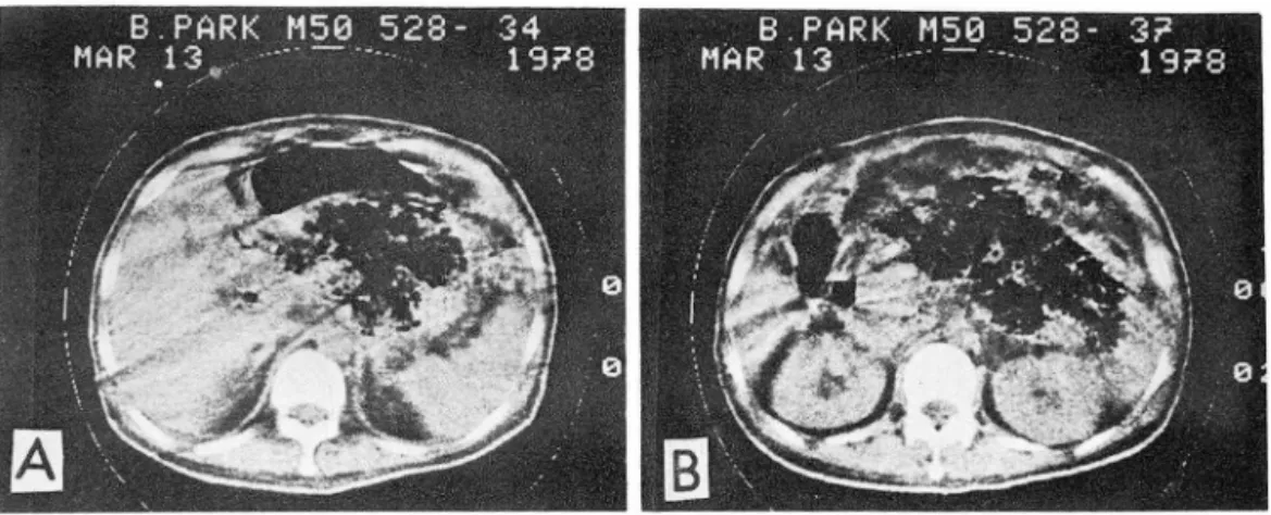
f숱例 1 : 爛↑월{生增大를 보안 急{生牌嚴갖 52歲, 男子
Table llL CT Findings of the Pancreatitis
Chronic
pan따 eatitis
Acute
P때 creatitís
3
2 6 - 126 -
9
6
5 0 Loss of peripancreatic
fat planes
thickening of the anterior
pararen외 fascia margin of the pancreas
smooth serrated EMI CT 5005를 利用하여 때l하位에서 등상 l홉職。1
位置하는 部位를 中心으로 13rnm 間隔j즈로 上下로 8切 片 (Slice) 정도 振影하였으며 必강동에 딱라 또 Gastrog- rafin을 投與하고 造影챔행을 뤘빠하였다. J!P 大部分 의 파遇 CT 振影前에 Gastrografin 400cc정 도를 投與 한 後 15"'20分後에 Buscopan 1 ml를 Rñ 肉注짧하고 얀시 Gastrografin 300cc를 짧與하여 몹와 陽管。l 造 影햄j 로 充滿되 도록 한 後에 Jli}影함으로써 牌嚴파 몹職 t홈;및 十二技1m파의 區別을 容易하게 하고 또 陽管。l 圖塊료 펴認될 可能性을 꾀l 除하였 다. 造影 t땀꿇은 65% Urografin 80rv10Ccc를 經l젠版急、速注入 하였다.
對깅없이 완 2H꺼j 中 急、性|停魔*이 12{꺼U, 慢{生牌職씻。1 9찌였으며 ‘땀者들의 年都分布는 쉰:性|停職씻안 강l遇 30 1~ 4 f71J , 40代 3例, 5011;; 3{깨, 601\; 및 70代가 各各 1 찌j였고, f'강 lil際廠갖안 짧遇 3이'~ 11JÚ, 4011;; 4f71J. 5이t 31JÚ, 6Cfl:: 1例였다. 性5}1; 分布는 뭇子가 17f71J, 女子가 4 찌]이 었다 (Table 1).
9후嚴의 크기 는 뼈:i뭘性 l싼大를 보안것 이 急{生안 쩔遇 는 11例이 었으나, 慢{生안 짧遇에 는 없었으며 , 部分的
t염大를 보인 것 은 急、{生안 따遇 H깨가 觀察되 었 다. ↑웰 性안 찮週 양質쫓縮을 보인것이 7{列이고 표常인 것이 2例이었다(Table
n ).
J젠탬指)J1J혐의 消갓 또는 ~lìËj生 f풋潤像 (所땀 dirty fat) 을 보인 例는 3例였으며 , 前’i''T fjjA줬9갯)J잉펄 (thicken
ing of anterior pararenal fascia) 가 뽑明된 것 은 急 性이 6例, ↑월{生이 l例였고, 隊職採은 不規則하거 나 銀
法 2) 方
fiÄ 績 J1I.
Table N. Complications of the Pancreatits
Chronic pancreatitis Acute
pancreatitis
1/9
nU nU nU nU nU --
7/12
?ι 이‘
1l 1i
5
Fig. 1. Acute panαeatitis with diffuse moderate enlarge- ment of pancreas and loss of peripancreatic fat plane.
1 1 No. of complication
Type of complication pseudocyst
lesser sac
ant. pararenal space post. pararenal space subhepatic
greater omentum intrapancreatic abscess
fat necrosis
O O 2
3
Fig. 3. Chronic pancreatitis with parenchymal atrophy and mild dilatation of p따lcreatic duct.
한 低密度을파 1훈質破짧들이 보이고 同|랴에 周圍n읍 Jlij R룡의 *~1生훌‘따l으로 因한 所땀 “dirty fat" 像이 證明 (Figure 1).
13 日前부터 題痛파 I멤&카 發生하였으며, 臨*웠흉 上 血淸 arnylase가 430 Sornogi Unit로 上昇되 었 다.
CT上 牌魔의 펀없f율{生增大와 周圍짧)Jjj層의 消失이 보안 다.
~19~ 2: 部分的썩大를 보안 (Figure 2).
主訴는 上記 t뿔、者와 類!니하였고. CT上 牌職 頭공ß.
특허 홍힘狀突起의 뺑大릎 보인다.
E융例 3: j용{生쫓佈{生牌職* 51歲, 女子(Figure 3).
~素에 間歐的언 心홈곰ß痛 및 右댐.u脫部痛이 있었으 - 며 • CT上 靜職의 賢質쫓縮파 牌職管:의 輕微한 짧張을 보인다.
훌例 4: 急、{生懷死{生牌職씻 50歲, 男子 (Figure 4).
25 日前쉰;1生出血性牌職씻 疑心下에 試驗|랩 R흉한바 있
~며 • CT上 極허 뺑大된 牌驗內에 多數의 大小不規씨IJ 촬、{生牌廳쏘 35歲, 男子
A,B: Acute pancreatitis with focal en1argement of head of panαeas , especia1ly uncinate pro ∞ ss.
Fig.2.
Fig. 4. Necrotic - hemorrhagic pan다eatitis.
A: The vo1ume of pancreas is noticeab1y increased with mu1tip1e,large and small irregu1ar 10w densities
B: In the 10wer scan, the massive destructive change in huge pancreas can be seen, which is associated with 10ss of peripancreatic fat p1ane by infIammatory infùtration, so caIled “dirty fat".
판마.
효例 5: 假i生製햄파 牌魔짤의 石.깐化를 보안 ↑장{生隊 磁Ø( 53歲, 男子(Figure 5).
약 20年前부터 間歐的안 心商部痛이 있 었으며 , CT 上 多數의 結節{生石짜沈힘이 隊廠管을 짜라 證明되 고 頭部에 境界가 分明한 짧R包性짤化가 있고, 그 雙에도 石f沈휩이 보안마 · 짧U댄內 低密많域의 X級 吸收係數 {直는 2.25 EMI Unit로서 물의 l냈收率파 거 의 -致한 마.
Fig. S. Chronic pancreatitis with pseudycost at head of pancreas and ca1cifications. Note 1inearly ar- ranged ca1cific deposits ∞nforming to the course of main pancreatic duct (Wirsung) and ca1cifica- tions of the media1 wall of pseudocyst.
fff.例 6 : 合快距을 보인 急{生牌魔갖 40歲, 男子 (Figure 6).
10티前 交通事故로 來院한 恩者로서 CT上 左씨n:F핏 部{立에 境界가 分明한 커 마란 分葉形 낌~n包睡짧가 보이 고 (Fig.6A), 이 는 마廠下部뿐만 아니 라 (Fig.6B). IJ、
網앓 및 前샘fj,')효까지 홉犯하고 있마 (Fig.6C). 手術結 果 CT所見과 同一하게 牌廠뽑部 中心에서 íl!!i컸되어 Ø(liËi生뿔뼈가 小網짧, H'l 廠下部 빛 前I용fj,')센에 서 證明 되었마.
N-
考 察R후職은 臨"*的으로나 -般的인 放射綠學的 檢곁쪼方法 으로는 등?斷이 어려운 職器이었마. CT나 超音波 B~斷r 機가 開發되 기 前에 는 牌職의 出血, 假{生짧睡 빛 g¥魔 外峰3월織씻고} 隊廳 g짧 ro동을 포항한 牌職*의 合tJtliË을 直接 影像化해서 양斷할 수 있는 方法。1 없였으며, 單 純R흥部振影이 나, 바륭造影)險효로 나타나는 애매 한 直 찮 또는 間接 所見만으로 핑斷하였고14' 低張住十二指 陽造影術은 際職增大의 間짚所見을 나타낼 뿐 隊職睡 塊의 種類에 따른 特異所見을 나타내는-경은 아니냐 假{生짧陣의 따遇는 67"-'86%의 높은 ~斷率을 보안
다9)
Selenium-75에 依한 際廠의 jjÍ(찌f*싸同{立元素走호는 R월魔똥‘댄에 對하여 약 50%의 높은 댐陽性率을 나다내 기 해문에 적합치 옷한 檢좁法으로 認定되었마 12)
평行{生牌R엉管造影術. 1l1H앓폐J1lJK造影術 빛 經皮n:F)jIj道 - 128 -
F뀔.6. Acute pancreatitis with extrapancreatic pseudy- cyst.
A: A huge well circumscribed lobulated cystic mass at the region of left lobe of liver is noted.
B: In the lower scan, the pseudocyst is seen in subhepatic region
C: The lower scan revels the pseudocyst extend- ing in to lesser sac and left an terior pararenal space.
造影術등은 뤘練펀 手技가 ~、꽃하고 또 훔害的인 ;險훌 法이 어 서 急、{生뽑魔갖 ,떤者와 같은 重恩者에 게 는 適合
치 않다 14)
Jln훌훌‘띤‘에 對한 超音波~~斷은 f윷f生牌廳Ø{)면者의 境 J‘
遇 解廳합質이 쫓縮되고 超音波檢홉에 fr}j害가 되 는 陽 管까스를 뺑加시 키 는 1傷閒鎭l1E이 好짧하기 때 운에 蘇 廳의 影像을 觀察하기가 正常A 보다 더욱 어렵고, 進 行펀 慢{生薦職*의 特徵的안 所見안 石K化도 證明하
기가 어렵다4)
CT에 依한 後짧部훌댄, 특히 勝職훌댄、에 對한 쓸斷 은 睡傷안 탤遇 그 確率이 거 의 90%에 달하고, 뿜홉뿜홍 의 形態, 位置 및 크기 뿐 아니 라 X~ 吸收{直도 알수 있고, 周圍組織파 써〈巴節의 病變까지도 좋j 別할수있다.
Siegelman등191 은 急性牌廠씻의 該斷은 病歷, 理學的 檢좁, 血淸 amyla~e의 V則定等으로 뚫斷될 수도있으나,
週切한 治擔를 하기 위한 씀斷을 엠기에는 어려운 점 이 있고 CT에 依해서 R¥R앓고} 그 周園의 fluid collec- tion 有無를 아는 것 이 큰 도움이 된다고 하였 다.
Mendez등14)은 最近 4年間의 經驗에 바 추어 CT는 急、性降職갖 o총 周圍組織, 특히 後服땐으로의 病變의 波
及 經路를 아는데 가장 正確한 方法이 라고 하였으며 , 또한 牌廳周圍홉犯, 峰2동織Ø{, 出血, 假i生짧睡및 8없셨 을 包含한 合까.1fË을 調효하는데 있어서, 超홉波양斷을 包含한 다은 어 떤 方法보마도 優秀고, 핑홍廠~‘[),、에 對 한 ←-次的안 양짧[方f똥이 되 어 야 한마고 행告하였다.
또한 이 들은 非표힘 所見을 보안 쏠、{生輕職갖 ‘댄者 32名의 CT所兒을 分析한 結果 뻐漫{生 牌職뺀大와 함 께 周圍뼈)J;Ij消失을 나타낸 것이 가장 많은 所見이었다 고 한마. 그러 고 蘇廳周圍훔犯의 好짧部位는 左빼前 l쯤 뿔뿔친과 小網짧이 고, 下fjß顆般前方까지 波及되 는 例도 있다고 한마.
Siegelman등 19)도 fluid collection이 探知할 수 있는 睡塊를 나타 낼 수 있고, 계속되는 熱파 痛l1E의 原因이 될 수 있기 해운에 降職*의 重몇한 所見이라고 했으,
며, 역시 小網靈과 前賢佛뾰이 가장 好發部位바고 하 였다.
Dembner퉁2)은 牌廳*時 씻뾰이 자주 後外測으로波 及되 어 쁨뽑院에 미 치 는데 100例 中 10Ø1J에 서 뿜흉職t당 大와 憐孩組織의 破壞빛 左맨、IJ샘짧院으로의 波及이 認 定되었으며, 이와같은 左測뽑않따로의 波及을 새로운 CT所見이 라고 報告하였다.
著者들의 境遇도 急、{生牌廳Ø{ 12例中 11例에 서 牌魔 의 濟漫性 增大를 보였£며 , 1例에 서 는 部分的 增大를 보였고 9例에서 周圍8릅)J;Ij消失이 觀察되었으며 4 Ø1J 에서 는 小網慶파 前땀홉1양Ø{l1E이 波及되어 있음을 보았다.
Ferrucci동.)은 漫{生牌嚴Ø{ 50例의 CT所見을 分析한 結果 石%化 8例, 흉質쫓縮 7Ø1]' 牌廳管織폈 2Ø1J. 그 리고 假性짧陣이냐 廳%이 15例였마고 하였으며, 가장 많은”所見3로 .Ø<l1Ei生 浮睡이 나 織維性 硬化에 依한牌 - 129 -
職t엠大와 睡塊效果라고 報告혔마.
著者들의 穩遇는 慢{生勝훌훌갖 뾰者 9例中 7例에서 牌 職의 養縮 所見올 보았고 增大되에 있는 例는 볼 수없 었다. 이 는 아마도 慢性牌職*에 對한 CT所見A로 牌 職쫓縮을 重要視하여 이 같은 所見에 합당한 ‘띤者들만 주로 선택되었기 혜문인 것으로 생각완마.
@후職假{生廳睡은 X썼吸收系數가 O單位前後안 境界가 分明하고 眼度가 均等한 圓形안 低密度뼈覆인데 反하 여 降魔拖의 壞死로 因한 低密度部는 大體的으로 中心 部에 &置하고 두껍 고 不規則한 뿔을 가졌으며 漸次的
o 로 睡塊組織으로 移行된다 1 ,4)
Kressel동9) 은 牌職假性짧睡 12例의 CT所見파 超音 波談斷所見을 分析한 結果 CT는 合供뾰이 없는 정우 가 8例中 6{7JJ. 感뽕판 정 우가 4例中 4例, 超音波~~斷I 機에 依한 경우 合 ÐHE이 없는 경우가 8例中 7Ø1J. 感 꼈완 정우가 4Ø1J中 2例가 ~~斷됨으로써 選7JIJ險흉와 追
~檢훌를 하는데 는 超音波양斷機가 가장 좋은 方法。1 며. CT로써 手術的 계획을 세우는데 더욱 精密한 解 해學的 知織을 얻을수 있어 서로 補따的 投힘을 한마 고 報告했다.
石:[J(~좁은 대 개 際魔管內에 생 기 고 Wirsunga;;管 을 따라 結節{生 짧狀 石次化像을 보인다 Ferrucci둥 은 假性짧健파 同伴왼 3例를 觀察하였마고하며, 著者 들은 牌廳管파 R¥職頭部의 假{生羅睡 윤좋을 따라 *Jí(狀 의 結節性 石;JJ(~홉을 招來한 1例를 經驗하였다 Ring 둥은 牌廳石짜化는 90% 以上이 알콜{生 隆職갖 ,만者에 서 觀察되었ξL며 대개 題痛을 앓은지 5"'10年後에 냐 타났마고 하였다.
Mendez둥13)은 450例中 9例에 서 牌職內 ~氣를 證明 하고 CT는 牌廠略찌의 첫 앓斷뿐만 아니 라 ~jJË의 周 園로의 波及올 아는데 도 큰 利點。l 있으켜 牌職內 호 氣를 證明하는 것 이 該斷에 가장 理잔!的안 標示라고하 였으며, 著者들이 經驗한 2例에서도 同-한 生됐을 하 게되었다.
Fishman둥5)은 牌職管의 據;;~은 牌廳3합의 二次的안 所見으로 알려 져 왔으나, 勝職管의 搬張。1 確認된 8例 中 4例가 慢{生牌廳씻 ,思者였마고 報告하였다.
Pistolesi풍16)은 急、f生牌職*中 壞死{生出血性解廳갖, 際職8農傷, 假性짧뚫睡破짧고l} j(6f隔洞씻을 隨伴한 際職{生 復水등의 境遇는 .!f!.期양斷에 依한 適切한 處置없이는 뿔、者의 死亡率이 100%에 달하으로 CT의 ~斷的 投剩 이 더 욱 重要하마고 週調하였다.
V.
結 짧1977年 10月부터 1980年8月까지 本大學附屬病院에 서 施行한 願部 CT 872例中 짧짜 및 手術上 牌짧갖」ζ 로 確認된 21例를 分析檢討하여 마음과 같은 結果를 얻었다.
1. 對象이펀 많者 21 Ø1J中 急、性牌職갖 恩者는 12例였 고, 慢{生解職갖 思者는 9例였마.
2. 恩者들의 年敵分布는 急、{生牌職*안 상중遇 30代 4 例. 40代 3例. 50代 3例. 60代 및 70代가 各各 1 例였 고, 慢性牌職갖안 뚫遇 30代 1例. 40代 4例. 50代 3例,
60代 1例였다. 性7JIJ 分布는 男子 17{J1J, 女子 4例였다. 3. 急{生解職씻인 첼遇 爛↑용{生행大 9例, 部分的뺑大 1 例, 周圍9읍防層消失 9例, 前댐f용映~EJJ 6ØU. 平 m한牌 嚴緣 5例가 觀察되 었다.
4. f용{生牌廳*인해 는 1핫質쫓縮 7例, 표常크기 2Ø1
J.
周圍服防層消失 3{J1J, 前’뽑행陳RE淳H끼U. zr: m한 牌職緣
2例, 銀짧狀 g¥顧緣 6例가 觀察되었다.
5. 合í1f-jJË은 急、{生牌職갖에서 7例, ↑용{生牌職씻에서 1 例가 觀察되었고, 이들 8Ø1J 中 假{生짧睡 6例. ~ili 2Ø1J.
順I얘빼 "E 3{7]이었 A며, 假性製睡形成部位는 小網裵 2 例, 前 l뿜홉峰 21J1J. 後 l홉佛뼈, 院左~lIJ Rt葉下部 및 大陽‘
媒l훈이 各各 1例씩이었고, 解廠內에도 2例 觀察되었마,
그러 고 合뺨뾰은 慢性牌職*에 서 의 假{生覆睡 1 例를 除 外하고 모두 急、|生牌職*과 同伴되었다.
REFERENCES
1‘ Bu tzelaar R.M.J .M.
,
et. al. : Computer tomography in acute pancreatitis. Acta Radiologica Diagnosis 79. (7978) Fasc. 3.2. Dembner A.G.
,
et. al. A new computed tomσgraphic sign of pancreatitis. Aj R 733:477-479
,
September 7979.
3. Fawcitt R.A., et. al. Computed tomography in pancreatic disease. 8ritish journal of Radiology.
57: 7-4, 7978.
4. Ferrucci J .T., et. al. : Computed body tomography in chronic pancreatitis. Radiology 730: 7 75-782
,
january 7979.
5. Fishman A., et. al. Significance of a dilated pan- creatic duct on CTexamination. Aj R 733:225-227
,
August 7979.
6. Haaga J .R.
,
et. al. : Definite role of CT scanning of the pancreas. Radiology 724:723-730,
September 7977.7. Kirkpatrick R.H., et. al. Scanning techniques in computed body tomography. AjR 730,-7069-7075
,
- 130 -
june 7978.
8. Kollins 5.A. : Computed tomography of the liver
,
spleen
,
and pancrea아 Seminars in Roentgenology,
Vol. XIII
,
No. 3,
july 7978.9. Kressel H.Y., et. al. : CT scanning and ultrasound in the evaluation of pancreatic pseudocysts. A prelimi- nary comparison. Radiology 726:753-757
,
january7978.
10. Kuhns L.R.
,
et. al. : Localization of the head of the pancreas using the junction of the left renal vein and the inferior vena cava. journal of computer assisted tomography 2:770.772,
April 7978.11. Levitt R.G.
,
et. al. Complementary use of ultra- sound and computed tomography in studies of the pancreas and kidney. Radiology 726:749.752,
january 7978.
12. MacCarty R.L., et. al. Retrospective comparison of radionuclide scans and computed tomography of the liver and pancreas. Aj R 729:23.28, july
7977.
13. Mendez G. and Isikoff M.B. : Signiηicance of intra- pancreatic gas demonstrated by CT. A review of
ninecases. AjR 732:59-62
,
january 7979.14. Mendez G., Isikoff M.B.,and Hill M.C.: CTofacute pancreatitis. Aj R 735:463-469
,
September 7980.15. Nacianceno 5.E.
,
et. al. Pancreatic pseudocyst simulating dilated biliary duct system on computed tomography. Radiology 734: 765-766, january 7980.16. Pistolesi G.F.
,
et. al. Computed tomography in surgical pancreatic emergencies. journal of com- puter assisted tomography. 2:765-769,
April 7978.17. Ring E.J., et. al. : Differential diagnosis of pancreatic calcification. AjR 777:445-442
,
February 7973.18. 5iegelmann F .E., et. al. CT demonstration of the splenic vein - pancreatic relationship: The pseudodi- lated pancreatic duct. AjR 729:77.27
,
july 7979.19. 5iegelman 5.5., et. al. : CT of fluid co/lections as- sociated with pancreatitis. AjR 734:7727-7732
,
june 7980.
20. 5tanley R.J.
,
5agel 5.5.,
and Levitt R.C. : Computed tomographic evaluation of the pancreas. Radiology 724:775-722,
September. 7977.- 131 -