3 )INTRODUCTION
Cellular interaction is achieved by cell surface m olecules, and play s a pivotal role in the communication betw een multiple my elom a (MM) cells, which are m alignant B cells, and bone m arrow strom al cells (BM SCs) ( 1,2) . Variou s surface m olecules are involved in the growth , activation , and homing action of immune cells by regulating cell-cell interaction s. The maj ority of my elom a cells are located in the bone m arrow (BM) ,
Correspondence: Inpyo Choi, Laboratory of Immunology, Korea Research Institute of Bioscience and Biotechnology, Eoun-Dong 52, Yusong, Taejon 305-333, Korea
Tel: 82-42-860-4223, Fax:82-42-860-4593 E-mail: ipchoi @mail.kribb .re.kr
becau se BM SCs present a variety of surface molecules and trap my elom a cells. This strongly suggests that cell surface m olecules are important for the growth and tum origenesis of B cells.
The developm ent of B lineage cells proceeds through several stages m arked by the rearrangement and expression of the immunoglobulin genes. Mature B cells are generated by functional V (D)J rearrangem ent of the IgH and IgL genes in the bone m arrow and target secondary lymphoid tissues (3) . M ature B cells then differentiate into m em ory B cells and lymphoblasts by antigen stimulation . The lymphoblasts enter germinal centers, where they undergo inten sive proliferation and somatic hypermutation in Ig genes, which leads to affinity m aturation . Cells that have hom ed to the BM
Ge n e r a t i o n a n d c h a r a c t e r i z a t i o n o f 1H8 m o n o c l o n a l a n t ib o d y a g a i n s t h u m a n b o n e m a r r o w s t r o m a l c e ll s
Hyung Sik K an g, Inpyo Choi
Laboratory of Immunology, Korea Research Institute of Bioscience and Biotechnology, Yusong, Taej on, Korea
= Abs t r ac t =
Background : Bone marrow stromal cells (BM SCs) express many cell surface molecules, which regulate the proliferation and differentiation of immune cells within the bone marrow .
Methods: To identify cell surface molecules, which can regulate cell proliferation through cell interaction, monoclonal antibodies (MoAbs) against BM SCs were produced. Among them, 1H8 MoAb , which recognized distinctly an 80 kDa protein, abolished myeloma cell proliferation that was induced by co-culturing with BM SCs.
Resu lts: IL-6 gene expression was increased when myeloma or stromal cells were treated with 1H8 MoAb . In addition, the expression of IL -6 receptor and CD40 was up-regulated by 1H 8 treatment, suggesting that the molecule recognized by 1H8 MoAb is involved in cell proliferation by modulating the expression of cell growth-related genes. Myeloma cells contain high levels of reactive oxygen species (ROS), which are related to gene expression and tumorigenesis. Treatment with 1H8 decreased the intracellular ROS level and increased PAG antioxidant gene concomitantly . Finally , 1H8 induced the tyrosine phosphorylation of several proteins in U266.
Conclu sion : Taken together, 1H8 MoAb recognized the cell surface molecule and triggered the intracellular signals, which led to modulate gene expression and cell proliferation.
Key W ords: stromal cells, myeloma cells, cell surface molecule, 1H8 monoclonal antibody
differentiate into long-lived mem ory cells. The pro- liferation and differentiation of B cells are regulated by a close interaction betw een hem atopoietic cells and the BM m icroenvironm ents. Within the BM , these cells receive the signals, which lead to proliferation and terminal differentiation(4-6) . BM SC s stimulate the proliferation and differentiation of my elom a cells via cell-cell interaction and the secretion of soluble mediators (2) . The contact betw een granulocyte-m acrophage pro- genitor and strom al cells is inhibited by antibodies, which suppress the function s of the cell surface m olecules (7-9) .
B cells are activated by several cytokines and differentiate into plasm a cells, and the apoptosis of B cells is blocked by the stimulation via CD40 antigen in a germ inal center . Durie et al. ( 10) dem on strated that CD 40 triggers IL -6 secretion and prom otes the cell survival of MM cells. The up -regulation of IL -6 in MM cells was found to be induced by CD40 and CD40 ligand interaction ( 11) . The stimulation of CD 40 antigen by the CD 40 m onoclonal antibody (MoAb) or CD40 ligand induced the up -regulation of IL -6 and prom oted cell proliferation in IL -6-dependent my elom a cells ( 12) . To investigate new cell surface m olecules, which regulate the function s of my eloma cells and BM SC s, MoAbs again st BM SCs were generated and characterized during this study . 1H 8 M oAb regulated cell proliferation and the gene expression of my elom a cells and BM strom al cells.
MATERIALS AND METHODS 1. R e a g en t s
Anti-IL -6 poly clonal antibody , monoclonal anti-IL -6 receptor antibody (MP 18), and IL -6 cDNA were kindly provided by Dr . T . Taga (O saka University , Japan) . Propidium iodide (PI), HA T (Hypoxanthine, Am in - opterin , and Thym idine), DMEM , and RPMI 1640 were obtained comm ercially from GIBCO (Gaithersburg, U SA) . RN ase and TUNN EL assay kits were purchased from Boehringer Mannheim (M annheim , Germany) . [-3 2P]ATP was purchased from Amersham International
(Braun schweig, Germ any) .
2 . C ell cu lt u r e
U 266, IM -9 cells, WIL -2 N S, and CE SS cell lines, w ere obtained from the Am erican Type Culture Collection (Rockville, U SA) and BM SCs w ere obtained as described previously ( 13) . Cells were cultured at 3 7℃ , 5% CO2 in RPMI 1640 supplem ented w ith 10%
fetal bovine serum (FB S, Hy clone, Logan , U SA), 100 U/
㎖ penicillin, and streptomycin (GIBCO). The cells were m aintained in RPMI 1640 supplem ented with 10% FB S.
3 . P r ep a r at ion of m y elom a c ell s
Fresh my elom a cells (MMB s) were purified from bone m arrow aspirates of MM patients as described previou sly ( 14) . Briefly , m ononuclear cells were isolated by differential centrifugation s on Ficoll-Hypaque den sity solution ( 1.077 g/cm- 2) . Mononuclear cells were washed tw ice w ith H ank s balanced salt solution (HB SS), without calcium and magnesium . Fresh myeloma cells w ere purified by removing T cells by rosetting w ith am inoethyl isothiouronium brom ide (AET)-treated sheep RBC . Adherent cells w ere depleted by 2 h adherence to a 24 well culture plate (Costar, Cambridge, U SA) at 3 7℃ , in RPMI 1640 supplemented with 10% FB S. The percentage of fresh my elom a cells was evaluated by cytoplasm ic immunofluorescence u sing anti- or anti-light chain antibodies directly coupled to fluorescein (DAKO , U SA) (>90%) . The cells were cultured for 3 day s at a den sity of 1x 10⁶ cells/㎖ in RPM I 1640 supplem ented w ith 5% FB S. Supernatants w ere then harvested and stored at -20℃ . To prevent direct cell-to-cell contacts between strom al cells and fresh my elom a cells, inner m embrane filter chamber s (0 .45 μm , M illicell CM , M illipore, Bedford, U SA) w ere u sed within the 24-well culture plates. A fter 3 day s of co-culture, fresh my elom a cells were harvested, and analyzed for IL -6 receptor expression by flow cytom etry (Becton Dickin son , Sunnyvale, U SA) . To exam ine the effects of cell proliferation , 1× 10⁴ cells w ere resu spended in 100 μl of culture m edium supplem ented w ith 2 .5% FB S, and incubated in a 96-well culture plate for 48h . Cell
proliferation was then m easured by [3H ]thymidine incorporation .
4 . P r od u ct ion of m on o clon a l a n t ib ody ( 1H 8 ) t o B M S C s
1) Im m u n iz a t io n
Stromal cells ( 1x 10⁷ cells) isolated from the BM aspirates of MM patients w ere immunized into the intraperitoneum of a BALB/C fem ale m ouse (5 week s old) . Boost was tw ice perform ed every two week s into the sam e site. Five day s later, a little blood w as collected from an ey e and assayed by cell ELISA to determine the activity and amount of antibody .
2 ) C e ll f u s io n
The splenocytes of the immunized mice were isolated by teasing and centrifuged at 1,200 rpm for 5 m in in serum -free DMEM medium . Erythrocytes of the splenocytes were ly sed in 0 .17 M N H4Cl at 37℃ for 5 m in , and then w ashed tw ice w ith cold DMEM . These splenocytes w ere mixed w ith the SP2/0-Ag 14 cell lines in the ratio of 5 :1 and centrifuged . The pellet was carefully resu spended and kept at 37℃ for 5 min . 1 ㎖ of prew arm ed 50% PEG was added drop by drop for 1 m in and then 40 ㎖ of serum -free DMEM w as added gently . This su spension w as centrifuged at 1,200 rpm for 5 m in . The cell pellet was diluted to 1-2x 10⁶/㎖
w ith HA T (Hypoxantine, Am inopterin , and Thym idine) m edium containing 10% FB S, dispen sed into the w ells of 96 well microtiter plates, and then incubated at 37℃ in a hum idified CO2 incubator for two week s.
3 ) S in g le c e ll c lo n ing o f hyb rid o m a
To obtain a clone of hybridom a cells, single-cell cloning by lim iting dilution was perform ed . Hybridom a cell clones w ere diluted to 5 cells/㎖ w ith HA T m edium containing 10% FB S and the 100 ㎕ each was added into the each w ell of a 96 well m icrotiter plate. 100 ㎕ of the splenocytes ( 1-2× 10⁵ cells/㎖) of a nonimmunized m ouse were added into each well as the feeder cells.
One w eek later, the culture supernatant of host w ells w as concentrated 10-fold by ultrafiltration (Amicon) .
4 ) S c re e n in g o f mo no c lo n a l a nt ib o d y ( 1H8 ) t o B MS C
Screening of 1H 8 m onoclonal antibody w as perform ed by cell ELISA . BM SCs w ere harvested and resu spended in serum -free RPMI 1640 . The cells were plated at a den sity of 5× 10⁴ cells/well and blocking buffer ( 1% of bovine serum album in in PB S) added for 30 m in . The plates were then washed three times by centrifugation and then w ith washing buffer (PB ST ; 0 .05% Tw een 20 in PB S, pH 7 .4) . 10 ㎕ of 2 .5% glutaraldehy de was added and incubated for 30 m in at room temperature (RT) . A fter three w ashings w ith PB ST, 100 ㎕ of the culture supernatant w as serially diluted in dilution buffer (0.05 M Tris-Cl, 1 mM MgCl2 6H20, 0 .15 M N aCl, 0 .92% N aN3, 1% B SA , and 0.05% Tw een 20, pH 8.1) w as added into the wells for 2 h at R T and then washed three tim es with PB ST . 100 ㎕ of a solution containing goat anti-m ou se IgG-alkaline phosphatase ( 1:1.000- diluted in dilution buffer) w as added to each w ell for 2 h at RT . After three w ashings, 100 ㎕ of sub strate buffer (0.05 M N aHCO3, 10 mM MgCl2 6H20, pH 9.8) containing 1 ㎎/㎖ of p -nitropheny l phosphate was added into each of the w ells for 20-30 m in . O .D at 495 nm w as m easured by an ELISA reader (Titertek Multiskan M CC/340, Finland) after the addition of 50 ㎕ of 1 N H2SO4.
5 ) C la s s d e t e r m in a t io n o f mo no c lo n a l a nt ib o d ie s
The class of monoclonal antibodies was determ ined by u sing the Ouchterlony double immunodiffu sion assay follow ed . Culture supernatant from clones of hybridom a in 0.0 1% PB S was added to the center well of an 1%
agarose gel plate and other standard antibodies (Sigm a, IgA , IgM , IgG 1, IgG2a, IgG2b , IgG 3) w ere added into the outer rings of the plate. Culture supernatant and standard antibody w ere incubated in a humidified atm osphere overnight at R T . Staining w as done using 0 .5% Coom assie brilliant blue R -250 (Sigm a) and destaining u sing 50% m ethanol/ 10% acetic acid .
6 ) P u rif ic a t io n o f mo n o c lo n a l a nt ib o d y
A 10 week old BALB/c mou se was u sed to obtain a large am ount of monoclonal antibody from cloned cells,.
Ten day s after prim ing w ith an intraperitoneal inj ection of Freund's incomplete adj uvant (IFA), 5× 10⁶ cells of m onoclone in 0 .5 ㎖ PB S w ere intraperitoneal inj ected . A s much ascitic fluid w as drawn as possible w ith a 18G needle approxim ately 1-2 w eeks after inj ecting the cells, when the ascitic fluid had built up . The fluid w as incubated at 37℃ for 1 h and kept at 4℃ overnight to remove precipitates. The fluid was centrifuged at 5,000
× g for 20 min to separate the oil layer and the cell pellet . The fluid was m ixed w ith PB S at a ratio of 1:1, and then purified u sing an anti-m ou se IgM column . The eluted m onoclonal antibody w as identified by m easuring O .D at 280 nm and concentrated .
5 . F low cy t om et r ic a n a ly s i s
IL -6 receptor expression of U 266 cells w as analyzed by staining with anti-IL -6 receptor antibody (MT 18) or anti-CD40 antibody in HB SS containing 3% FB S and 0 .1% N aN3, for 30 m in at 4℃ . Fluorescein isothio- cyanate (FITC)-conj ugated anti-m ouse Ig w as u sed as the secondary antibody . After incubation for 30 m in on ice, the cells w ere w ashed and analyzed using a flow cytom eter . The levels of IL -6 receptor and CD 40 expression were expressed as the m ean fluorescence inten sity (MFI) .
6 . R ev er s e t r a n s cr ip t ion - p oly m er a s e ch a in r ea ct ion (R T - P C R )
My elom a cells were harvested by centrifugation , w ashed w ith ice-cold phosphate buffered saline (PB S) , and ly sed w ith NP -40 ly sis buffer . Total cytoplasm ic RN A was isolated by acid guanidinium thiocyanate phenol-chloroform extraction . cDN A w as produced for u se in PCR u sing murine m oloney leukem ia viru s reverse tran scriptase (MMLV RT) . PCR w as carried out w ith Taq polymerase in a DN A therm al cy cler (Perkin -Elm er Cetu s, N orw alk , CT) for 30 cy cles of, 1min at 94℃ , 1 m in at 55℃ , and 2 m in at 72℃ . The amplified PCR products were electrophoresed on a 0 .8 % agarose gel
(Sigm a) . The sequences of the oligonucleotides were as follow s; for IL -6, sen se oligonucleotide, 5'-A TGAA CTCCTTCTCCA CAA GCGC -3'; antisen se oligonucleo- tide, 5'-GAAGAGCCCTCA GGCTGGA CTG -3'; for Pro- liferation-associated gene (PA G), sense oligonucleotide, 5'-CTTTGGTATCAGACCCGAA G-3'; antisen se oligonu - cleotide, 5'-TT TGGCTTTGGGA CA TCA -3'; and for β -actin, sense oligonucleotide, 5'-GTGGGGCGCCCCA G GCA CCA -3'; antisen se oligonucleotide, 5'-CTCCTTAA TGTCACGCA CGATTTC-3'.
7 . N or t h er n b lot a n aly s i s
My elom a cell total cytoplasm ic RN A was isolated u sing the guanidinium isothiocy anate and cesium chloride m ethod by ultracentrifugation . Samples were separated on a 1% agarose-form aldehyde gel, tran sferred to ny lon m embranes (Genescreen Plu s, N EN) , and hybridized w ith a [3 2P]-labeled PAG cDN A probe prepared by nick tran slation . The hybridization was detected by autoradiography .
8 . P r ep a r at ion of B M S C p la s m a m em b r a n e
BM SCs ( 1× 10⁸) w ere w ashed w ith PB S and ly sed w ith ly sis buffer (7.5 mM sodium phosphate, pH 7 .5, 1 mM EDTA , 1 mM PM SF , 10 μg/㎖ leupeptin) for 10 m in at 4℃ . A fter centrifugation at 500 rpm for 5 m in , the supernatant was centrifuged again at 15,000 rpm for 30 min . The pellet w as collected and the concentration of protein w as determ ined u sing Bradford reagent (BioRad) .
9 . W e s t er n b lot an a ly s i s
The BM SC plasm a m embrane fraction w as electro- phoretically separated and then transferred from a gel to a nitrocellulose membrane (N CM), which was then incubated with blocking buffer (3% B SA in PB S) at R T for 1 h and incubated w ith monoclonal antibody at RT for 2 h . A fter three w ashings with TB ST (50 mM Tris.
pH 7 .4, 150 mM N aCl, 0 .05% Tween 20), 100 ㎕ of 1:5,000 diluted alkaline phosphatase conj ugated goat-anti- m ou se IgM was added at RT for 1 h . A fter three w ashings w ith TB ST, N CM was m ixed with the
sub strates for alkaline phosphatase (66 ㎕ of NB T, 33 ㎕ of BCIP in 10 ㎖ of alkaline phosphatase buffer) .
10 . IL - 6 E L I S A
Poly styrene 96 well microtiter plates w ere coated overnight at 4℃ w ith 100 ㎕ of purified m onoclonal anti IL -6 antibody ( 1 ㎍/㎖) . After three washings with PB ST , the plates w ere blocked w ith 1% B SA in PB S at RT for 2 h, and after a further three washings w ith PB ST , 100 ㎕ of hum an IL -6 tw o-fold diluted in dilution buffer ( 1% B SA in PB S) was added for 2 h at RT . The plates w ere w ashed 3 tim es with PB ST , 100 ㎕ of biotinylated poly clonal antibody (250 ng/㎖) w as added and incubation w as continued at R T for 2 h . Plates were
then washed 3 tim es w ith PB ST, 100 ㎕ of 1:5,000 diluted streptavidin -HRP was added and incubation continued at R T for 2h . A fter three washings w ith a PB ST, 100 ㎕ of HRP sub strate buffer (5 ㎕ H2O2 and 4 ㎎ Orthopheny diam ine in 10 ㎖ dilution buffer) w as added and plates w ere re-incubated at RT for 20-30 m in . The reaction was stopped by adding 20 ㎕ of 1M H2SO4 solution and the O .D at 495 nm was measured .
1 1. IL - 6 b ioa s s a y
IL -6 activity in the culture supernatants of the fresh my elom a cells, and in the sera of patients w ith MM w as assay ed using an IL -6 dependent murine hybridoma subclone, the B 9.55 cell line . 5× 10³ B 9.55 cells per
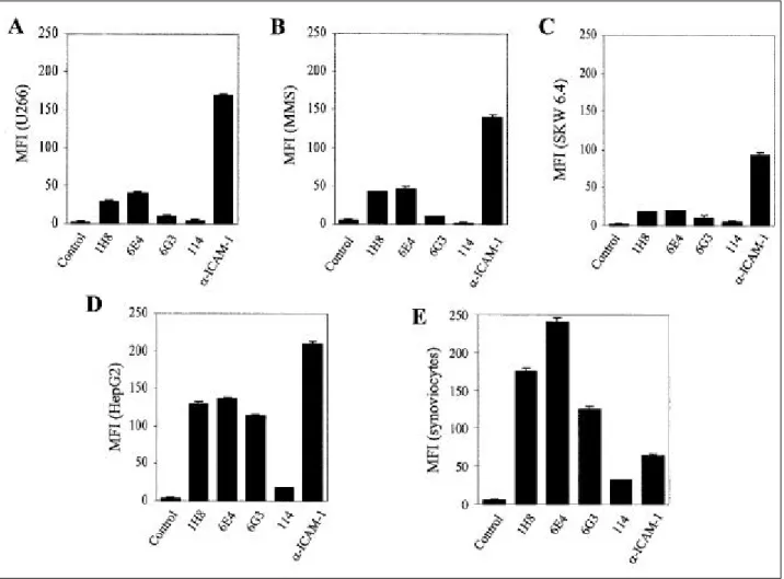
Fig . 1 . The e x p re s s io n of 1H8 in v a rio us h um a n ce lls by flow cyto me t ric a na lys is . Va rio us h u ma n c e lls w e re s ta ine d w it h MoAbs a g a inst BMS C s , a nd F ITC c o nj ug a te d a nt i-mo us e Ig w a s us e d a s a s e c o nd a ry a nt ibo dy . Aft e r 3 0 m in o n ic e , t he c e lls w e re w a s he d a nd a na lyz e d by flow cyt o m et ry . MF I; me a n fluo re s c e nc e int e ns ity , A; U2 6 6 , B;
BMS C fro m MM pa t ie nts , C ; S KW 6 .4 , D; He pG 2 , E ; s ynov io cyte s fro m rhe u ma t o id a rt h rit is pa t ie nt s .
well were seeded in 96-well flat-bottom microtiter plates in 100 ㎕ of RPMI 1640 supplemented with 10% FBS, and 100 ㎕ of serial dilutions of test samples were added. The plates were incubated for 72 h in a humidified 5% CO2 incubator at 37℃. Cells were pulsed with 0.5 μCi of [3H] thymidine (specific activity; 84.8 Ci/mmol, New England Nuclear, Boston, USA) per well for the last 6 h of the incubation time, and were harvested onto glass fiber filter papers using an automated cell harvester (Inotech, Brieger, Zurich). The amount of radioactivity incorporated into the DNA was determined using a liquid scintillation counter (Beckman, LS 6000A). In all assays, recombinant human IL-6 was used as the internal standard. One unit of IL-6 was defined as the amount inducing half-maximal prolifera- tion.
RESULTS
1. P r odu ct ion an d s cr een in g of 1H 8 MoAb a g ain st B M S C
To investigate whether cell surface molecules promote cell proliferation through cellular interaction, a co-culture
of human myeloma cells with BMSCs was performed.
U266 cell proliferation increased on co-culturing with BMSCs compared with the culture of U266 cells alone, but the inner cell membrane chamber, which blocked direct cell-to-cell contact inhibited U266 cell proliferation (data not shown) . This result suggested that the cell surface molecules of BMSCs and myeloma cells are involved in the proliferation of myeloma cells. To identify surface molecules, which may regulate cell growth, MoAbs against BMSCs obtained from an MM patient were raised. Screening the positive MoAbs using BMSC ELISA showed that four MoAbs (IgM) had high reactivity to BMSCs compared with the control (data not shown). Flow cytometric analysis was performed to assess whether these MoAbs stain other human cell lines, (Fig. 1). Among these, 1H8 MoAb highly stained
Fig . 2 . We ste rn blot of MoAbs a ga inst BMS C in U266 ce lls . The me mbra ne fra ct ion of BMS Cs wa s e le ct ro- phoretica lly s e pa rate d a nd t ra nsfe rre d from a ge l to a nit roce llulos e me mbra ne (NCM), w hich was incubate d w it h the MoAbs a ga inst BMS Cs at room te mpe rature for 2 h. Afte r three wa s hings , a lka line phos phata s e conjugate d goat a nti- mous e IgM wa s a dde d at RT for 1 h. Afte r a furthe r t hre e wa s hings , NCM wa s mixe d w it h t he s ubstrate s for a lka line phos phata s e .
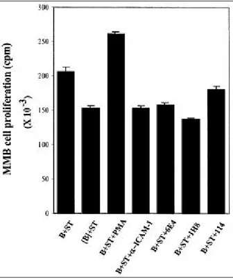
Fig . 3 . Effe cts of MoAbs a ga inst BMS Cs on fre s h mye loma ce ll prolife ration afte r co-culture w ith BMS C.
BMS Cs (ST) we re plate d at a de ns ity of 2x 10⁴ ce lls/
㎖. Afte r 12 h incubation, fres h mye loma ce lls (B) we re a dde d at a de ns ity of 5x 10⁴ ce lls/㎖. Cocult ure wa s use d in t he a bs e nce (B) or pre s e nce ([B]) of a n inne r ce ll me mbra ne filte r cha mbe r. Afte r 3 days , ce ll prolife ration wa s me a s ure d us ing[3H]-thymidine incorporat ion.
synoviocytes and HepG2 cells, and was moderately reactive to SKW 6.4, BMSCs, and U266 cells. ICAM-1 MoAb was used as positive control expressed in all cell lines analyzed. However, the staining patterns of the various cells with ICAM- 1 MoAb was obviously different from those stained with 1H8, indicating that 1H8 did not recognize ICAM-1. Immunoblots were preformed using BMSC extract to identify the specific proteins recognized by these anti-BMSC antibodies (Fig.
2). 1H8 recognized distinctly an 80 kDa molecule, indicating that it is different from opther antibodies.
2. E f f ect s of 1H 8 on cell pr olif er at ion an d IL - 6 p r odu ct ion of h u m an m y elom a cells
Next, to assess whether the antibodies could inhibit myeloma cell proliferation induced by BMSCs, these antibodies were added to primary myeloma cells, which were co-cultured with BMSC. Blocking the co-culture between these cells with an inner cell membrane chamber ([B]) inhibited fresh myeloma cell proliferation (Fig. 3). It is been known that IL-6 is one of the growth factors involved and that the up-regulation of IL-6 in fresh myeloma cells is induced via CD40 stimulation 1 1).
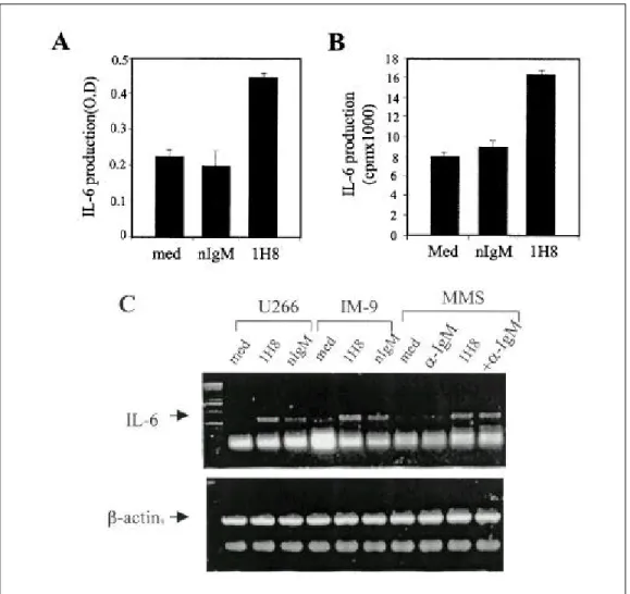
Fig . 4 . Effects of 1H8 on IL-6 expres s ion in mye loma ce lls a nd BMS Cs . 1 ug/㎖ of 1H8 or norma l mous e IgM (nIgM, is otype cont rol) we re coate d in 24-we ll cult ure plate w ith P BS at RT for 1 h, a nd t he n wa s he d w it h P BS thre e time s . U266 or IM-9 ce lls we re a dde d a nd culture d for 48 h. The cult ure s upe rnata nts we re ha rveste d a nd a s saye d for IL-6 production us ing a n IL-6 ELISA (A) a nd a n IL-6-de pe nde nt B9.55 bioa s s ay (B). U266, IM-9 or BMS Cs we re st imulate d by 1H8 a lone or by a ddit iona l cros s-linking w it h a nt i-IgM a nt ibody (+-IgM) for 6 h, a nd cytopla s mic RNA wa s is olate d. RT-PCR wa s pe rforme d a s de s cribe d in Mate ria ls a nd Met hods (C).
As shown in Fig. 4, the effects of 1H8 on the IL-6 ex- pression of BMSCs and myeloma cells were investigated.
IL-6 production by U266 cells was significantly increased by 1H8 treatment as determined by IL-6 ELISA (Fig. 4-A) and IL-6 bioassay (Fig. 4-B). The IL-6 gene expressions of U266, IM9, and BMSCs were also increased by 1H8 treatment (Fig. 4-C). In addition, IL-6 receptor and CD40 expression in U266 cells was analyzed by flow cytometry . As shown in Fig. 5, 1H8 treatment increased the IL-6 receptor and CD40 expression of U266 cells.
3. 1H 8 in h ib it s R OS p r odu ct ion f r om h u m an m y elom a cells
Many reports have shown that ROS modulates cellular functions including gene expression, survival, apoptosis, and tumorigenesis (15-19). Previous data including the regulation of proliferation and IL-6 gene expression of myeloma cells suggest that 1H8 can modulate ROS level in myeloma cells. The intracellular ROS levels of myeloma cells were compared to those of other B cells.
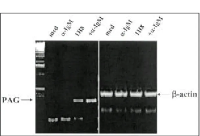
As shown in Fig. 6, endogenous ROS levels were much higher in U266 (A) or IM-9 (B) than in WIL-2 NS (C) or CESS cells (D) . Interestingly, 1H8 treatment sup- pressed the ROS level of U266 cells (E). Intracellular ROS levels are known to be controlled by an intra- cellular antioxidant pool, and PAG, which is well-known as a thio-specific antioxidant gene, is fairly ubiquitouslyexpressed and induced to higher levels by serum stimulation and oxidative stress. The expression of PAG was determined in U266 cells, and PAG was found to be induced by 1H8 in U266 cells, and that this was increased by cross-linking 1H8 with anti-mouse IgM (Fig. 7).
4 . 1H 8 in du ce s pr ot ein ph osph or y lat ion of h u m an m y elom a cells .
Protein phosphorylation plays a crucial role in the regulation of cell growth, cytokine expression, and cell cycle (20,2 1). U266 were treated with 1H8 and cell ex- tracts were immunoprecipitated with anti-phosphotyrosine antibody followed by immunoblot with same antibody . Fig . 5 . Effe cts of 1H8 on the expre s s ion of IL-6 re ce ptor a nd CD40 on U266 ce lls . U266 ce lls
we re cocult ure d w ith BMS Cs in the a bs e nce (me dia ) or pre s e nce of MoAbs (114 a nd 1H8) a ga inst BMS Cs . Afte r 3 days of cocult ure , U266 ce lls we re sta ine d w it h a nti-IL-6 re ce ptor a ntibody (MT18) a nd a nt i-huma n CD40 a ntibody. FITC-conjugate d a nt i-mouse Ig wa s use d a s a s e conda ry a nt ibody.
Immunoblot showed that several phosphoproteins such as 60, 65, and > 100 kDa proteins were tyrosine phosphory- lated time-dependently in 5 to 30min (Fig. 8), confirming that 1H8 delivers intracellular signaling through its binding molecule.
DI SCUSSI ON
BMSCs produce soluble factors and express cell
surface molecules, which regulate the proliferation and differentiation of hematopoietic cells. Thus, these cell surface molecules play a central role in the communi- cation between myeloma and BMSC. Most malignant plasma cells are bound to BMSCs via cell surface molecules, which are locally located in the BM and rarely circulate in the peripheral blood. Moreover, the expression of cell surface molecules in malignant plasma cells is related to the regulation of immune cells. This Fig . 6 . Effe cts of 1H8 on int ra ce llula r ROS leve ls . Huma n B ce lls we re incubate d w it h
50 M of 2', 7'-dichloro- fluore s ce in dia cetate (DCFH-DA) for 5 min at 37℃ . The ce lls we re was he d tw ice a nd a na lyz e d by flow cytomet ry. A, U266 ; B, IM-9; C, Wil-2 NS ; D, CES S . Ope n a nd clos ed histogra ms re pre s e nt unsta ine d a nd sta ine d ce lls , re s pect ive ly. U266 Ce lls we re incubate d w it h 1H8 for 20 min (E) a nd intrace llula r ROS leve ls we re a na lyz ed a s de s cribe d a bove .
means that cell-cell interactions via cell surface molecules plays a crucial role in the proliferation of
malignant plasma cell ( 1) .
In an attempt to identify the cell surface molecules of BMSCs, we generated several monoclonal antibodies against intact BMSCs. These antibodies recognized different cell surface molecules based on Facscan and immnoblot analysis. In addition, these antibodies blocked B cell proliferation induced by co-culturing with BMSCs.
Among these antibodies, we characterized a 1H8 monoclonal antibody, which recognized an 80 kDa molecule.
Multiple myeloma is a B cell neoplastic disease, which results from malignant transformation of a single clone of plasma cells in the BM. Many soluble factors such as IL-3, IL-6, IL-8, TGF-β (22), and GM-CSF (23) are known to be involved in the progression of MM. In addition, adhesion molecules including CD54, CD56, CD29, VLA-4, and LFA are also known to have some role in cell proliferation and in the trapping of myeloma cells in the BM environment (6) . IL-6 is a well-known growth factor for myeloma cells, although there have been some controversial reports on the mode of its action i.e., paracrine (14) or autocrine (24,25).
To test whether 1H8 regulated the functions of myeloma cells and stromal cells, the regulation of IL-6 expression by 1H8 was examined. 1H8 increased IL-6 expression in myeloma cells and BMSCs. In addition, it increased IL-6 receptor and CD40 expression in U266 cells cocultured with BMSCs. Urashima et al. (11) demonstrated that the up-regulation of IL-6 was induced by the stimulation of CD40 in MM cells, and Westendorf et al. (12) reported that the activation of CD40 induced the up-regulation of IL-6 to and stimulated IL-6-dependent myeloma cell proliferation.
However, 1H8 inducing IL-6 production showed inhibitory effects on myeloma cell proliferation in the case of co-culturing with MMSCs. This may have been due to the epitope of IH8 being different from the binding site for the antigen for 1H8 and its ligand.
Alternatively, the stimulating effects of co-culturing with MMSCs may require multiple interactions between different surface molecules including the antigen for 1H8, which by itself can induce IL-6 production. The Fig . 7 . Effe cts of 1H8 on the expre ss ion of PAG in
U266 ce lls . U266 ce lls we re incubated w it h 1 ug/㎖ of 1H8 for 20 min, a nd wa s hed w it h P BS , a nd the n 1 ug/
㎖ of a nti-mous e IgM wa s a dde d. Afte r 6 h incubation, t he ce lls we re ha rve ste d a nd cytopla s mic RNA wa s is olate d. PAG ge ne expre s s ion wa s a na lyz e d by RT-PCR.
Fig . 8 . Effe cts of 1H8 on the phos phorylat ion of s e- ve ra l prote ins of U266 ce lls . U266 ce lls we re stimulate d w ith 1 ug/㎖ of 1H8 at va rious t ime s . The ce ll lys ate s we re the n immunopre cipitate d w it h a nt i- phos photyros ine a nt ibody. P hos phorylate d prote ins we re s e pa rate d on a 10% S DS-PAGE a nd dete cte d by immunoblot us ing a nti-phos photyros ine a ntibody.
identification of antigen for 1H8 and the physiological ligand for the antigen are necessary to resolve this structural and functional issue.
Intracellular ROS level is believed to be important for tumorigenesis, cell survival and gene regulation.
Recently, it has been reported that ROS is also a critical factor for cellular signaling. Endogenous ROS levels in other B lymphocytes such as WIL-2, SKW6.4, are CESS were different from those of U266 cells, suggested that elevated ROS function is a regulatory factor of cell survival and gene expression in myeloma cells. 1H8 can modulate ROS level by regulating PAG antioxidant gene expression. However, it did not affect cell apoptosis at whatsoever (data not shown).
Immunoblot with anti-phosphotyrosine antibody indicated that 1H8 can induce and deliver transient (5- 10 min) and sustained (>30 min) signals into the myeloma cells, suggesting that 1H8 can induce diverse intracellular signals. Identification and studies upon the functional roles of these phosphoproteins are required to understand the intracellular signaling pathways triggered by 1H8.
In summary, 1H8 effects intracellular signaling by modulating protein phosphorylation, ROS level, and gene expression. It provides a tool for the studying the regulation of B cell proliferation and B cell function.
REFERENCES
1. Ahsmann EJ, Lokhorst HM, Dekker AW, Bloem AC:
Lymphocyte function-associated antigen-1 expression on plasma cells correlates with tumor growth in multiple myeloma. Blood 79; 2068-2075, 1992
2. Kaisho T, Ishikawa J, Oritani K, Inazawa J, Tomizawa H, Muraoka O, Ochi T, Hirano T: BST-1, a surface molecule of bone marrow stromal cell lines that facilitates pre-B-cell growth. Proc Natl Acad Sci USA 91; 5325-5329, 1994
3. Hallek M, Bergsagel PL, Anderson KC: Multiple myeloma: Increasing evidence for a multistep trans- formation process. Blood 91; 3-2 1, 1998
4. Caligaris-Cappio F, Bergui L, Gregoretti MG, Gaidana G, Gaboli M. Schena M, Zallone A, Marchisio PC:
Role of bone marrow stromal cells in the growth of human multiple myeloma. Blood 77; 2688-2693, 1991 5. Hamilton MS, Ball J, Bromidge E, Frankin, IM: Surface antigen expression of human neopastic plasma cells includes molecules associated with lymphocyte recirculation and adhesion. Br. J. Hamatol. 78; 60-65,
1991
6. van Riet I, Waele MD, Remels L, Lacor P, Schots R, Camp BV: Expression of cytoadhesion molecules (CD56, CD54, CD 18 and CD29) by myeloma plasma cells. Br. J. Haematol. 79; 42 1-427, 1991
7. Simmons PJ, Torok-Storb, B: Identification of stromal cell precursors in human bone marrow by a novel monoclonal antibody, STRO-1. Blood 78; 55-62, 1991 8. Teixido J, Hemler ME, Greenberger JS, Anklesaria, P:
Role of beta 1 and beta 2 integrins in the adhesion of human CD34hi stem cells to bone marrow stroma. J.
Clin. Invest . 90; 358-367, 1992
9. Clark BR, Gallagher JT, Dexter TM: Cell adhesion in the stromal regulation of haemopoiesis. Baillieres Clin.
Haematol. 3; 619-652, 1992
10. Durie FH, Foy TM, Masters SR, Laman JD, Noelle RJ:
The role of CD40 in the regulation of humoral and cell-mediated immunity. Immunol. Today 15; 406-411, 1994
11. Urashima M, Chauhan D, Uchiyama H, Freeman GJ, Anderson KC: CD40 ligand triggered interleukin-6 secretion in multiple myeloma. Blood 85; 1903-1912, 1995
12. Westendorf JJ, Ahmann GJ, Armitage RJ, Spriggs MK, Lust JA, Greipp PR, Katzmann JA, Jelinek DF: CD40 expression in malignant plasma cells. Role in stimulation of autocrine IL-6 secretion by a human myeloma cell line. J. Immunol. 152; 117-128, 1994 13. Yamasaki K, Taga T, Hirata Y, Yawata H, Kawanishi
Y, Seed B, Taniguchi T, Hirano T, Kishimoto T:
Cloning and expression of the human interleukin-6 (BSF-2/IFN-beta 2) receptor. Science 241; 825-828, 1988
14. Klein B, Zhang XG, Jourdan M, Content J, Houssiau F, Aarden L, Piechaczyk M, Bataille R: Paracrine rather than autocrine regulation of myeloma-cell growth and differentiation by interleukin-6. Blood 73; 517-526, 1989
15. Kowluru A, Metz SA : Ceramide-activated protein phosphatase 2A activity in insulin-secreting cells. FEBS Lett. 4 18; 179-182, 1997
16. Polyak K, Xia Y, Zweler JL, Kinzler KW, Vogelstein B: A model for p53-induced apoptosis. Nature 18;
300-305, 1997
17. Rieber MS, Rieber M: Sensitization to DNA damage by okadaic acid or bromodeoxyuridine involves unequal effects on melanoma cell adhesion and differentiation.
DNA and Cell Biol. 16; 12 1-125, 1997
18. Rodrigues CMP, Fan F, Ma X, Kren BT, Steer CJ: A novel role for ursodeoxycholic acid in inhibiting apoptosis by modulating mitochondrial membrane perturbation. J. Clin. Invest. 101; 2790-2799, 1998 19. Zezschwitz CV, Vorwerk H, Tergau F, Steinfelder HJ:
Apoptosis induction by inhibitors of Ser/Thr phos- phatases 1 and 2A is associated with transglutaminase activation in two different human epithelial tumor lines.
FEBS Lett. 4 13; 147-151, 1997
20. Shuai K, Schindler C, Prezioso VR, Darnell JE: Acti- vation of transcription by IFN-gamma: Tyrosine phosphorylation of a 91-kD DNA binding protein.
Science 258; 1808-1812, 1992
2 1. Gj ertsen BT, Doskeland, SO: Protein phosphorylation in apoptosis. Biochim. Biophys. Acta. 1269: 187-199, 1995 22. Lagneaux L, Delforge A, Dorval C, Bron D, Stryckmans P: Excessive production of transforming growth factor-
beta by bone marrow stromal cells in B-cell chonic lymphocytic leukemia inhibits growth of hematopoietic precursors and interleukin-6 production. Blood 82;
2379-2385, 1993
23. Zhang XG, Bataille R, Jourdan M, Saeland S, Banche- reau J, Mannoni P, Klein, B: Granulocyte-macrophage colony stimulating factor synergizes with interleukin-6 in supporting the proliferation of human myeloma cells.
Blood 76; 2599-2605, 1990
24. Kawano M, Hirano T, Matsuda T, Taga T, Horii Y, Iwato K, Asaoku H, Tang B, Tanabe O, Tanaka H, Kuramoto A, Kishimoto, T: Autocrine generation and requirement of BSF-2/IL-6 for multiple myelomas.
Nature 332; 83-85, 1988
25. Schwab G, Siegall CB, Aarden LA, Neckers LM, Nordan, RP: Characterization of an interleukin-6 mediated autocrine growth loop in the human multiple myeloma cell line, U266. Blood 77; 587-593, 1991
FOOTNOTES
1. This work was supported by a grant from the HAN proj ect (HSM0100033) at MOST, Republic of Korea.
2. Abbreviations used: BM, bone marrow; BMSC, bone marrow stromal cells; MM, multiple myeloma; MoAb, monoclonal antibody; ROS, reactive oxygen species;
PAG, Proliferation-associated gene