심바스타틴과 아토바스타틴이 치료적인 농도에서 TPC-1 세포의 NIS 발현에 미치는 영향
김태균1·정혜숙2·윤창신2·고정해1·전혜정1·권민정1·이순희1·김미경2,3·박정현1,2
인제대학교 의과대학 부산백병원 내분비대사내과1, 백인제 임상의학 연구소2, 메리놀병원 내분비대사내과3
Received: 11 March 2010, Accepted: 28 May 2010 Corresponding author: Jeong Hyun Park
Department of Internal Medicine, Pusan Paik Hospital, College of Medicine, Inje University, Gaegeum-dong, Busanjin-gu, Busan 614-735, Korea
Tel: +82-51-890-6074, Fax: +82-51-894-0145, E-mail: [email protected]
The Effect of Atorvastatin and Simvastatin on NIS Expression of the TPC-1 Cell under the Therapeutic Blood Concentrations
Tae Kyoon Kim1, Hye Sook Jung2, Chang Shin Yoon2, Jung Hae Ko1, Hae Jung Jun1, Min Jung Kwon1, Sun Hee Lee1, Mi Kyung Kim2,3, Jeong Hyun Park1,2
Department of Internal Medicine1, Pusan Paik Hospital, College of Medicine, Inje University, Busan; Molecular Therapy Lab.2, Paik Memorial Institute for Clinical Research, Inje University, Busan; Department of Internal Medicine3, Marynoll Medical Center, Busan, Korea
Background: Although so many experimental trials have been done to improve the redifferentiation and responsiveness of radioio- dide therapy, they have not yet yielded any satisfactory results. As statins inhibit both farnesylation and geranylgeranylation, they have been reported to have an antineoplastic and redifferentiation effect in experimental and clinical studies. In this study, we investigated the relationship between statins and the alteration of the NIS expression and, TPC-1 cell apotosis to evaluate the possibility of using statins as adjuvant therapeutic agents for papillary thyroid cancer.
Methods: We used the TPC-1 cell lines for our experiments. Cell viabilities were measured by CCK-8. The degrees of apoptosis and, the expressions of NIS mRNA and NIS protein were measured by flow cytometry, semi quantitative RT-PCR and Western blot assay.
Results: Increased levels of NIS mRNA and NIS protein were observed under therapeutic blood concentrations (concentrations of simvastatin: 20, 50, 80 nM, concentrations of atorvastatin: 50, 80,110 nM), but the dose-response relationship was only manifested within simvastatin. The TPC-1 cells showed a concentration dependent decrease of viability and an increase of apoptosis not under therapeutic blood concentrations, but under excessively high concentrations (after treatment with 10-50 μM of atorvastatin and with 1-10 μM of simvastatin).
Conclusion: The results of this study show that effective therapeutic blood concentrations of simvastatin and atorvastatin can give a favorable effect on the NIS expression under effective therapeutic blood concentrations. Therefore, we demonstrated the possibility that simvastatin and atorvastatin might have an important role as adjuvant therapeutic agents to improve the responsiveness of ra- dioiodide therapy for papillary thyroid cancer. Further studies are needed to clarify this issue. (Endocrinol Metab 25:192-198, 2010) Key Words: Thyroid Papillary Cancer, TPC-1 cell, Statin, NIS
서 론
잘분화된갑상선유두암들은갑상선여포세포의기저막(basolat- eral membrane)에 sodium iodide symporter (NIS)가잘발현되어 있어갑상선절제술후방사선요오드치료시방사선요오드의섭 취를증가시켜잔존 갑상선암이나재발성, 그리고, 전이성갑상선
암의치료효과증대에중요한역할을하고있다는것이잘알려져 있다[1]. 갑상선유두세포암중일부는조기탈분화(early dedifferen-
tiation) 과정을거쳐보다더공격적으로변하여빠른성장을하거
나주위조직으로의침윤이증가하는경향을보이는데, 이런탈분화 과정을거친갑상선세포암은 NIS의발현이없거나감소하여갑상선 절제술후방사선동위원소치료효과를감소시키며, TSH 억제치료 에저항성을 보여궁극적으로는환자의나쁜 예후에영향을주게
된다[2]. 이러한분화도가감소된갑상선암에대해항암치료보조
약제로서분화도및방사성요오드섭취능의호전을위한여러약 제들을이용한시도들이있어왔지만 만족할만한효과를보이는
약제는없었다. 최근활발한연구가이루어지고있는스타틴의경우 콜레스테롤생합성억제효과이외에세포막의유지, 신호전달, 단백 질생합성, 세포주기에영향을미치는 mevalonate 경로를억제하여 Ras의 farnesylation과 Rho의 geranylgeranylation을억제하고, 종양 세포의발생, 성장, 분화도의조절, 전이의억제및종양세포의고사 를유도시켜[3] 장기사용하면혈액암, 췌장, 전립선, 대장암, 유방암
및흑세포암세포주의위험이감소된다는보고가있다[4].
따라서, 저자들은심바스타틴과아토바스타틴을이용하여갑상선 유두세포암의분화도회복을통해 sodium iodide symporter (NIS) 의발현이실제치료적인혈중유효농도에서이루어지는지를확인 하여향후방사성요오드치료의보조치료약제로서뿐만아니라, 다른 고형암에서와같이 갑상선 유두세포암에서도 TPC-1 세포의 자멸사를유도하여항암치료보조제로서스타틴적용가능성을확 인하고자하였다.
대상 및 방법
1. 재료
심바스타틴과 아토바스타틴은 BIOMOL Research Laboratories, Inc. Plymouth Meeting (PA, USA)로부터구매하여사용하였고, 세 포배양에필요한 Dulbecco’s Modified Eagle’s Medium (DMEM)과 우태아혈청등은 GIBCO (CA, USA)에서구매하였다. Cell Counting Kit-8 (CCK-8)은 Dojindo Laboratories (Kumamoto, Japan)에서구입 하였으며, Apoptosis marker인 Annexin V-FITC와 Propidium Io- dide (100 μg/mL)는 BD bioscience (CA, USA)에서구입하였다. 반정 량적역전사중합효소연쇄반응을시행하기위해 RNeasy Mini kit 는 QIAGEN (Hilden, Germany)에서, Accupower RT/PCR premix와 human NIS primers는 Bioneer (Daejeon, Korea)에서구매하였다. CelLyticTM MT Mammalian tissue Lysis/Extraction Reagent는 Sigma (MO, USA)에서, 1차 Anti-sodium/Iodide symporter (NIS) monoclo- nal Antibody는 Chemicon (CA, USA)에서, 이차항체인 Goat Anti- Mouse antibody는 Biorad (CA, USA)에서구입하였으며, 단백정량 에사용된 BCATM Protein Assay Reagent Kit는 PIERCE (IL, USA)에 서구입하였다. 그외에사용된화학약품들은 Sigma-Aldrich (MO, USA)에서구입하였다.
2. 세포주 및 배양 조건
갑상선유두암세포주(TPC-1 세포: 충남의대내과송민호교수님
기증, Passage 15)는 10% 우태아혈청을첨가한 DMEM 배양액을사
용하여 37℃, 5% CO2표준가습배양기에서배양하였다. 배양액은
2-3일마다교환하였으며세포가 90-95% 정도자라면 계대배양을 하였다. 정해진 농도의스타틴 시약을 만들기 위하여 스타틴과 DMSO를일정한비율로녹였으며, 스타틴처리직전우태아혈청을
첨가하지않은 DMEM 배양액과혼합하였다. 6-well plate에 1 × 10⁵/
well 세포를분주하여 70% 정도자랐을때정해진농도의스타틴을
처리하여 주었으며, 세포 사멸을 보기위한 실험에서는 96-well plate에 2 × 10³/well 세포를분주하여 2일후에정해진농도의스타 틴을처리하였다.
3. 세포생존율 측정(Cell Viability Assay)
스타틴이세포사멸에미치는영향을알아보기위해 CCK-8을이
용한발색분석법을사용하였다. 96-well plate에각 well당 2 × 10³ 개씩의세포를 분주하고 2일 후정해진농도의 스타틴을 함유한
DMEM 배양액으로갈아준후 48시간동안세포배양기에서배양
하였다. 배양액을제거하고 PBS 용액으로 2번세척한후 CCK-8 100 μL씩첨가하였다. 세포배양기에서 1-4 시간동안배양후, 발색정도 를측정하기위해 Microplate reader (SLT, Austria)에서 450 nm의파 장으로흡광도를측정하였으며, 각실험은독립적으로 3회반복실 행하여평균치와표준편차를구하였다.
4. 세포자멸사 분석(Flow Cytometry)
세포자멸사분석은 Apoptosis marker인 Annexin V-FITC와 Prop- idium Iodide (100 μg/mL)로반응시킨후유세포분석기(FACS: fluo- rescence activated cell shorting, Becton-Dickinson, San Jose, CA, USA)를이용하여확인하였다. 반응이끝난세포는 single cell로만 들기위해서 Trypsin-EDTA를처리하고, FACS tube에모아서 1,500 rpm에서 5분간 원심분리하였다. 상층액은제거하고 1 × binding buffer (140 mM NaCl, 10 mM HEPES [pH 7.4], 2.5 mM CaCl2, store at 2-8℃) 1 mL을넣고세포를풀어준뒤 1,500 rpm으로 5분간원심 분리하였다. 상층액을제거한뒤 Annexin V-FITC 3 μL와 Propidium Iodide 10 μL를 15분간반응시켰다. 그후 FACS buffer (1% FBS, 0.1
% NaN3) 300 μL를넣고 FACS로분석하였다.
5. 반정량적 역전사 중합효소 연쇄반응(Semiquantitative RT-PCR) TPC-1 cell에서 RNA 분리는 RNeasy Mini kit를이용하여시행하 였다. 농도조건은심바스타틴과아토바스타틴 40 mg을건강한성 인에서경구투여하였을때의최고혈중농도와최저혈중농도를(심 바스타틴의혈중유효농도는각각 20-50 nM이었고, 아토바스타틴 에서는 50-80 nM) 기준으로정하였다[5]. 6-well plate에배양된갑상 선유두암세포를각각의정해진농도의스타틴에 1시간동안반응 시켰다. 반응이끝난후 PBS로 3회세척하고즉시 Buffer RLT 350 μL 를넣어 pipette으로 RNA를분리하였다. 분리된 RNA는 Eppendorf tube에넣고 30초간 vortex 시행하였다. 70% ETOH 300 μL를추가 하여 pipette으로잘섞은뒤총 700 μL를 collection tube내 RNeasy spin column에넣고상온에서 15초간 10,000 RPM으로원심분리하 였다. Collection tube에모인액을제거한뒤 700 μL Buffer RW1을
spin column에넣고 15초간 10,000 RPM으로원심분리하였다. Col- lection tube에모인액을제거한뒤 500 μL Buffer RPE를 spin col- umn에넣고 15초간 10,000 RPM에서원심분리하고, 다시 collection tube에모인액을제거한뒤 500 μL Buffer RPE를 spin column에넣 고 2분동안 10,000 RPM에서원심분리하였다. 그후새 collection tube에서 30 μL RNase free water를 spin column에넣고 1분간 10,000 RPM으로원심분리한후 Nano-drop ND-1000 spectropho- tometer를이용하여 RNA를정량하였다.
역전사과정은 42℃에서 1시간, 중합효소 연쇄반응과정은 95℃ 에서 15분간예비변성을하고 95℃에서 2분, 40.1℃에서 1분, 72℃에 서 1분을 40주기를시행한후마지막확장은 72℃에서 10분간한다 음 4℃에서보관하였다.
Semiquantitative RT-PCR에필요한 human NIS primer의염기서 열은 Forward primer: 5′-TCTCTCAGTCAACGCCTCT-3′, Reverse primer: 5′-ATCCAGGATGGCCACTTCTT-3′, 증폭된생성물의크기 는 299 bp이며, GAPDH mRNA를 internal control로사용하였다. 만 들어진 PCR 생성물은 1.2% agarose gel에서전기 영동하여 LAS- 3000 (Fujifilm, Japan)에서사진을찍어확인하고, Multi Gauge 3.0
(Fujifilm, Japan)을이용하여 densitometry 정량을시행하였다. 각 실험은독립적으로 3회반복하여시행하였다.
6. 웨스턴블롯(Western blot) 분석
웨스턴블롯시농도 조건은 semiquantitative RT-PCR을시행할 때와동일하게정하였다[5].
스타틴이처리된세포를 PBS 용액으로 2회세척하고단백질을분
리하기위해서 Mammalian tissue lysis buffer 100 μL를넣고세포를 융해시켰다. 12,000 RPM, 4℃에서 10분간원심분리하여상층액만 을깨끗한 eppendorf tube에옮기고사용하기전까지 -70℃에보관 하였다. Western blot 실험을시작하기전 BCATM protein assay kit로 단백질을정량하였다. 10% SDS-PAGE에한 well당 100 μg을로딩하 여전기영동을하고 10 V에서 PVDF membrane에옮긴후 5% skim milk로 blocking하였다. 1차 Anti-Sodium/Iodide symporter (NIS) monoclonal antibody (1:1,000)를 4℃에서 12시간냉장반응시키고, 2차 Goat Anti-Mouse antibody (1:200)를실온에서 1시간반응시킨 뒤 AP-conjugated development kit Biorad (CA, USA)로발색하였다.
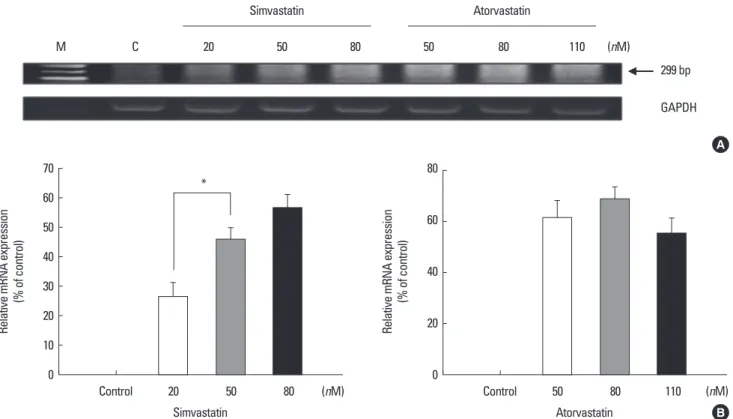
Fig. 1. Human NIS mRNA expression after treatment of simvastatin and atorvastatin. A. After treatment of each statins for 48 hr, human NIS mRNA expression treated with simvastatin was detected in a dose dependent manner by semiquantitative RT-PCR. But, human NIS mRNA expression treated with atorvastatin showed the only increase of mRNA expression in a dose independent manner. Treated concentrations of statins were decided by the range of effective blood concentrations which normal healthy adults ingest simvastatin or atorvastain 40 mg P.O. (C: control). B. These graphs show the increased percent of relative mRNA expression in a dose dependent manner after treatment of simvastatin. But, atorvastain showed the only increased percent of relative mRNA expression in a dose independent man- ner. These densitometric result were measured by Multi Gauge 3.0. (concentrations of simvastatin: 20, 50, 80 nM, atorvastatin: 50, 80,110 nM).
Relative mRNA expression (% of control) 70 60 50 40 30 20 10 0
Control 20
Simvastatin
50 80 (nM)
*
M C 20 50 80 50 80 110 (nM)
299 bp
GAPDH
Simvastatin Atorvastatin
A
B Relative mRNA expression (% of control)
80
60
40
20
0
Control 50
Atorvastatin
80 110 (nM)
결 과
1. 반정량적 역전사 중합효소 연쇄반응(Semiquantitative RT-PCR)
건강한성인에서스타틴 40 mg 경구투여시얻어지는치료적인
혈중유효농도는심바스타틴에서는 23.9-81.2 nM, 아토바스타틴에 서는 48.3-119.6 nM이었다[5]. 6-well plate에배양된 TPC-1 세포에 각각심바스타틴 20, 50, 80 nM을, 아토바스타틴 50, 80, 110 nM의 농도로투여하여, 스타틴투여전과투여 1시간후 NIS mRNA의발 현정도를확인하기위하여 semi-quantitative RT-PCR을이용하여 이를확인하였다. 스타틴투여 1시간후심바스타틴에서농도의존
적인 NIS mRNA 발현 증가가 관찰되었지만, 아토바스타틴에서는
NIS mRNA 발현이관찰되었으나, 농도의존적이진않았다(Fig. 1).
2. 웨스턴 블롯(Western blot)
10 cm petri dish에배양된 TPC-1 세포에반정량적 semiquantita-
tive RT-PCR을시행할때와동일한농도의스타틴을투여하여(심바
스타틴의농도는 20, 50, 80 nM, 아토바스타틴의농도는 50, 80, 110 nM이었다)[5], 스타틴투여전에비해투여 48시간후 NIS 단백발현 의변화를측정하였다. 심바스타틴의경우농도의존적인 NIS 단백
질의증가가관찰되었지만, 아토바스타틴의경우 NIS 단백질이발현
되었으나농도의존적인 NIS 단백질발현의증가는관찰되지않았
다(Fig. 2).
3. 심바스타틴과 아토바스타틴 처리 후 농도에 따른 TPC-1 세포의 생존변화
치료적인 혈중유효농도에서스타틴의 TPC-1 세포생존변화에
미치는영향을관찰하였으나아무런변화가관찰되지않았으며, 보 다높은고농도에서심바스타틴과아토바스타틴을 48시간동안처
리하고 CCK-8을통해측정하였을때모든세포주에서세포생존율
의농도의존적감소가관찰되었다(Fig. 3).
4. 스타틴 처리 후 각 세포의 농도에 따른 유세포 분석
TPC-1 세포를대조군과각각 일정한고농도의 스타틴을투여한
군으로나누어살아있는세포, 고사된세포및죽은세포로나누어 농도에따른세포분획의변화를관찰하였다. 대조군에비해스타틴 투여군에서농도에따라살아있는세포의비율이감소되면서고사 된세포의비율이점차로증가되어농도에따른세포자멸사의진행 을관찰할수있었다(Fig. 4).
Fig. 2. Human NIS protein expression after treatment of simvastatin and atorvastatin. A. After treatment of each statins for 48 hr, human NIS protein expression treated with simvastatin was detected in a dose dependent manner by Western blot. But, human NIS protein expression treated with atorvastatin showed the only increase of protein expression in a dose independent manner. Treated concentrations of statins were decided by the range of effective blood concentrations which normal healthy adults ingest simvastatin or atorvastain 40 mg P.O. (C: control). B. These graphs show the increased percent of relative protein expression in a dose dependent manner after treatment of simvastatin. But, atorvastain showed the only increased percent of relative protein expression in a dose independent manner.
These densitometric result were measured by Multi Gauge 3.0. (concentrations of simvastatin: 20, 50, 80 nM, atorvastatin: 50, 80,110 nM).
Relative protein expression (% of control) 800
600
400
200
0 Control
Simvastatin
(nM)
*
80 50
20
A
B Relative protein expression (% of control)
350 300 250 200 150 100 50 0
Atorvastatin
(nM)
Control 50 80 110
M 20 50 80 50 80 110 (nM)
97 kDa
β-actin 70 kDa
Simvastatin Atorvastatin
고 찰
본연구에서가장의미있는소견은실제치료적용량인심바스타 틴 40 mg과아토바스타틴 40 mg을투여했을때유효혈중농도(건
강한성인에서스타틴 40 mg 경구투여시얻어지는혈중유효농도
는심바스타틴에서는 23.9-81.2 nM, 아토바스타틴에서는 48.3-119.6 nM) 내에서 human NIS mRNA와단백질의발현이다. 심바스타틴의 경우 20, 50, 80 nM, 아토바스타틴의경우 50, 80, 110 nM에서 TPC-1
세포의생존에어떠한영향을주지않고 human NIS mRNA와단백
질의발현이각각관찰되었다. 또한, 심바스타틴의경우치료적인유
효혈중농도에서 human NIS mRNA와단백질의농도의존적인발
현증가가관찰되었지만, 아토바스타틴의경우 human NIS mRNA와 단백질모두농도비의존적인발현이관찰되었다. 즉, 심바스타틴과 아토바스타틴은치료적인농도에서 human NIS mRNA와단백질의 발현을유도하여추후방사성요오드치료의보조치료약제로서 적용될수있는가능성을제시하였다.
Fig. 3. Viability of TPC-1 cell and depending on both simvastatin and atorvastatin concentrations. TPC-1 cell were exposed to the indicated concentrations of simv- astatin (1-10 μM) or atorvastatin (10-50 μM) for 48 hr, and cell viability was measured by CCK-8 assay. Cell viability decreased in a dose dependent manner. A. Sim- vastatin, B. Atorvastatin (data shown are mean ± SD) (P < 0.05).
Cell viability (% of control)
Simvastatin (μM) 140
120
100
80
60
40
20
0
Control 1 3 5 7 10 A
Cell viability (% of control)
Atorvastatin (μM) 120
100
80
60
40
20
0
Control 10 20 30 40 50 B
Fig. 4. Induction of apoptosis in TPC-1 cell treated with simvastatin and atorv- astatin. A. After treatment of simvastatin and atorvastatin for 48 hr, TPC-1 cells were double stained with Annexin V and PI staining for 15 min and followed by FACS analysis. Induction of apoptosis in TPC-1 cell treated with each agent was increased by concentration. B. These graphs show the increase of apopto- sis in a dose dependent manner. (C: control, S1: Simvastatin 1 μM, S5: Simvas- tatin 5 μM, S10: Simvastatin 10 μM, A10: Atorvastatin 10 μM, A20: Atorvas- tatin 20 μM, A30: Atorvastatin 30 μM).
B
A
0 1 2 3 4
10 10 10 10 10
0 1 2 3 410101010 10FL2-Height
FL1-Height Control
0 1 2 3 4
10 10 10 10 10
0 1 2 3 410101010 10FL2-Height
FL1-Height S1
0 1 2 3 4
10 10 10 10 10
0 1 2 3 410101010 10FL2-Height
FL1-Height S5
0 1 2 3 4
10 10 10 10 10
0 1 2 3 410101010 10FL2-Height
FL1-Height S10
0 1 2 3 4
10 10 10 10 10
0 1 2 3 410101010 10FL2-Height
FL1-Height A10
0 1 2 3 4
10 10 10 10 10
0 1 2 3 410101010 10FL2-Height
FL1-Height A20
0 1 2 3 4
10 10 10 10 10
0 1 2 3 410101010 10FL2-Height
FL1-Height A30
% of Cell apoptosis by simvastatin
Apoptosis (%)
120 100 80 60 40 20
0 (nM)
(P < 0.05)
C S1 S5 S10
% of Cell apoptosis by atorvastatin
Apoptosis (%)
120 100 80 60 40 20
0 (nM)
(P < 0.05)
C A10 A20 A30
1998년 Jhiang 등[9]은형질전환된 mouse model의갑상선종양 에서, 2004년 Venkateswaran 등[10]은배양된갑상선세포에서갑상 선유두암의암화과정중 RET/PTC-1 재배열이있을경우 NIS 발현 감소와방사성요오드농축능의감소를보고하였다. 2003년 Knauf 등[11]은 RET/PTC 종양단백질이 rat PCCl3 갑상선 세포에서 Shc/
Ras/MAPK 신호전달체계를통해 NIS mRNA level을낮추며 MAP/
ERK kinase (MEK) (MAPK kinase) inhibitors를사용할경우 NIS mRNA level이회복됨을보고하였다. 하지만, 최근 Romei 등[2]에의
하면 RET/PTC 재배열이있는환자군에서 NIS의발현감소는관련
성이 없음을주장하기도하였다. 한편 Vadysirisack 등[12]은 RET/
PTC-1과 MEK를발현하는 rat PCCI3 갑상선세포주에서 MEK를억
제할경우 NIS 단백이증가함을보여주었다. 그러므로, 갑상선유두
암의암화과정에서나타나는분자유전학적이상에는 RET/PTC 재
배열, Ras 돌연변이, BRAF 돌연변이등의여러과정이 mitogen-acti- vated protein kinase (MAPK)를활성화하는데관여하며[13], 이러한 신호전달체계가 NIS 발현억제와관련성이있는것같다. 저자들은 스타틴이 Isoprenylation과 Ras를억제하여 Ras-Raf-MAPK 3/1 전달 체계를억제하므로[14] 스타틴의투여와 Ras-Raf-MAPK 3/1 전달체 계억제를통한 NIS의발현증가는어느정도상관관계가있을것으 로생각하고있다. 하지만저자들의이러한주장및연구결과들을 뒷받침할가능성있는기전들에대해아직밝혀진바는없으며, 아 직밝혀지지않은수많은신호전달체계억제기전들에대한추가적 인연구가더시행되어야한다.
한편실제상용하는용량에서스타틴이 TPC-1 세포증식에미치
는영향을보았을때에아무런변화가없었지만, 상당히높은농도 에서는(심바스타틴의경우 1-10 μM, 아토바스타틴의경우는 10-50 μM) 농도의존적인세포사멸이관찰되었다. 이는정상인이스타틴
40 mg을복약할경우의치료적인혈중농도보다(심바스타틴에서는
23.9-81.2 nM, 아토바스타틴에서는 48.3-119.6 nM)[5] 상당히높은
농도에서 TPC-1 세포의사멸이이루어졌음을의미한다. 이결과는
미분화갑상선세포주의생존분석을알아보기위해심바스타틴을 이용하여시행된기존의연구결과와비슷하며실제임상에서사용
되기에는농도적인측면에서한계를보여준다[6]. Wang 등[7]에의하
면 Lovastatin에의한미분화 갑상선암의고사(apoptosis)는주로 Rho family의 geranylgeranylation 과정억제를통하여유도된다고 보고하였으며, Laezza [8]은 Lovastatin의선택적인 farnesylation 억 제가 K-ras-transformed thyroid cell의고사와증식억제를유도한다 고보고하였다. 미분화갑상선암에대한이들의실험결과를토대
로생각해볼때스타틴의 TPC-1 세포에대한세포사멸기전역시
이들의연구결과와비슷한기전으로이루어질가능성이있지만아
직까지 TPC-1 세포에대해이루어진연구및결과에대한보고는없
다. 한편, 저자들은스타틴이동일한고농도조건에서정상세포에 어떠한영향을 미치는지확인하기 위해대조군실험으로서 HEK
293 세포에대한세포사멸실험을추가적으로시행하였다. 하지만
HEK 293 세포에서도역시농도의존적인세포사멸이 관찰되었다
(data not shown). 그러므로, 실제 임상에서 TPC-1 세포의사멸을 목적으로스타틴이갑상선유두암치료의보조요법으로서사용되 기에는여러가지한계점이있다.
저자들은이번연구를통해스타틴이상용하는용량에서는비록 직접적인세포고사를유도하지는않지만, NIS의발현을증가시킴으 로써방사성요오드치료효과의증대를통한치료적효과를기대 해볼수있을것으로생각한다. 하지만, 이번연구와관련된한계점 은첫째, TSH의증가가 NIS의발현증가에영향을미치지만, 실제 갑상선암의방사성요오드치료시 TSH를상승시킴에도불구하고
요오드섭취가안되는경우가있으므로스타틴에의한 NIS 발현증
가가 TSH 유무에따라어떤변화를보이는지에대한추가적인연구
가필요한상황이다. 둘째, 스타틴-MAP kinase pathway-human NIS
mRNA와단백질발현과의연관성에대해향후추가적인실험실적
인입증이필요하다. 셋째, 심바스타틴에서는관찰되는용량의존적
인 NIS 발현증가현상이아토바스타틴에서는관찰할수없는데, 이
는심바스타틴보다아토바스타틴의콜레스테롤저하능이더강력하 기때문이라고생각된다. 하지만, 이를확인하기위해서는보다 더 낮은농도에서 아토바스타틴을통한 추가적인실험이이루어져야 할것같다.
결 론
심바스타틴과 아토바스타틴은 치료적인 유효 혈중 농도에서
TPC-1 세포의 NIS mRNA와단백질의발현증가를보여주어향후
갑상선암의방사성요오드치료시보조치료제로서의가능성을제 시해주었다. 이결과는스타틴이갑상선유두세포암의분화에영향 을미치는 Ras-Raf-MAPK pathway와아직밝혀지지않은수많은신 호전달체계억제를통해유도되는것으로사료된다. 하지만, 현재까 지이러한가능성에대한실험이나기전에대해보고된바는없으 며, 향후이러한가능성과이들의인과관계를명확히하기위해추 가적인연구가필요하다고생각한다.
참고문헌
1. Min JJ, Chung JK, Lee YJ, Jeong JM, Lee DS, Jang JJ, Lee MC, Cho BY:
Relationship between expression of the sodium/iodide symporter and 131I uptake in recurrent lesions of differentiated thyroid carcinoma. Eur J Nucl Med 28:639-645, 2001
2. Romei C, Ciampi R, Faviana P, Agate L, Molinaro E, Bottici V, Basolo F, Miccoli P, Pacini F, Pinchera A, Elisei R: BRAFV600E mutation, but not RET/PTC rearrangements, is correlated with a lower expression of both thyroperoxidase and sodium iodide symporter genes in papillary thyroid
cancer. Endocr Relat Cancer 15: 511-520, 2008
3. Boudreau DM, Yu O, Miglioretti DL, Buist DS, Heckbert SR, Daling JR:
Statin use and breast cancer risk in a large population-based setting. Cancer Epidemiol Biomarkers Prev 16:416-421, 2007
4. Chan KK, Oza AM, Siu LL: The statins as anticancer agents. Clin Cancer Res 9:10-19, 2003
5. Shitara Y, Sugiyama Y: Pharmacokinetic and pharmacodynamic alterations of 3-hydroxy-3-methylglutaryl coenzyme A (HMG-CoA) reductase inhib- itors: drug-drug interactions and interindividual differences in transporter and metabolic enzyme functions. Pharmacol Ther 112:71-105, 2006 6. Choi HJ, Kim TY, Kim EY, Kim WG, Kim WB, Shong YK: Effects of
simvastatin on the growth and invasion of anaplastic thyroid cancer cells lines. J Korean Endocr Soc 23:238-244, 2008
7. Wang CY, Zhong WB, Chang TC, Lai SM, Tsai YF: Lovastatin, a 3-hy- droxy-3-methylglutaryl coenzyme A reductase inhibitor, induces apoptosis and differentiation in human anaplastic thyroid carcinoma cells. J Clin En- docrinol Metab 88:3021-3026, 2003
8. Laezza C, Fiorentino L, Pisanti S, Gazzerro P, Caraglia M, Portella G, Vi- tale M, Bifulco M: Lovastatin induces apoptosis of k-ras-transformed thy- roid cells via inhibition of ras farnesylation and by modulating redox state.
J Mol Med 86:1341-1351, 2008
9. Jhiang SM, Cho JY, Furminger TL, Sagartz JE, Tong Q, Capen CC, Maz- zaferri EL: Thyroid carcinomas in RET/PTC transgenic mice. Recent Re- sults Cancer Res 154:265-270, 1998
10. Venkateswaran A, Marsee DK, Green SH, Jhiang SM: Forskolin, 8-Br-3,5- cyclic adenosine 5-monophosphate, and catalytic protein kinase A expres- sion in the nucleus increase radioiodide uptake and sodium/iodide symport- er protein levels in RET/PTC 1-expressing cells. J Clin Endocrinol Metab 89:6168-6172, 2004
11. Knauf JA, Kuroda H, Basu S, Fagin JA: RET/PTC-induced dedifferentia- tion of thyroid cells is mediated through Y1062 signaling through SHC- RAS-MAP kinase. Oncogene 22:4406-4412, 2003
12. Vadysirisack DD, Venkateswaran A, Zhang Z, Jhiang SM: MEK signaling modulates sodium iodide symporter at multiple levels and in a paradoxical manner. Endocr Relat Cancer 14: 421-432, 2007
13. Nikiforov YE: Thyroid carcinoma: molecular pathways and therapeutic tar- gets. Mod Pathol 21: S37-S43, 2008
14. Piotrowski PC, Kwintkiewicz J, Rzepczynska IJ, Seval Y, Cakmak H, Ari- ci A, Duleba AJ: Statins inhibit growth of human endometrial stromal cells independently of cholesterol availability. Biol Reprod 75:107-111, 2006