locking condylar plates have been developed to treat these frac- tures8,9). However, poor knee function, prolonged confinement to bed and high mortality rate have been noted even after operative treatment in elderly patients1,2,5,10).
Primary total knee arthroplasty (TKA) has been advocated as a treatment modality in patients with distal femoral fractures who already have a painful arthritic knee1,10-15). Most of them have been treated using a hinged prosthesis1,10-12,14). However, there are concerns about the high rate of loosening and mechanical failure of this type of prosthesis10,13). Yoshino et al.15) reported on 3 cases of posterior-stabilized (PS) TKAs. But, PS femoral com- ponent also requires an intercondylar box cut for cam and post mechanism and this procedure may affect the fracture stability in case of juxta articular fractures13). In et al.13) treated 3 cases using cruciate-retaining (CR) augmentable total knee implants with stem extension.
The Medial Pivot Knee (Wright Medical Technology Inc., Ar- lington, TN, USA) was developed specifically to replicate normal knee kinematics and reduce polyethylene wear by providing more conforming surface shapes16,17). Posterior cruciate ligament
Primary Total Knee Arthroplasty for Simple Distal Femoral Fractures in Elderly Patients with Knee Osteoarthritis
Nam-Yong Choi, MD
1, Jong-Min Sohn, MD
2, Sung-Gil Cho, MD
1, Seung-Chan Kim, MD
3, and Yong In, MD
31Department of Orthopaedic Surgery, St. Paul’s Hospital, Seoul; 2Department of Orthopaedic Surgery, Incheon St. Mary’s Hospital, Incheon; 3Department of Orthopaedic Surgery, Seoul St. Mary’s Hospital, The Catholic University of Korea College of Medicine, Seoul, Korea
pISSN 2234-0726 · eISSN 2234-2451
Knee Surgery & Related Research
Purpose: Primary total knee arthroplasty (TKA) can be an alternative method for treating distal femoral fractures in elderly patients with knee osteoarthritis. The purpose of this study was to evaluate the clinical and radiographic results in patients with knee osteoarthritis who underwent TKA with the Medial Pivot prosthesis for distal femoral fractures.
Materials and Methods: Eight displaced distal femoral fractures in 8 patients were treated with TKA using the Medial Pivot prosthesis and internal fixation. The radiographic and clinical evaluations were performed using simple radiographs and Hospital for Special Surgery (HSS) knee scores during a mean follow-up period of 49 months.
Results: All fractures united and the mean time to radiographic union was 15 weeks. The mean range of motion of the knee joint was 114.3o and the mean HSS knee score was 85.1 at the final follow-up.
Conclusions: Based on the radiographic and clinical results, TKA with internal fixation can be considered as an option for the treatment of simple distal femoral fractures in elderly patients who have advanced osteoarthritis of the knee with appropriate bone stock.
Keywords: Knee, Distal femoral fracture, Osteoarthritis, Arthroplasty, Medial Pivot
Received March 20, 2013; Revised (1st) May 19, 2013; (2nd) July 8, 2013;
Accepted July 15, 2013
Correspondence to: Yong In, MD
Department of Orthopaedic Surgery, Seoul St. Mary’s Hospital, The Catholic University of Korea College of Medicine, 222 Banpo-daero, Seocho-gu, Seoul 137-701, Korea
Tel: +82-2-2258-2838, Fax: +82-2-535-9834 E-mail: [email protected]
Introduction
Distal femoral fractures in the elderly usually occur as a result of low energy trauma, and are difficult to treat because of osteo- porosis and pre-existing osteoarthritis1,2). Traditional methods for internal fixation of distal femoral fractures have included the 95-degree angled blade plate fixation, dynamic condylar screw fixation and retrograde nailing3-7). More recently, anatomic
141
This is an Open Access article distributed under the terms of the Creative Commons Attribution Non-Commercial License (http://creativecommons.org/licenses/by-nc/3.0/) which permits unrestricted non-commercial use, distribution, and reproduction in any medium, provided the original work is properly cited.
Copyright © 2013 KOREAN KNEE SOCIETY www.jksrr.org
(PCL) can either be retained or sacrificed during the soft tissue balancing procedure without femoral box cutting18). Femoral stem can be easily extended with minimal constraint as per the requirement. We are of the opinion that the Medial Pivot Knee offers advantages of preservation of femoral bone stock and flex- ibility in the decision of PCL treatment with the use of the same component in TKA for distal femoral fractures.
The purpose of this study was to evaluate the clinical and ra- diographic results in patients with knee osteoarthritis who un- derwent TKA with the Medial Pivot prosthesis for distal femoral fractures.
Materials and Methods
Between January 2006 and December 2009, 8 displaced distal femoral fractures in 8 patients were treated with TKA using the Medial Pivot prosthesis. All patients were females and the average age at the time of surgery was 76.8 years (range, 65 to 89 years) (Table 1). All fractures had occurred after fall to the ground dur- ing walking. All patients had been treated conservatively for ad- vanced osteoarthritis in the affected knee and considering TKA.
Among them, 2 patients had been scheduled for TKA. No patient had a history of previous knee surgery. We explained the treat- ment modalities and possible complications. All patients were scheduled for TKA for the treatment of distal femoral fractures and knee osteoarthritis at the same time and gave their written informed consent. The Arbeitsgemeinschaft für Osteosynthe- sefragen/Orthopaedic Trauma Association (AO/OTA) clas- sification19) was used to classify the fractures. All fractures were caused by low-energy trauma. Three patients had type A or extra articular fractures (two cases of A1 fractures and one case of A2
fracture). Two patients had type B or condylar fractures (two cases of B2 fractures). Three patients had type C or supracondy- lar fractures with an intercondylar extension (three cases of C1 fractures).
All operations were performed under general endotracheal anesthesia. Patients were positioned supine on a radiolucent operation table. A standard midline skin incision in conjunction with medial parapatellar approach was used. According to the fracture type, the incision was extended proximally. Preliminary deep medial collateral ligament (MCL) release was performed at the tibial attachment site using a periosteal elevator in all pa- tients. Displaced fractures were addressed first with reduction and application of bone clamps to the femoral shaft and a pelvic bone clamp to the condyles. Temporary Kirschner wire fixation was performed to maintain the fracture reduction during TKA procedure. Distal femoral cut was performed using the intramed- ullary femoral cutting guide. Anterior and posterior condylar cuts and chamfer cuts were made while holding the clamps. A proximal tibial cut was made using the extramedullary cutting guide. Femoral stems were used in 6 patients depending on the fracture stability. PCL was resected to achieve soft tissue balance in 5 patients who had a tight flexion gap after bone cutting. Flex- ion-extension gap balance was achieved in all patients. Femoral and tibial components were implanted with cement. To promote fracture union, bone cement was not filled into the femoral med- ullary canal. Additional fixation was performed using Kirschner wires, Dall-Miles cables, and screws depending on the fracture stability after prosthesis implantation. The mean tourniquet time was 76 minutes (range, 55 to 95 minutes) and the mean blood loss during operation was 365 mL (range, 220 to 500 mL).
After surgery, a long leg splint was applied and it was worn for
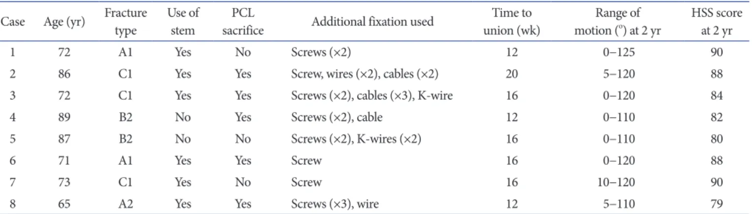
Table 1. Study Population Data (n=8) Case Age (yr) Fracture
type Use of
stem PCL
sacrifice Additional fixation used Time to
union (wk) Range of
motion (o) at 2 yr HSS score at 2 yr
1 72 A1 Yes No Screws (×2) 12 0−125 90
2 86 C1 Yes Yes Screw, wires (×2), cables (×2) 20 5−120 88
3 72 C1 Yes Yes Screws (×2), cables (×3), K-wire 16 0−120 84
4 89 B2 No Yes Screws (×2), cable 12 0−110 82
5 87 B2 No No Screws (×2), K-wires (×2) 16 0−110 80
6 71 A1 Yes Yes Screw 16 0−120 88
7 73 C1 Yes No Screw 16 10−120 90
8 65 A2 Yes Yes Screws (×3), wire 12 5−110 79
All patients were female and they had Kellgren-Lawrence grade IV osteoarthritis on injured knee.
The mechanism of injury was fall down in all cases.
PCL: posterior cruciate ligament, HSS: Hospital for Special Surgery, K-wire: Kirschner wire.
1 to 6 weeks depending on the fracture stability. On the second postoperative day, the drain was removed and passive range of motion exercise was performed twice daily for 30 minutes each time using the continuous passive motion (CPM) machine to 90o of knee flexion. Partial weight bearing using crutches was allowed from 1 to 6 weeks after surgery.
Clinical and radiographic evaluations were made at 6 weeks and 3 months after surgery. If there was no evidence of bony union at the postoperative 3-month visit, the patient was followed up monthly till the fracture united. The patients were followed up for 1 year after surgery and then yearly thereafter. Clinical re- sults were evaluated using Hospital for Special Surgery (HSS) knee scores20) and range of motion. Radiographic evaluation was performed using anteroposterior (AP), lateral, and merchant radiographic views of the knee. Evidence of fracture union, posi- tioning and maintenance of prosthesis, and the lower extremity
alignment were carefully examined. Fracture union was assessed using the findings of cortical or trabecular bridging and disap- pearance of fracture lines.
Results
The mean follow-up period was 49 months (range, 17 to 62 months). There were no cases of infection or perioperative death.
All fractures united and the mean time to radiographic union was 15 weeks (range, 12 to 20 weeks). The mean femorotibial an- gle at the final follow-up was valgus 5.7o (range, valgus 4o to 8o).
The Medial Pivot prosthesis was found to be easily adaptable for stem extension without additional bone cutting. Three patients with supracondylar fractures (AO/OTA classification A) (Fig.
1) and 3 patients with supra-intercondylar fractures (AO/OTA classification C) (Fig. 2) were treated using a stem extension. Two
Fig. 1. Preoperative radiographs (A, B) of a 72-year-old female patient show an Arbe- itsgemeinschaft für Osteosynthesefragen/
Orthopaedic Trauma Association type A1 distal femoral fracture. Medial Pivot total knee arthroplasty was performed with the use of a stem. Postoperative radiographs (C, D) show bony union with a well-main- tained prosthesis (case 1).
Fig. 2. Preoperative radiographs (A, B) of a 72-year-old female patient show an Arbe- itsgemeinschaft für Osteosynthesefragen/
Orthopaedic Trauma Association type C1 distal femoral fracture. Medial Pivot total knee arthroplasty was performed with the use of a stem. Additional fixations were per- formed using Dall-Miles cables and screws.
Postoperative radiographs (C, D) show bony union and the prosthesis is in good alignment with the stem (case 3).
patients who had condylar fractures (AO/OTA classification B) were treated without using a stem extension (Fig. 3, 4). There was no angular deformity, malunion, or shortening on the AP and lateral knee radiographs of all cases. The components were well maintained without evidence of loosening. All knees were stable anteroposteriorly and mediolaterally regardless of PCL retention or sacrifice. The mean postoperative flexion contracture of the knee joint was 2.5o (range, 0o to 10o). At the final follow-up, the mean range of motion of the knee joint was 114.3o (range, 105o to 125o) and the mean HSS knee score was 85.1 (range, 79 to 90).
Discussion
In the present study, TKA was a viable option for the treatment of distal femoral fractures in the elderly who have knee osteoar- thritis. Getting relief of the arthritic pain and improvement of the knee function at the time of fracture treatment was considered as
the advantages of TKA over osteosynthesis in our patients. TKA can be performed after bony union through the osteosynthesis procedure in these patients. However, the elderly patients should be under anesthesia for additional procedures. It also costs mon- ey and time. In our patients, all fractures united with good range of motion of the knee and HSS scores. The advantages of the Me- dial Pivot prosthesis that we discovered were ease of stem exten- sion without additional bone resection and flexibility in flexion gap balancing with the use of the same components.
Distal femoral fractures usually have a bimodal age distribu- tion1). In the younger age group, comminuted fractures are usu- ally caused by traffic accidents and are best treated with anatomi- cal reduction and internal fixation2,4). In contrast, distal femoral fractures in elderly patients are often simple fractures and are caused by minor trauma1,2,4). Thin cortices, osteoporosis, a wide intramedullary canal, and osteoarthritis of the knee make stable fixation difficult to achieve particularly in infirm women who Fig. 3. Preoperative anteroposterior and lat- eral radiographs (A, B) of an 89-year-old fe- male patient show an Arbeitsgemeinschaft für Osteosynthesefragen/Orthopaedic Trauma Association classification type B2 distal femoral fracture. Medial Pivot total knee arthroplasty was performed without the use of a stem. Postoperative radiographs (C, D) show bony union with a well-main- tained prosthesis (case 4).
Fig. 4. Preoperative anteroposterior and lateral radiographs of an 87-year-old female patient (A, B) show an Arbeitsgemeinschaft für Osteosynthesefragen/Orthopaedic Trauma Association classification type B2 distal femoral fracture. Medial Pivot total knee arthroplasty was performed without the use of a stem. Postoperative radiographs (C, D) show bony union with a well-main- tained prosthesis (case 5).
have medical problems1,9). The patients included in this study were the typical cases of distal femoral fractures. All patients were diagnosed with osteoporosis. Seven of 8 patients were more than 70 years of age. Only 1 patient had a mechanically unstable frac- ture of the distal femur (AO/OTA classification A2, A3, C2, and
C3)9,19). However, surgical decision for TKA cannot be made with
only radiographic evidence of knee osteoarthritis in these elderly patients. All patients in our study had been suffering from the arthritic pain of the knee and wanted to address the knee arthritis at the same time if possible. If the elderly people are in a high- velocity accident, mechanically unstable distal femoral fractures with severe comminution would develop certainly. In such a case, a standard TKA with stem extension might be impractical and other treatment options using a hinged knee prosthesis or constrained prosthesis would be a better alternative13). However, most of the distal femoral fractures in the elderly patients are sta- ble fractures and can be treated with a standard TKA prosthesis.
The Medial Pivot prosthesis is a newer concept design16,17). AP stability is achieved by the ball and socket configuration of the medial compartment. Translation in the lateral compartment is unrestricted. It does not roll back as in the post and cam mecha- nism of the PS prosthesis. In vivo fluoroscopic analysis of the Me- dial Pivot prosthesis has shown that the medial femoral condyle remains fully constrained and posterior translation occurs in the lateral compartment, as called for by the design16). The femoral preparation procedure only requires condylar and chamfer cuts regardless of whether or not the stem is extended. We thought that the Medial Pivot prosthesis has considerable merits compa- rable to those of the CR augmentable femoral component for its use in TKA for the treatment of distal femoral fractures in the elderly who have knee osteoarthritis13).
Technical considerations include the decision regarding the use of a femoral stem according to the fracture stability. In our study population, two patients had AO/OTA type B2 fractures. After bone cuts and trial component insertion, we felt that the frac- ture was mechanically stable. Hence, we decided not to use the femoral stem. As a result of implanting the femoral component with cement and performing additional fixation with screws and cables, good fracture stability was achieved. Both patients were treated uneventfully. However, we believe that the femoral stem should be used for mechanically unstable fractures. Other con- siderations include balancing the PCL depending on the flexion gap. The Medial Pivot prosthesis is different from the CR aug- mentable implant since it allows for flexibility in the decision of PCL treatment. With the use of the Medial Pivot prosthesis, PCL can be excised depending on the flexion gap tightness without
violating the joint stability. In our study, PCL was sacrificed in 5 patients who had flexion gap tightness. Good fracture stability was achieved in all patients at the final follow-up regardless of PCL resection.
This study has several limitations. First, there was a small number of patients. Second, there was no control group for the comparison. If a retrospective cohort study could have been performed with a large number of patients, it would have shown more decisive conclusions than those of this study. However, dis- tal femoral fractures are relatively uncommon in the elderly and this study included a 4-year consecutive case series at a University Medical Center. Long-term durability of the implant is another concern. The longevity of the insert and component loosening are major possible causes of failure of the implant. Longer follow- up is required to determine the true efficacy of this type of treat- ment. Nevertheless, we think that the longevity of the prosthesis in the patients included in this study would be the same as that in the patients who underwent primary Medial Pivot TKA for osteoarthritis of the knee.
Conclusions
Based on the clinical and radiographic results, TKA with the Medial Pivot prosthesis can be considered as an option for the treatment of minimally comminuted distal femoral fractures in elderly patients who have advanced osteoarthritis of the knee with appropriate bone stock.
Conflict of Interest
No potential conflict of interest relevant to this article was re- ported.
References
1. Bell KM, Johnstone AJ, Court-Brown CM, Hughes SP. Pri- mary knee arthroplasty for distal femoral fractures in elderly patients. J Bone Joint Surg Br. 1992;74:400-2.
2. Marks DS, Isbister ES, Porter KM. Zickel supracondylar nailing for supracondylar femoral fractures in elderly or infirm patients: a review of 33 cases. J Bone Joint Surg Br.
1994;76:596-601.
3. Higgins TF, Pittman G, Hines J, Bachus KN. Biomechani- cal analysis of distal femur fracture fixation: fixed-angle screw-plate construct versus condylar blade plate. J Orthop Trauma. 2007;21:43-6.
4. Mize RD, Bucholz RW, Grogan DP. Surgical treatment of displaced, comminuted fractures of the distal end of the fe- mur. J Bone Joint Surg Am. 1982;64:871-9.
5. Muckley T, Wahnert D, Hoffmeier KL, von Oldenburg G, Frober R, Hofmann GO. Internal fixation of type-C distal femoral fractures in osteoporotic bone: surgical technique. J Bone Joint Surg Am. 2011;93 Suppl 1:40-53.
6. Petsatodis G, Chatzisymeon A, Antonarakos P, Givissis P, Papadopoulos P, Christodoulou A. Condylar buttress plate versus fixed angle condylar blade plate versus dynamic con- dylar screw for supracondylar intra-articular distal femoral fractures. J Orthop Surg (Hong Kong). 2010;18:35-8.
7. Schatzker J, Mahomed N, Schiffman K, Kellam J. Dynamic condylar screw: a new device. A preliminary report. J Or- thop Trauma. 1989;3:124-32.
8. Kregor PJ, Stannard JA, Zlowodzki M, Cole PA. Treatment of distal femur fractures using the less invasive stabilization system: surgical experience and early clinical results in 103 fractures. J Orthop Trauma. 2004;18:509-20.
9. Weight M, Collinge C. Early results of the less invasive sta- bilization system for mechanically unstable fractures of the distal femur (AO/OTA types A2, A3, C2, and C3). J Orthop Trauma. 2004;18:503-8.
10. Appleton P, Moran M, Houshian S, Robinson CM. Distal femoral fractures treated by hinged total knee replacement in elderly patients. J Bone Joint Surg Br. 2006;88:1065-70.
11. Anderson SP, Matthews LS, Kaufer H. Treatment of juxtaar- ticular nonunion fractures at the knee with long-stem total knee arthroplasty. Clin Orthop Relat Res. 1990;(260):104-9.
12. Freedman EL, Hak DJ, Johnson EE, Eckardt JJ. Total knee replacement including a modular distal femoral component
in elderly patients with acute fracture or nonunion. J Orthop Trauma. 1995;9:231-7.
13. In Y, Koh HS, Kim SJ. Cruciate-retaining stemmed total knee arthroplasty for supracondylar-intercondylar femoral fractures in elderly patients: a report of three cases. J Arthro- plasty. 2006;21:1074-9.
14. Wolfgang GL. Primary total knee arthroplasty for intercon- dylar fracture of the femur in a rheumatoid arthritic patient:
a case report. Clin Orthop Relat Res. 1982;(171):80-2.
15. Yoshino N, Takai S, Watanabe Y, Fujiwara H, Ohshima Y, Hirasawa Y. Primary total knee arthroplasty for supracon- dylar/condylar femoral fracture in osteoarthritic knees. J Arthroplasty. 2001;16:471-5.
16. Schmidt R, Komistek RD, Blaha JD, Penenberg BL, Maloney WJ. Fluoroscopic analyses of cruciate-retaining and medial pivot knee implants. Clin Orthop Relat Res. 2003;(410):139- 47.
17. Shakespeare D, Kinzel V, Ledger M. Achieving ligament sta- bility and correct rotational alignment of the femur in knee arthroplasty: a study using the Medial Pivot knee. Knee.
2005;12:419-23.
18. Bae DK, Song SJ, Cho SD. Clinical outcome of total knee ar- throplasty with medial pivot prosthesis a comparative study between the cruciate retaining and sacrificing. J Arthroplas- ty. 2011;26:693-8.
19. Fracture and dislocation compendium. Orthopaedic Trauma Association Committee for coding and classification. J Or- thop Trauma. 1996;10 Suppl 1:1-154.
20. Insall JN, Ranawat CS, Aglietti P, Shine J. A comparison of four models of total knee-replacement prostheses. J Bone Joint Surg Am. 1976;58:754-65.