of the total TKA patients1-3). Most of the fractures around the prosthesis occur in the supracondylar region of the femur during daily living activities, although those in the femur, tibia, and pa- tella have also been reported4-6).
Supracondylar fractures of the femur following TKA can be treated either conservatively or surgically. However, the conserva- tive approach has been associated a variety of problems, such as increased pain and limited ambulation7). Metal plate fixation after open reduction may result in nonunion8). Intramedullary nail- ing may not be indicated depending on the type of TKA. Thus, efforts have been made to develop new treatment methods for supracondylar fractures of the femur following TKA.
Minimally invasive plate osteosynthesis for distal femoral frac- tures has recently been introduced as a promising technique that is effective for preserving periosteal blood supply and bone perfusion and minimizing soft tissue dissection while providing satisfactory clinical and radiological outcomes9). In this study, we evaluated the clinical and radiological outcomes of minimally invasive plate osteosynthesis for the treatment of distal femoral
Minimally Invasive Plate Osteosynthesis for
Periprosthetic Distal Femoral Fractures after Total Knee Arthroplasty
Chul-Wung Ha, MD, Oog Jin Shon, MD, Seung Wan Lim, MD, and Kang Hyun Park, MD
Department of Orthopaedic Surgery, Yeungnam University Medical Center, Daegu, Korea
Purpose: To evaluate the outcomes of the treatment of distal femoral fractures using minimally invasive plate osteosynthesis following total knee arthroplasty (TKA).
Materials and Methods: From July 2008 to October 2011, 14 patients were treated with minimally invasive plate osteosynthesis for periprosthetic fractures following TKA. The mean duration of follow-up was 19.2 months and the mean age was 69.7 years. Lewis and Rorabeck classification was used to categorize the type of fracture. Pre- and postoperative range of motion, femorotibial angle, and Knee society score, time to bony union, and complications were evaluated.
Results: The mean range of motion was 108.4 degrees preoperatively and 107.3 degrees postoperatively. No significant difference was observed in the pre- and postoperative mean range of motion. The average time to bony union was 3.9 months. The knee society score was 82.6 points preoperatively and 78.9 points postoperatively. The mean femorotibial angle was changed from 6.1 degrees valgus postoperatively to 4.6 degrees valgus postoperatively. There was no complication during the follow-up.
Conclusions: Minimally invasive plate fixation for distal femur fractures after TKA showed good results. Minimally invasive plate osteosynthesis is a recommendable treatment method for periprosthetic fractures.
Keywords: Knee, Arthroplasty, Periprosthetic femoral fracture, Minimally invasive surgery pISSN 2234-0726 · eISSN 2234-2451
Knee Surgery & Related Research
Received August 21, 2013; Revised (1st) January 10, 2014;
(2nd) January 14, 2014; Accepted February 3, 2014 Correspondence to: Oog Jin Shon, MD
Department of Orthopaedic Surgery, Yeungnam University Medical Center, 170 Hyeonchung-ro, Nam-gu, Daegu 705-717, Korea Tel: +82-53-620-3640, Fax: +82-53-628-4020
E-mail: [email protected]
Introduction
The incidence of degenerative arthritis of the knee has been increasing due to the longer life expectancy and growing senior population. Accordingly, the numbers of patients who undergo total knee arthroplasty (TKA) and develop postoperative peri- prosthetic fractures have been on the rise as well. Recent reports estimated that periprosthetic fractures occurred in 0.6%–2.5%
27
This is an Open Access article distributed under the terms of the Creative Commons Attribution Non-Commercial License (http://creativecommons.org/licenses/by-nc/3.0/) which permits unrestricted non-commercial use, distribution, and reproduction in any medium, provided the original work is properly cited.
Copyright © 2014 KOREAN KNEE SOCIETY www.jksrr.org
fractures following TKA and investigated the efficacy of the sur- gical technique.
Materials and Methods
A total of 16 patients who had been treated for periprosthetic fractures following TKA at our institution between July 2008 and October 2011 were retrospectively reviewed. Patients who had been transferred from other clinics after TKA were excluded in this study. Of the 16 patients, 2 patients who had Rorabeck et al.10) classification type 3 fractures were excluded from the study because a revision TKA was determined necessary in them. The remaining 14 patients were available for a mean follow-up of 19.2 months (range, 10 to 37 months) until bony union was achieved.
There were 2 males and 14 females with a mean age of 69.7 years (range, 52 to 78 years) (Table 1). The cause of TKA was degenera- tive osteoarthritis in 11, rheumatoid arthritis in 2, and posttrau- matic osteoarthritis in 1 patient.
The fractures were type 1 in 2 patients and type 2 in 12 patients when categorized according to the Rorabeck et al.10) classification, whereas type 33-A1 in 10 patients and type 33-A2 in 4 patients according to the Orthopaedic Trauma Association (OTA) classi- fication. The cause of fracture was slip and fall in 12 patients and a car accident in 2 patients. The affected side was the right side in 7 patients and left side in 7 patients. Concomitant vascular or
nerve damage was not observed in any of the patients.
Clinical and radiological results were analyzed at the last follow- up. Clinical results were obtained by a questionnaire designed to assess the knee range of motion (ROM), functional score, and Knee Society score during outpatient clinical visits or telephone interviews. Radiological results were assessed using anteroposte- rior and lateral radiographs obtained every month until the 6th postoperative month and every three months thereafter: the time to bony union and femorotibial angle were assessed and the dis- tance between the anterior flange and the fracture line was mea- sured to identify the cause of failure according to the presence of notching.
Bony union was defined as the absence of fracture site tender- ness and false motion, absence of pain on full weight-bearing, and the presence of bridging of three of the four cortices on the anteroposterior and lateral radiographs.
All the operations were performed by a single surgeon under general or spinal anesthesia using ZPLP (Zimmer periarticular locking plate; Zimmer Inc., Warsaw, IN, USA) in 8 patients and LCP-DF (Locking Compressing Plate-Distal Femur; Synthes, Solothurn, Switzerland) in 6 patents. The patient was placed in the supine position on the operating table and the unaffected side was lowered to facilitate identification of the trans-lateral view during surgery. An indirect supracondylar towel bump was placed for reduction. In the distal area, direct reduction and fixa- Table 1. Patients’ Demographic Data
No. of patients
Type of fracture
Age (yr) Sex Follow-up (mo)
Bone union (mo)
Cause of
fracture Notching Distance from anterior flange to fracture line Rorabeck et al.10) (mm)
classification OTA classification
1 2 33-A1 78 F 12 4 S/D O 0
2 1 33-A2 55 F 30 4 S/D O 0
3 2 33-A1 68 F 10 5 S/D O 0
4 2 33-A1 73 F 10 4 S/D O 3
5 1 33-A1 72 M 12 3 TA O 5
6 2 33-A1 52 M 15 5 S/D × 67
7 2 33-A1 67 F 33 5 S/D × 4
8 2 33-A1 67 F 15 5 S/D × 158
9 2 33-A2 68 F 10 5 S/D × 107
10 2 33-A2 72 F 10 3 S/D × 26
11 2 33-A2 83 F 24 3 TA × 18
12 2 33-A1 66 F 37 3 S/D × 65
13 2 33-A1 77 F 33 4 S/D × 48
14 2 33-A1 78 F 18 2 S/D × 34
OTA: orthopaedic trauma association, S/D: slipped down, TA: traffic accident.
tion were performed through a lateral parapatellar arthrotomy.
In the proximal area, a long incision was made for screw fixation of the LCP-DF, whereas several small incisions were made us- ing a jig system for fixation of each screw in cases of ZPLP. After reduction under traction, C-arm fluoroscopy was used to verify the reduction state. The knee was flexed on the towel bump placed beneath the femoral supracondyle and the distal femur was hyperextended. When anatomical reduction was consid- ered obtained by adjusting the axis, rotation, and length, a metal plate was placed to be over the fracture site to the metaphysis. If necessary, the metal was contoured to fit the bone shape using a plate bender. To maintain the length, Steinmann pins were used for temporary fixation in the proximal and distal regions. The first screw was fixated to be in parallel with the joint surface, and more than 4 screws were used in the region distal to the fracture line and more than 3 screws in the proximal region.
Continuous passive motion exercises of the hip and knee were initiated within 5–7 days after surgery when pain was subdued.
Active motion exercises were allowed within 1 to 2 weeks after surgery. Partial weight-bearing with crutches was permitted at 6 weeks after surgery depending on the type of fracture and was gradually increased according to the degree of osseous bridging on radiographs.
For statistical analysis, if the data had a normal distribution, a parametric test was used. Otherwise, a non-parametric test was used. The pre- and postoperative clinical results and the imme- diate postoperative and last follow-up radiological results were compared using a paired t-test. The distance between the fracture line and the anterior femoral flange according to the presence of notching was compared using the Mann-Whitney test. A p-value
<0.05 was considered statistically significant.
Fig. 1. (A) A 67-year-old woman injured in a slip and fall accident without notching. (B) A 68-year-old woman injured in a slip and fall accident with notching.
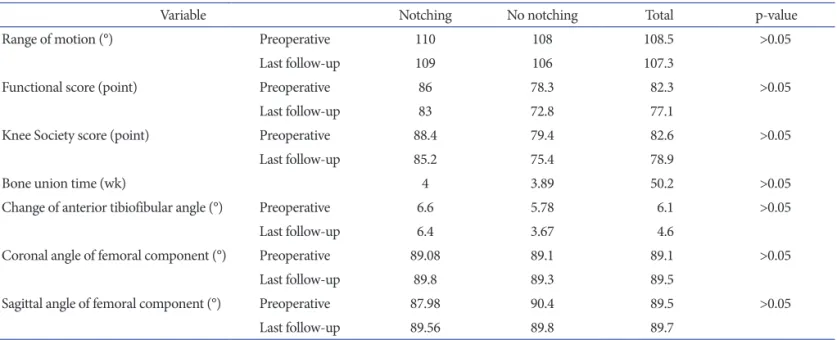
Table 2. C omparative Results between the Notching Group and the No Notching Group
Variable Notching No notching Total p-value
Range of motion (°) Preoperative 110 108 108.5 >0.05
Last follow-up 109 106 107.3
Functional score (point) Preoperative 86 78.3 82.3 >0.05
Last follow-up 83 72.8 77.1
Knee Society score (point) Preoperative 88.4 79.4 82.6 >0.05
Last follow-up 85.2 75.4 78.9
Bone union time (wk) 4 3.89 50.2 >0.05
Change of anterior tibiofibular angle (°) Preoperative 6.6 5.78 6.1 >0.05
Last follow-up 6.4 3.67 4.6
Coronal angle of femoral component (°) Preoperative 89.08 89.1 89.1 >0.05
Last follow-up 89.8 89.3 89.5
Sagittal angle of femoral component (°) Preoperative 87.98 90.4 89.5 >0.05
Last follow-up 89.56 89.8 89.7
Results
The mean ROM was decreased from 108.4°±14.6° (range, 80°
to 130°) preoperatively to 107.3°±20.3° (range, 70° to 130°) at the last follow-up, but the change was not statistically significant (p>0.05). The functional score and Knee Society score decreased between the preoperative and last follow-up examinations from 82.3±7.3 points to 77.1±8.6 points and from 82.6±9.8 points to 78.9±11.6 points, respectively, but no statistical significance was observed in the changes (p>0.05).
Radiographic bony union was observed at 3.9±0.9 months (range, 2 to 5 months) after surgery. The femorotibial angle was decreased from 6.1°±0.9° valgus preoperatively to 4.6°±2.8° val- gus postoperatively, but the change was not statistically signifi- cant (p>0.05).
The mean distance between the fracture line and the anterior femoral flange (Fig. 1) was measured as 58.4±18.9 mm in patients
without notching (N=9) and 1.8±4.6 mm in patients with notch- ing (N=5), showing statistically significant difference according to the presence of notching (p=0.011). When the patients were subdivided according to the presence of notching, there was no significant difference in radiological results between groups (Table 2). According to the presence of notching, there are radiographs after TKA state, preoperatively, and immediately postoperatively in Fig. 2.
At the last follow-up, complications, such as infection, nerve or vascular damage, fixation failure, and component loosening, were not noted.
Discussion
Periprosthetic fractures after TKA are complicated by delayed union, nonunion, or metal failure in 25% to 75% of the cases4,8,11). Most of the fractures occur due to combined action of rotational
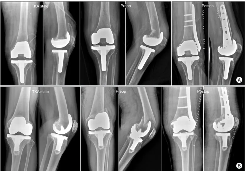
Fig. 2. (A) Radiographs obtained from a 72-year-old woman without notching after total knee arthroplasty (TKA), preoperatively (Preop), and im- mediately postoperatively (Postop). (B) Radiographs obtained from a 73-year-old woman with notching after TKA, preop, and immediately postop.
and axial force in the event of a slip and fall, an accident, or a fall12,13).
The treatment can be challenging due to following reasons: 1) most of the fractures occur in the elderly patients; 2) osteoporo- sis, periprosthetic osteolysis, and limited capacity for bony union are present in most cases; and 3) instrument insertion is difficult due to the implanted joint prosthesis8,14-18). A variety of treatment methods have been introduced to overcome these difficulties and the common principles of those methods are to secure stable fixation and facilitate early joint movement. Thus, a treatment method should be determined by the prospect of maintaining proper ROM after surgery, preserving mechanical axis of the fe- mur, and achieving internal fixation for bony union.
Treatments for periprosthetic fractures following TKA can be broadly divided into conservative and surgical. Conservative treatment is a noninvasive approach that carries a lower risk of infection. However, it has been associated with a high incidence of nonunion, does not allow for early joint exercises, and re- quires a prolonged period of bed rest. Accordingly, bed sore and cardiorespiratory dysfunctions have become major problems in the elderly patients treated conservatively. Culp et al.12) reported that ROM was reduced in 15 of the 30 patients after conservative treatment and Harlow and Hofmann19) reported that surgical in- tervention was necessitated in 29 out of 142 patients after a con- servative treatment; thus, they recommended surgical approaches for periprosthetic fractures.
Different surgical measures may be employed according to the stability of TKA. In general, Rorabeck et al.10) classification type 3 fractures are treated with revision TKA due to the presence of implant instability, whereas type 1 and 2 fractures have been treated with open reduction and metal fixation, increasing the chances of nonunion, breakage of an internal fixation device, and infection due to excessive soft tissue dissection. On the other hand, retrograde intramedullary nailing may not be feasible if the intercondylar region of the femoral component is narrow, a fracture line is extended to the lateral cortex due to the difficulty of securing strong fixation with locking screws, or the knee pros- thesis has a box20-22).
Recently, locking compression metal plates have been intro- duced as an alternative to these metal plates and screws. Surgical techniques using the new metal plates that exhibit high biome- chanical strength facilitates firm fixation through indirect reduc- tion of major bone fragments without anatomical fracture reduc- tion and bony union through preservation of blood supply to bone fragments. In the meantime, there has been advancement in minimally invasive plate osteosynthesis technique that mini-
mizes soft tissue damage. This technique enables strong fixation of distal fragments, allows for multiple screw fixation for insuf- ficient bone fragments, and provides strong resistance to varus force. Nayak et al.9) reported that bony union was obtained in all cases and satisfying knee function score was achieved in 93.5%
after minimally invasive plate osteosynthesis for distal femoral fractures, and Phillips and Christie23) showed satisfying results.
In our study, there was no significant change in the ROM of the knee, functional score, and Knee Society score after minimally invasive plate osteosynthesis for periprosthetic fractures follow- ing TKA. Bony union was achieved without additional surgery.
The femorotibial angle was not significantly changed after sur- gery. Kregor et al.24) reported that minimally invasive plate osteo- synthesis resulted in a mean of 90° of ROM and bony union in all cases (N=13), and bone grafting was required in 8%. In the study by Kolb et al.25) the mean postoperative ROM was 102°, bony union was obtained in the total 23 cases, and varus malalignment was noted in 4%. In our study, bony union was obtained in all pa- tients as was in the above-mentioned studies and the postopera- tive complication rate was lower.
In our study, we paid attention to the presence of notching as a risk factor for fracture because it is responsible for most of the fractures after TKA. The distance between the anterior femoral flange and the fracture line was shorter in patients with notch- ing than those without. It is our understanding notching blocks load transfer from the femoral metaphysis to diaphysis, resulting in concentration of the load on the femoral epicondyle. Thus, care should be taken during TKA to prevent a fracture caused by notching.
Although minimally invasive osteosynthesis using locking compression plates yielded satisfying results in all of the 14 pa- tients, we think the results should be confirmed in further studies involving larger study populations. In addition, the influence of metal plates from different manufacturers on the study results should also be taken into consideration.
Conclusions
The distance from the fracture line to the anterior femoral flange was shorter when notching was present in a periprosthetic fracture after TKA. Minimally invasive plate osteosynthesis can be considered as a promising surgical treatment technique that provides good results without any complications
Conflict of Interest
No potential conflict of interest relevant to this article was re- ported.
References
1. Aaron RK, Scott R. Supracondylar fracture of the fe- mur after total knee arthroplasty. Clin Orthop Relat Res.
1987;(219):136-9.
2. Wick M, Muller EJ, Muhr G. Supracondylar femoral frac- tures in knee endoprostheses: stabilizing with retrograde interlocking nail. Unfallchirurg. 2001;104:410-3.
3. Lachiewicz PF. Periprosthetic fracture between a con- strained total knee arthroplasty and a long-stem total hip arthroplasty: treatment with a novel device. J Arthroplasty.
2007;22:449-52.
4. Merkel KD, Johnson EW Jr. Supracondylar fracture of the femur after total knee arthroplasty. J Bone Joint Surg Am.
1986;68:29-43.
5. Oni OO. Supracondylar fracture of the femur following Attenborough stabilized gliding knee arthroplasty. Injury.
1982;14:250-1.
6. Shepperd JA, Franklin A. Supracondylar fracture of the femur following Attenborough stabilized knee arthroplasty treated by a long-stem prosthesis plus internal fixation. In- jury. 1984;16:65-6.
7. Chen F, Mont MA, Bachner RS. Management of ipsilateral supracondylar femur fractures following total knee arthro- plasty. J Arthroplasty. 1994;9:521-6.
8. Figgie MP, Goldberg VM, Figgie HE 3rd, Sobel M. The re- sults of treatment of supracondylar fracture above total knee arthroplasty. J Arthroplasty. 1990;5:267-76.
9. Nayak RM, Koichade MR, Umre AN, Ingle MV. Minimally invasive plate osteosynthesis using a locking compression plate for distal femoral fractures. J Orthop Surg (Hong Kong). 2011;19:185-90.
10. Rorabeck CH, Angliss RD, Lewis PL. Fractures of the fe- mur, tibia, and patella after total knee arthroplasty: decision making and principles of management. Instr Course Lect.
1998;47:449-58.
11. Bogoch E, Hastings D, Gross A, Gschwend N. Supracondy- lar fractures of the femur adjacent to resurfacing and MacIn- tosh arthroplasties of the knee in patients with rheumatoid arthritis. Clin Orthop Relat Res. 1988;(229):213-20.
12. Culp RW, Schmidt RG, Hanks G, Mak A, Esterhai JL Jr,
Heppenstall RB. Supracondylar fracture of the femur fol- lowing prosthetic knee arthroplasty. Clin Orthop Relat Res.
1987;(222):212-22.
13. Sisto DJ, Lachiewicz PF, Insall JN. Treatment of supracon- dylar fractures following prosthetic arthroplasty of the knee.
Clin Orthop Relat Res. 1985;(196):265-72.
14. Engh GA, Ammeen DJ. Periprosthetic fractures adjacent to total knee implants: treatment and clinical results. Instr Course Lect. 1998;47:437-48.
15. Felix NA, Stuart MJ, Hanssen AD. Periprosthetic fractures of the tibia associated with total knee arthroplasty. Clin Orthop Relat Res. 1997;(345):113-24.
16. Healy WL, Siliski JM, Incavo SJ. Operative treatment of dis- tal femoral fractures proximal to total knee replacements. J Bone Joint Surg Am. 1993;75:27-34.
17. Keenan J, Chakrabarty G, Newman JH. Treatment of supra- condylar femoral fracture above total knee replacement by custom made hinged prosthesis. Knee. 2000;7:165-70.
18. Kim KI, Egol KA, Hozack WJ, Parvizi J. Periprosthetic frac- tures after total knee arthroplasties. Clin Orthop Relat Res.
2006;446:167-75.
19. Harlow ML, Hofmann AA. Periprosthetic fractures. In: Scott WN, ed. The Knee. St Louis, MO: Mosby-Year Book; 1994.
p1405-17.
20. Cordeiro EN, Costa RC, Carazzato JG, Silva Jdos S. Peri- prosthetic fractures in patients with total knee arthroplasties.
Clin Orthop Relat Res. 1990;(252):182-9.
21. McLaren AC, Dupont JA, Schroeber DC. Open reduction internal fixation of supracondylar fractures above total knee arthroplasties using the intramedullary supracondylar rod.
Clin Orthop Relat Res. 1994;(302):194-8.
22. Rolston LR, Christ DJ, Halpern A, O’Connor PL, Ryan TG, Uggen WM. Treatment of supracondylar fractures of the femur proximal to a total knee arthroplasty: a report of four cases. J Bone Joint Surg Am. 1995;77:924-31.
23. Phillips JE, Christie J. Undisplaced fracture of the neck of the femur: results of treatment of 100 patients treated by single Watson-Jones nail fixation. Injury. 1988;19:93-6.
24. Kregor PJ, Hughes JL, Cole PA. Fixation of distal femoral fractures above total knee arthroplasty utilizing the Less Invasive Stabilization System (L.I.S.S.). Injury. 2001;32 Suppl 3:SC64-75.
25. Kolb W, Guhlmann H, Windisch C, Marx F, Koller H, Kolb K. Fixation of periprosthetic femur fractures above total knee arthroplasty with the less invasive stabilization system:
a midterm follow-up study. J Trauma. 2010;69:670-6.