How to Minimize Rotational Conflict between Femoral &
Tibial Component in Total Knee Arthroplasty
–The Use of Femoro-Tibial Axial Synchronizer (Linker)–
Jai-Gon Seo,1 Young-Wan Moon,1 Sang-Min Kim,1 and Sang-Hoon Park2
1Department of Orthopaedic Surgery, Samsung Medical Center, Sungkyunkwan University School of Medicine, Seoul;
2Department of Orthopaedic Surgery, National Health Insurance Service Ilsan Hospital, Goyang, Korea.
Received: January 3, 2014 Revised: May 1, 2014 Accepted: May 26, 2014
Corresponding author: Dr. Sang-Hoon Park, Department of Orthopaedic Surgery, National Health Insurance Service Ilsan Hospital, 100 Ilsan-ro, Ilsandong-gu,
Goyang 410-719, Korea.
Tel: 82-31-900-0540, Fax: 82-31-900-0343 E-mail: [email protected]
∙ The authors have no financial conflicts of interest.
© Copyright:
Yonsei University College of Medicine 2015 This is an Open Access article distributed under the terms of the Creative Commons Attribution Non- Commercial License (http://creativecommons.org/
licenses/by-nc/3.0) which permits unrestricted non- commercial use, distribution, and reproduction in any medium, provided the original work is properly cited.
Purpose: The purpose of this study was to investigate the correlation between ro- tational axes of femur and tibia with the use of Linker. Materials and Methods:
This study was carried out from August 2009 to February 2010 on 54 patients (106 knees), who were diagnosed with simultaneous bilateral total knee arthro- plasty. With the use of postoperative computed tomography scans, it was investi- gated how much the rotational angle of femoral and tibial components matched.
Results: The tibial component was internally rotated for the femoral component at an angle of 0.8°. The femoral component was externally rotated for the surgical transepicondylar axis (TEA) at an angle of 1.6 (range: from 4.8° of internal rota- tion to 7.9° of external rotation, SD=2.2°), and the tibial component was externally rotated for the surgical TEA at an average angle of 0.9 (range: from 5.1° of internal rotation to 8.3° of external rotation, SD=3.1°). Conclusion: The femoro-tibial syn- chronizer helped to improve the orientation and positioning of both femoral com- ponent and tibial component, and also increase the correlation of the rotational axes of the two components.
Key Words: Rotational axis, femoro-tibial synchronizer, extramedullary refer- ence, total knee arthroplasty
INTRODUCTION
The rotational conflict in total knee arthroplasty (TKA) was mainly caused by the rotational mismatch of femur and tibia.1-3 Malpositioning and malorientation caused by the rotational conflict are the main causes of anterior knee pain and stiff- ness after surgery in a clinical aspect. And femoro-tibial subluxation and polyeth- ylene (PE) wear give an impact even on the longevity of TKA.1,3-6 Even rotating flatform mobile bearing TKA, which helps to address such an issue, is still faced with difficulty in the subsurface wear. Studies on each rotation standard of femur and tibia actively been pursued so far. However, since the rotation of a knee joint includes factors coupled with both femur rotation and tibia rotation, it is better to realize rotational correlative relationship between the femoral and tibial compo-
NJ, USA)- were used for the operation. Patients were 68 years of age on average (range, 58‒79), and patients received informed consent from this medical center and were ap- proved by the Institutional Review Board of this medical center. Five days after the operation, their rotation axis was measured with the use of Transverse CT scans (X-Vigor, Toshiba Medical Inc., Tokyo, Japan), and clinical outcomes such as knee society score for 3 years after their operation and their radiologic outcomes with alignment precision were analyzed.
Femoro-tibial axial synchronizer (Linker)
Anteromedial limited parapatellar approach was used, and distal femoral block was set after soft tissue balancing. EM axis guider was used to check femur’s coronal axis and sag- ittal mechanical axis, thereby determining femoral compo- nent’s rotation. When transverse axis of distal femoral block nearly approached surgical transepicondylar axis (TEA), distal femoral resector was installed, and knee extension was made. This femoro-tibial axial synchronizer was designed to match the anteroposterior axis of femur and knee center, and is aimed at achieving femoro-tibial synchronization by us- ing the inter-connected instrument of distal femoral resector and proximal tibial resector (Fig. 1).
After anterior condylar skim cut of femur, distal femoral resector was installed (Fig. 2). With the consideration of screw home movement, knee extension was made. After that, a connector with a plane such as proximal tibial resec- tor was inserted into a slot of distal femoral resector, and axial synchronizer was installed (Fig. 3). The tail of proxi- mal tibial resector presenting femur’s coronal and sagittal axis was synchronized with tibia’s coronal & sagittal axis (Fig. 4). AP pin was inserted for fixation by making it par- allel with the AP plane of distal femoral resector. After that, proximal tibial resector was installed in the status of knee nents rather than to independently set rotational axis of
each component in order to actually address the rotational conflict.7,8
The rotation of femur and tibia appears at the last exten- sion of a joint knee in the form of screw home movement. It represents that tibia is relatively externally rotated for femur at the extension of a knee joint.7,9 Accordingly, the rotation angle of tibia for femur at the extension is determined by the rotation of femur and screw home movement. The rotation of femur varies depending on an operation, and screw home movement can happen differently in each knee. For such reasons, it is very difficult to synchronize the rotation of fe- mur and the rotation of tibia during TKA operation.
Therefore, femoro-tibial axial synchronizer instrument, which helps to synchronize the rotation of femur and the ro- tation of tibia when screw home movement occurs, was de- signed to address the problem, and the prospective study was performed after TKA operation to identify through CT wheth- er the rotation of femur and tibial components is matched.
MATERIALS AND METHODS
Study subjects
The prospective study had been performed from August 2009 to February 2010 on 66 patients (132 knees), who re- ceived bilateral TKA operation. Excluded were patients who had history of trauma to the pelvis or lower limb, a neuromuscular disorder, or prior surgery of the knee, hip, or pelvis, and more than 10 degrees of flexion contracture af- ter operation. Consequently, 54 patients or 106 knees were analyzed. All patients received TKA using extramedullary (EM) alignment by a single surgeon (JGS), and two types of prosthesis-PCS (Scorpio posterior-cruciate-sacrificing PCS, Stryker, NJ, USA)- and NRG (Scorpio NRG, Stryker,
Fig. 1. Proximal resector with connecting instrument. This tibial AP axis
synchronizer is composed of tail, caliper, and proximal tibial resector. Fig. 2. The photo of distal femoral resector. Linker is composed of distal femoral resector and proximal tibial resector at knee extension position.
tibial resector’s AP pin was parallel with femoral resector’s AP pin. After the removal of connector, the bone resection of proximal tibia and distal femur was performed, and a component was then inserted. As a result, femoral rotation matched with tibial rotation in the inter-related status, and planes were parallel even in coronal plane. Patella resurfac- ing was routinely performed, and implants became fixation with cement.
Evaluation of femoral and tibial rotation
By performing postoperative CT scans, we measured the rotation of femoral and tibial component after operation.
Transverse CT scans (X-Vigor, Toshiba Medical Inc., To- kyo, Japan) was used from tibial tuberosity to femoral distal metaphysic to take photos at 2.5 mm intervals. A patient’s lower extremities were maximally extended in supine posi- tion, and photos were taken under the condition of non-in- ternal or non-external rotation. Femoral component rotation- al axis was defined as a line linking the posterior margins of both pegs (Fig. 6), and tibial component was also defined extension, and Linker was completed. After detachment of
synchronizer, tibial resection was done, and tibial AP axis was then marked on the central part parallel with two AP pins. AP axis of tibial plate was synchronized with or paral- lel with the marked tibial AP axis (Fig. 5). After distal re- section of femur, component was inserted. This utilizes the vertical relationship of the femur rotation and the tibia rota- tion with AP axis. When femeur’s AP pin was parallel with tibia’s AP pin, femoral component has a parallel relation- ship with a tibial component.
Operative procedures
Modified antero-medial parapatellar incision of Insall was used to make an approach. After soft tissue balancing, ex- tramedullary technique was used to install distal femoral block. The connector to which proximal tibial resector was attached was inserted into distal femoral resector’s slot, and EM axis guider was then used to adjust femur’s coronal axis and sagittal alignment for synchronization. Then, a pin was inserted into proximal tibial resector. At this time, proximal
Fig. 3. Linker is a connecting instrument of distal femoral resector and proximal tibial resector. When femeur’s AP pin is parallel to tibia’s AP pin, femoral component has a parallel relationship with a tibial component. And resectional planes have the rectangularity with each other.
Fig. 5. Tibial plate is inserted as synchronized with marked tibial AP axis.
Fig. 4. With knee extension position, the synchronization of AP axes of fe- mur and tibia is confirmed. The tail of proximal tibial resector presenting fe- mur’s coronal & sagittal axis is synchronized with tibia’s coronal & sagittal axis.
Fig. 6. With the patient supine on the CT scanning table, lower extremities were stabilized in maximum extension in a plastic frame without any internal or external rotation. Femoral component rotational axis was defined as the line joining the posterior margins of both pegs of the femoral component.
1
Regarding ROM of postoperative 3 years, averages were 124.3±12.4 degrees, postoperative KSS score was 92.7±7.0 (62‒100). There was no implant-related revision during 3 years.
DISCUSSION
Component’s rotation in TKA is very important, and malro- tation gives an impact on flexion instability, tibiofemoral and patellofemoral kinematic problem, and clinical outcome.1,10,11 In this respect, rotation alignment of femoral component and tibial component is significant, and the correlation of the two component’s rotation is also important. Until now, there have been many studies on the rotation alignment, however, no absolute standards have yet been made.12-14
As a standard of femoral component’s rotational axis, there are many methods, such as posterior condylar axis, Whiteside anteroposterior axis, transepicondylar axis, and flexion gap rectangularity balancing, but each method has an issue of variation.8,15 Among various methods, the meth- od of making rotation with the focus of TEA has been known to have the least individual variation.2,6,11,16,17 Since TEA is the approximate value of flexion-extension axis and the ori- gin of both collateral ligaments, it is considered as the most valid reference in many studies.4,7,8,18 As a standard of tibial as a line linking posterior margin of keels (Fig. 7). In doing
so, the difference in the two component’s rotation was ana- lyzed. To evaluate inter-observer variability, three indepen- dent observers measured rotational axis of the femoral com- ponent and surgical TEA’s angle.
Statistical analysis
Rotation’s margin of error was set as about 5 degrees. This study was designed with more than 90% of statistical pow- er and less than 5 degrees of error margin. The sample size of this study was more than 104 knees, and 106 registered knees were investigated further for precise evaluation. We examined the results of this study through one-sample pro- portions test, and utilized two-way random effects model of interclass correlation coefficients (ICC) to analyze statisti- cal significance of inter-observer difference.
RESULTS
Comparative analysis of femoral component’s rotational axis and tibial component’s rotational axis showed that tibi- al component was internally rotated for femoral component at an average angle of 0.8°, and that all inter-observer re- peated observations had no statistical significance at ±2.1°.
The ICC about inter-observer variability was 0.96 indicat- ing excellent agreement.
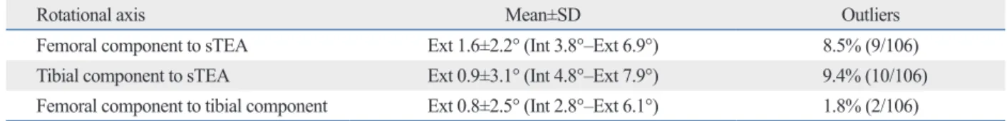
Femoral component was externally rotated for surgical TEA at an angle of 1.6° (range: from 4.8° of internal rota- tion to 7.9° of external rotation, SD=2.2°). In case of 97 out of 106 knees (91.5%). femoral component was rotated for surgical TEA at an angle of less than 5°. Tibial component was externally rotated for surgical TEA at an average angle of 0.9°, and outliers with more than 5 degrees were 6 knees, and variance was SD=3.1° (Table 1).
Mean alignment of the femoral component in the coronal plane was 89.5±2.5° (83.4‒97.2°) postop and mean align- ment of the tibial component was 90.3±2.1° (85.3‒94.1°) postop. And the average tibial component posterior inclina- tion was 4.8±2.1 (1.9‒8.1) degrees.
Table 1. The Results of Comparative Analysis on Femoral Component’s Rotational Axis and Tibial Component’s Rotational Axis
Rotational axis Mean±SD Outliers
Femoral component to sTEA Ext 1.6±2.2° (Int 3.8°–Ext 6.9°) 8.5% (9/106)
Tibial component to sTEA Ext 0.9±3.1° (Int 4.8°–Ext 7.9°) 9.4% (10/106)
Femoral component to tibial component Ext 0.8±2.5° (Int 2.8°–Ext 6.1°) 1.8% (2/106) sTEA, surgical tranepicondylar axis.
Fig. 7. Rotation of tibial component can be measured with the line connect- ing the posterior margin of keels.
1 2
and patellofemoral maltracking, thereby weakening anteri- or knee pain. Furthermore, it possibly answers the long-de- bated question about fixed bearing and rotating platform bearing.
Ideal gap space should be perpendicular to the mechani- cal axis and matched to the component size. Furthermore, gap space between femur and tibia should be parallel and femoral resectional plane should be correlated to the femo- ral surgical TEA. Linker is a connecting instrument of fe- mur and tibia. If it is used for bone resection, the femoral component has a parallel relationship with the tibial com- ponent in coronal plane, and coronal and rotational axes are matched to keep extension gap’s rectangularity. Therefore, ideal gap space can be made by using Linker. In addition, AP axes are synchronized at full extension before resection of lateral femoral condyle and knee extension is made at the time of installation of distal femoral block and proximal tibial resector to receive rotational alignment with consider- ation of screw home mechanism.
There were some limitations in the present study. First, it is difficult to evaluate the rotational alignment because scan- ning direction of CT scan and flexion contracture easily af- fect the accuracy of measurement. In addition, the interval (2.5 mm) was slightly large with which peg holes and pos- terior margin may not clearly be detected. Second, there were nocomparative group (without Linker). Third, we chose the size of tibial plate not to be overhanged, nevertheless, rota- tional alignment could easily be changed if surgeons empha- sized the coverage. Even though the use of a Linker can’t be an absolute standard of rotational alignment, rotational con- flict is likely to be minimized by synchronizing femur rota- tion with tibial rotation.
In conclusion, to address the rotational conflict of femur and tibia, femoro-tibial synchronizer (Linker) helped to im- prove the orientation and positioning of both the femoral component and the tibial component, and to increase the correlation of the rotational axis of the two components.
REFERENCES
1. Aglietti P, Sensi L, Cuomo P, Ciardullo A. Rotational position of femoral and tibial components in TKA using the femoral transepi- condylar axis. Clin Orthop Relat Res 2008;466:2751-5.
2. Nicoll D, Rowley DI. Internal rotational error of the tibial compo- nent is a major cause of pain after total knee replacement. J Bone Joint Surg Br 2010;92:1238-44.
3. Toms AD, Mandalia V, Haigh R, Hopwood B. The management
component’s rotation, medial one-third of tibial tuberosity proposed by Insall has most widely been used. However, since it is independent from femur’s transepicondylar axis, rotational axis of femur and tibia should be set respectively.
As described by Akagi, et al.,19,20 tibial component is ex- tremely inclined to make external rotation compared with new anteroposterior axis perpendicular to femur’s transepi- condylar axis, and is likely to have no rotational correlation with femur component. Self-alignment of a conforming mo- bile tibial insert method using floating insert was proposed.16 The method has an advantage of femoral component’s rota- tion and insert, and also correlated with tibial component rotation. However, according to Ikeuchi, et al.,10 self align- ment method has a problem which makes tibial component to have more internal rotation.2 In spite of component’s ro- tational axis and the importance of its orientation, standards related to the issue are not clear. Even though there is no problem in each rotation, rotation axis of each component can be different, which may lead to some problems.
Moreover, it is screw home movement that should addi- tionally be taken into account. The screw home movement is the phenomenon where external rotation of tibia for fe- mur occurs in normal knees at the extension of a knee joint, and it is seen as complex function of surface geometry, ten- sion of ligaments and muscle.7,9 Regarding the motion in TKA, some studies found that the screw home movement did not occur, but the study conducted by Ishii, et al.9 re- vealed that muscle’s activity significantly influenced screw home mechanism, more than ligaments did. If screw home movement occurs in knees after TKA operation, rotational congruency should be confirmed at the full extension of knees, and then femur and tibia’s rotational matching should be performed.
In fact, there are many other techniques in dealing with rotational coupling. In case of measured resection tech- nique, femur bone and tibia bone are independently resect- ed with uncertain standards. In case of gap technique, tibia rotation is fixed at medial 1/3 of tuberosity, bone resection is made, and then rotation is decided.5,13,14,16,17 In addition, there are many difficulties in determining standards about screw home movement’s variability and femoral external ro- tation. Because of difficulties, it is not easy to synchronize femoral component’s rotation and tibial component’s rota- tion, and it is necessary to determine the rotational axis of the two components with one absolute standard. As a solu- tion of rotational conflict, the femur and tibial rotational syn- chronization can reduce polyethylene wear, prevent stiffness
12. Suter T, Zanetti M, Schmid M, Romero J. Reproducibility of mea- surement of femoral component rotation after total knee arthro- plasty using computer tomography. J Arthroplasty 2006;21:744-8.
13. Vanin N, Panzica M, Dikos G, Krettek C, Hankemeier S. Rota- tional alignment in total knee arthroplasty: intraoperative inter- and intraobserver reliability of Whiteside’s line. Arch Orthop Trauma Surg 2011;131:1477-80.
14. Victor J. Rotational alignment of the distal femur: a literature re- view. Orthop Traumatol Surg Res 2009;95:365-72.
15. Won YY, Cui WQ, Baek MH, Yun TB, Han SH. An additional reference axis for determining rotational alignment of the femoral component in total knee arthroplasty. J Arthroplasty 2007;22:
1049-53.
16. Matziolis G, Pfiel S, Wassilew G, Boenicke H, Perka C. Kinemat- ic analysis of the flexion axis for correct femoral component placement. Knee Surg Sports Traumatol Arthrosc 2011;19:1504-9.
17. Merican AM, Ghosh KM, Iranpour F, Deehan DJ, Amis AA. The effect of femoral component rotation on the kinematics of the tib- iofemoral and patellofemoral joints after total knee arthroplasty.
Knee Surg Sports Traumatol Arthrosc 2011;19:1479-87.
18. Harvie P, Sloan K, Beaver RJ. Three-dimensional component alignment and functional outcome in computer-navigated total knee arthroplasty: a prospective, randomized study comparing two navigation systems. J Arthroplasty 2011;26:1285-90.
19. Akagi M, Mori S, Nishimura S, Nishimura A, Asano T, Hamani- shi C. Variability of extraarticular tibial rotation references for to- tal knee arthroplasty. Clin Orthop Relat Res 2005:172-6.
20. Akagi M, Oh M, Nonaka T, Tsujimoto H, Asano T, Hamanishi C.
An anteroposterior axis of the tibia for total knee arthroplasty. Clin Orthop Relat Res 2004:213-9.
of patients with painful total knee replacement. J Bone Joint Surg Br 2009;91:143-50.
4. Hirschmann MT, Konala P, Amsler F, Iranpour F, Friederich NF, Cobb JP. The position and orientation of total knee replacement components: a comparison of conventional radiographs, trans- verse 2D-CT slices and 3D-CT reconstruction. J Bone Joint Surg Br 2011;93:629-33.
5. Matziolis G, Krocker D, Weiss U, Tohtz S, Perka C. A prospec- tive, randomized study of computer-assisted and conventional to- tal knee arthroplasty. Three-dimensional evaluation of implant alignment and rotation. J Bone Joint Surg Am 2007;89:236-43.
6. Olcott CW, Scott RD. The Ranawat Award. Femoral component rotation during total knee arthroplasty. Clin Orthop Relat Res 1999:39-42.
7. Lee DH, Seo JG, Moon YW. Synchronisation of tibial rotational alignment with femoral component in total knee arthroplasty. Int Orthop 2008;32:223-7.
8. Seo JG, Moon YW, Lim JS, Park SJ, Kim SM. Mechanical axis- derived femoral component rotation in extramedullary total knee arthroplasty: a comparison between femoral transverse axis and transepicondylar axis. Knee Surg Sports Traumatol Arthrosc 2012;
20:538-45.
9. Ishii Y, Terajima K, Koga Y, Bechtold JE. Screw home motion af- ter total knee replacement. Clin Orthop Relat Res 1999:181-7.
10. Ikeuchi M, Yamanaka N, Okanoue Y, Ueta E, Tani T. Determining the rotational alignment of the tibial component at total knee re- placement: a comparison of two techniques. J Bone Joint Surg Br 2007;89:45-9.
11. Thompson JA, Hast MW, Granger JF, Piazza SJ, Siston RA. Bio- mechanical effects of total knee arthroplasty component malrota- tion: a computational simulation. J Orthop Res 2011;29:969-75.