Received November 19, 2013, Revised November 10, 2014, Accepted for publication November 23, 2014
Corresponding author: Jun-Mo Yang, Department of Dermatology, Samsung Medical Center, Sungkyunkwan University School of Me- dicine, 81 Irwon-ro, Gangnam-gu, Seoul 135-710, Korea. Tel: 82-2- 3410-3541, Fax: 82-2-3410-3869, E-mail: [email protected] This is an Open Access article distributed under the terms of the Creative Commons Attribution Non-Commercial License (http://
creativecommons.org/licenses/by-nc/4.0) which permits unrestricted non-commercial use, distribution, and reproduction in any medium, provided the original work is properly cited.
ORIGINAL ARTICLE
Antibiotic Susceptibility of Staphylococcus aureus
in Atopic Dermatitis: Current Prevalence of Methicillin- Resistant Staphylococcus aureus in Korea and Treatment Strategies
Mi-Young Jung, Jong-Youn Chung, Hae-Young Lee, Jiho Park, Dong-Youn Lee, Jun-Mo Yang
Department of Dermatology, Samsung Medical Center, Sungkyunkwan University School of Medicine, Seoul, Korea
Background: Staphylococcus aureus is a well-known microbe that colonizes or infects the skin in atopic dermatitis (AD).
The prevalence of methicillin-resistant S. aureus (MRSA) in AD has recently been increasing. Objective: This study aimed to determine the antimicrobial susceptibility patterns in AD skin lesions and evaluate the prevalence of MRSA in Korea.
We also recommend proper first-line topical antibiotics for Korean patients with AD. Methods: We studied S. aureus-pos- itive skin swabs (n=583) from the lesional skin of infants, children, and adults who presented to our outpatient clinic with AD from July 2009 to April 2012. Results: S. aureus ex- hibited high susceptibility against most antimicrobial agents.
However, it exhibited less susceptibility to benzylpenicillin, erythromycin, clindamycin, and fusidic acid. The preva- lence of MRSA was 12.9% among 583 S. aureus isolates, and the susceptibility to oxacillin was significantly lower in in- fants in both acute and chronic AD lesions. Conclusion: S.
aureus from AD has a high prevalence of MRSA and multi- drug resistance, especially in infants. In addition, the rate of fusidic acid resistance is high among all age groups, and mu- pirocin resistance increases with age group regardless of le- sional status. This is the first study comparing the anti- microbial susceptibility rates of S. aureus isolates from AD
cases with respect to age and lesion status in Korea. (Ann Dermatol 27(4) 398∼403, 2015)
-Keywords-
Atopic dermatitis, Antimicrobial susceptibility, Methicillin- resistant Staphylococcus aureus, Staphylococcus aureus
INTRODUCTION
Atopic dermatitis (AD) is a common chronic inflammatory skin disorder that induces several symptoms including pruritus, dryness, and secondary cutaneous infection. The skin lesions of patients with AD are classified as acute or chronic according to the skin’s condition and cytokine ex- pression level in the lesional skin. Both types of atopic le- sions often impair skin barrier function and exhibit de- creased antimicrobial peptides expression and defective innate immune activity. Accordingly, patients with AD are more susceptible to colonization with bacteria, especially Staphylococcus aureus.
S. aureus is a well-known organism that colonizes and in- fects the skin in AD. The prevalence of skin colonization with S. aureus is much higher in patients with AD than that in healthy individuals: 75%∼100% of patients with AD exhibit S. aureus colonization on their lesional skin, while the bacterium is isolated from only 5%∼30% of in- dividuals without AD1-3. Among S. aureus strains, methi- cillin-resistant S. aureus (MRSA) is one of the most im- portant pathogens of community-acquired infections in many countries. Since MRSA was first reported in 1968 by Barrett et al.4, the prevalence of MRSA has increased grad- ually, especially in recent years. Growing evidence sug-
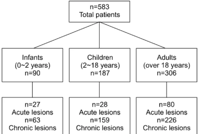
Fig. 1. Patient characteristics.
gests the skin of patients with AD may be a preferred res- ervoir for this pathogen.
Topical antimicrobial agents are cost-effective therapies for cutaneous bacterial infections. Several approved top- ical agents are used to treat skin infections, many of which are usually prescribed if bacterial skin infection is sus- pected in AD. Among them, mupirocin and fusidic acid are the most commonly used to treat cutaneous infection5. However, the percentage of isolates resistant to these top- ical antibiotics has been increasing.
As antibiotic therapy plays an important role in treating bacterial skin infections in AD, the present study eval- uated the antimicrobial susceptibility of S. aureus in pa- tients with AD and determined the prevalence of resistant strains, especially MRSA. We also determined whether there are differences in the resistance rates with respect to age, because previous studies show that MRSA is more prevalent in children than in adults6-8. Furthermore, we determined the prevalence of resistance rates with respect to the duration of skin lesions to provide helpful in- formation regarding the proper selection of topical anti- biotics according to lesional status in AD. The results also provide information about the current prevalence of MRSA in Korea.
MATERIALS AND METHODS
The study protocol was approved by the Samsung Medical Center institutional review board (2002-10-009) and con- ducted in accordance with the Declaration of Helsinki.
Written informed consent was obtained from all patients or their guardians.
Patients
We retrospectively collected data from patients with AD who were positive for S. aureus on a skin swab performed during their first visit. A total of 583 patients including 90 infants, 187 children, and 306 adults (135 acute and 448 chronic skin lesions) who visited our dermatology out- patient clinic from July 2009 to April 2012 were recruited (Fig. 1). The exclusion criteria included were the presence of impetigo, cellulitis, or fungal infection; and the presence of other inflammatory cutaneous disorder such as psor- iasis, seborrheic dermatitis, and pityriasis rosea. No pa- tients had a history of any treatment including antibiotics in the preceding weeks. We classified AD skin lesions as acute or chronic lesions: acute lesions were defined as er- ythematous eczematous skin lesions with oozing and crust, while chronic lesions included erythematous to brownish papules and lichenification with xerosis.
Isolation and identification of S. aureus from AD lesions
Skin swabs for culture were taken from the lesions (i.e., oozing sites in patients with acute skin lesions or the ante- cubital fossa in patients with intact skin) using sterile cot- ton tips (BBL CultureSwab Plus; Sparks, MD, USA).
Samples were inoculated onto blood agar plates, and S.
aureus isolates were identified by a coagulase slide test and catalase reaction.
Antimicrobial susceptibility
The antimicrobial susceptibility of each isolate was de- termined by the VITEK 2 system using the AST-P601 card panel (Biomerieux, Lyon, France). The tested antibiotics in- cluded benzylpenicillin, oxacillin, gentamicin, habekacin, ciprofloxacin, telithromycin, tigecycline, erythromycin, clindamycin, quinupristin/dalfopristin, linezolid, teicopla- nin, vancomycin, tetracycline, nitrofurantoin, fusidic acid, rifampicin, trimethoprim/sulfamethoxazole, and mupirocin.
Statistical analysis
Statistical significance was analyzed by the χ2 test or Fisher’s exact test with a Bonferroni correction where ap- propriate. Logistic regression analysis was used for multi- variate analysis. The level of significance was set at p<0.05.
Statistical analyses were performed by using SAS version 9.4 software (SAS Institute, Cary, NC, USA).
RESULTS
Antimicrobial susceptibility patterns of S. aureus in patients with AD
Most S. aureus isolates exhibited high susceptibility to most antimicrobial agents. The isolates exhibited less sus- ceptibility to benzylpenicillin, erythromycin, clindamycin,
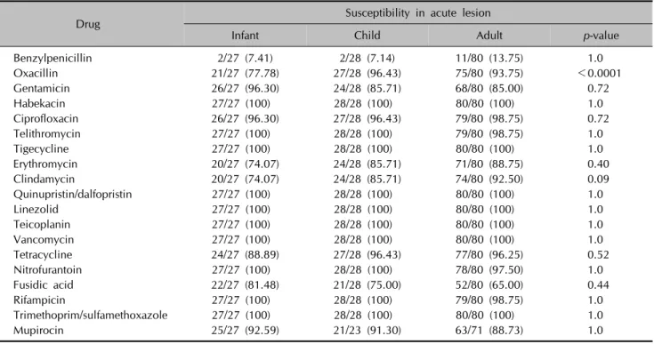
Table 1. Comparison of antimicrobial susceptibility of acute lesions between age groups
Drug Susceptibility in acute lesion
Infant Child Adult p-value
Benzylpenicillin 2/27 (7.41) 2/28 (7.14) 11/80 (13.75) 1.0
Oxacillin 21/27 (77.78) 27/28 (96.43) 75/80 (93.75) <0.0001
Gentamicin 26/27 (96.30) 24/28 (85.71) 68/80 (85.00) 0.72
Habekacin 27/27 (100) 28/28 (100) 80/80 (100) 1.0
Ciprofloxacin 26/27 (96.30) 27/28 (96.43) 79/80 (98.75) 0.72
Telithromycin 27/27 (100) 28/28 (100) 79/80 (98.75) 1.0
Tigecycline 27/27 (100) 28/28 (100) 80/80 (100) 1.0
Erythromycin 20/27 (74.07) 24/28 (85.71) 71/80 (88.75) 0.40
Clindamycin 20/27 (74.07) 24/28 (85.71) 74/80 (92.50) 0.09
Quinupristin/dalfopristin 27/27 (100) 28/28 (100) 80/80 (100) 1.0
Linezolid 27/27 (100) 28/28 (100) 80/80 (100) 1.0
Teicoplanin 27/27 (100) 28/28 (100) 80/80 (100) 1.0
Vancomycin 27/27 (100) 28/28 (100) 80/80 (100) 1.0
Tetracycline 24/27 (88.89) 27/28 (96.43) 77/80 (96.25) 0.52
Nitrofurantoin 27/27 (100) 28/28 (100) 78/80 (97.50) 1.0
Fusidic acid 22/27 (81.48) 21/28 (75.00) 52/80 (65.00) 0.44
Rifampicin 27/27 (100) 28/28 (100) 79/80 (98.75) 1.0
Trimethoprim/sulfamethoxazole 27/27 (100) 28/28 (100) 80/80 (100) 1.0
Mupirocin 25/27 (92.59) 21/23 (91.30) 63/71 (88.73) 1.0
Values are presented as number (%).
Fig. 2. Antimicrobial susceptibility patterns of Staphylococcus aureus in total patients.
and fusidic acid. In particular, isolates from chronic skin lesions showed low susceptibility to oxacillin. Among the tested antibacterial agents, S. aureus showed the highest resistance rate to benzylpenicillin followed by fusidic acid. There were no significant differences in susceptibility between acute and chronic skin lesions (Fig. 2).
Antimicrobial susceptibility of S. aureus in acute AD lesions
In acute cutaneous lesions, S. aureus had the lowest sus- ceptibility to benzylpenicillin. These isolates also exhibited low susceptibility to erythromycin, clindamycin, and fusi- dic acid. However, all S. aureus isolates were susceptible to vancomycin, habekacin, tigecycline, quinupristin/dalfo- pristin, linezolid, teicoplanin, and trimethoprim/sulfame- thoxazole.
Regarding the susceptibility rates among age groups, sus- ceptibility to oxacillin was significantly lower in the infant group (77.78% vs. 96.43% vs. 93.75% in the infant, child, and adult samples, respectively, p<0.0001). There were no significant differences in the susceptibility rates to other antimicrobial agents with respect to age group (Table 1).
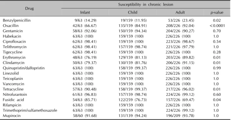
Antimicrobial susceptibility of S. aureus in chronic AD lesions
Similar to the results in patients with acute AD lesions, S.
aureus from chronic lesions exhibited the lowest suscepti-
bility to benzylpenicillin followed by fusidic acid, eryth- romycin, clindamycin, and oxacillin. Meanwhile, S. aur- eus exhibit 100% susceptibility to habekacin, linezolid, teicoplanin, vancomycin, and rifampicin.
The antimicrobial susceptibility to oxacillin was sig-
Table 2. Comparison of antimicrobial susceptibility of in chronic lesions between age groups
Drug Susceptibility in chronic lesion
Infant Child Adult p-value
Benzylpenicillin 9/63 (14.29) 19/159 (11.95) 53/226 (23.45) 0.02
Oxacillin 42/63 (66.67) 135/159 (84.91) 208/226 (92.04) <0.0001
Gentamicin 58/63 (92.06) 150/159 (94.34) 204/226 (90.27) 0.70
Habekacin 63/63 (100) 159/159 (100) 226/226 (100) 1.0
Ciprofloxacin 62/63 (98.41) 159/159 (100) 223/226 (98.67) 0.54
Telithromycin 62/63 (98.41) 157/159 (98.74) 221/226 (97.79) 1.0
Tigecycline 62/63 (98.41) 159/159 (100) 226/226 (100) 0.28
Erythromycin 48/63 (76.19) 129/159 (81.13) 203/226 (89.82) 0.01
Clindamycin 50/63 (79.37) 130/159 (81.76) 206/226 (91.15) 0.01
Quinupristin/dalfopristin 63/63 (100) 158/159 (99.37) 226/226 (100) 0.99
Linezolid 63/63 (100) 159/159 (100) 226/226 (100) 1.0
Teicoplanin 63/63 (100) 159/159 (100) 226/226 (100) 1.0
Vancomycin 63/63 (100) 159/159 (100) 226/226 (100) 1.0
Tetracycline 57/63 (90.48) 158/159 (99.37) 217/226 (96.02) 0.01
Nitrofurantoin 61/63 (96.83) 157/159 (98.74) 224/226 (99.12) 0.60
Fusidic acid 54/63 (85.71) 122/259 (76.73) 157/226 (69.47) 0.04
Rifampicin 63/63 (100) 159/159 (100) 226/226 (100) 1.0
Trimethoprim/sulfamethoxazole 63/63 (100) 159/159 (100) 224/226 (99.12) 1.0
Mupirocin 58/60 (91.68) 131/139 (94.24) 196/209 (93.78) 1.0
Values are presented as number (%).
Table 3. Methicillin-resistant Staphylococcus aureus isolation rates in all atopic dermatitis cases
Source Methicillin- resistant
Methicillin-
sensitive Total Acute lesion
Infant 6 (22.2) 21 (77.8) 27
Child 1 (3.6) 27 (96.4) 28
Adult 5 (6.3) 75 (93.7) 80
Chronic lesion
Infant 21 (33.3) 42 (66.7) 63
Child 24 (15.1) 135 (84.9) 159
Adult 18 (8.0) 208 (92.0) 226
Total 75 (12.9) 508 (87.1) 583
Values are presented as number (%) or number only.
nificantly lower in the infant group than the child and adult groups (p<0.0001). There were other significant dif- ferences in the antimicrobial susceptibility among the three age groups: benzylpenicillin (p=0.02), erythromycin (p=0.01), clindamycin (p=0.01), tetracycline (p=0.01), fusidic acid (p=0.04). The S. aureus isolates from the in- fant group were less susceptible to these antibiotics, ex- cept fusidic acid, which showed a lower susceptibility rate in the adult group (Table 2).
Isolation rates of MRSA in AD
Among the 583 S. aureus isolates, 75 (12.9%) were MRSA. The isolation rate of MRSA was significantly higher in infants with AD than that of adults or children with AD in both acute and chronic cutaneous lesions (Table 3).
Treatment response to oral antibiotics in the acute MRSA group
Among the 12 AD cases with acute skin lesions colonized with MRSA, 7 patients (4 infants, 1 child, and 2 adults) were treated with oral antimicrobial agents because of se- vere oozing over a large body surface area. Three patients were treated with a first-generation cephalosporin (i.e., cephradine) for two weeks, and four were treated with a second- or third-generation cephalosporin (i.e., cefurox- ime and cefpodoxime). All patients taking oral cepha- losporin antibiotics showed improvement of subjective
symptoms and good recovery of cutaneous lesions.
DISCUSSION
MRSA (i.e., oxacillin resistant) is a major pathogen in many infectious diseases. MRSA was historically consid- ered an important healthcare-acquired pathogen but has recently been regarded as a major cause of infection in normal populations without healthcare-associated risk fac- tors such as long-term admission periods and intensive care unit stay. So called “community-associated MRSA”
(CA-MRSA) strains are some of the most common patho- gens found in skin and soft tissue infections in many coun- tries9,10. One study reports the prevalence of CA-MRSA ranged from 15% to 75% among adults in 11 universi- ty-affiliated emergency departments throughout the United States11. MRSA strains account for 36%, 30%, and 23% of staphylococcal skin and soft tissue infections in North America, Latin America, and Europe, respectively12. Re- garding patients with AD, several studies have inves- tigated the incidence of MRSA isolated from AD skin le- sions13-16. Hoeger14 did not identify MRSA from patients with AD in a pediatric outpatient population in 2004.
However, in New Zealand, 2% of S. aureus isolates from pediatric AD cases were MRSA13. In addition, Niebuhr et al.15 found MRSA in 3% of S. aureus isolates in patients with AD. Up to 30% of S. aureus isolates from AD cases were reported to be MRSA in a Taiwanese study pop- ulation in 201116. Eczematous lesions in AD are known to be a source of transmission of S. aureus. The increasing in- cidence rates of CA-MRSA in skin and soft tissue in- fections raise concerns that AD skin is a favorable reser- voir for this drug-resistant organism. According to one study of the epidemiological characteristics of MRSA in Korea, 18.3% of S. aureus isolates in children with AD le- sions were MRSA17.
In the present study, 12.9% (75/583) of S. aureus isolates were MRSA. MRSA was found in both acute and chronic AD lesions but more so in chronic cutaneous lesions.
MRSA colonization rates are generally higher in acute skin lesions than chronic skin lesions. Our previous study of the colonization rate in AD also shows a higher S. aureus colonization rate (74%) in acute skins lesions than chronic skin lesions (38%)18. However, a large percentage of chron- ic AD skin lesions were colonized with MRSA (8.9% in acute lesions vs. 14.1% in chronic lesions, p>0.05). This might be explained by a history of repetitive topical anti- biotics administration in chronic AD.
Interestingly, in the present study, the prevalence of MRSA was higher in the infant group regardless of lesional status.
In one study, almost half of MRSA-positive children were
<5 years old, and children aged between 1 month and 2 years represented just over one-third of all MRSA-positive cases19. However, it is unknown why the resistance rate is higher in infants than other age groups. Therefore, the mo- lecular characteristics of MRSA strains by genotyping must be evaluated further to better understand about the resist- ance rate in infants.
The topical use of antimicrobial agents for the treatment of AD skin lesions is common and has advantages over sys- temic therapy with respect to cost-effectiveness and the absence of severe systemic side effects. However, the fre-
quent use of topical antibiotics promotes the development of resistant strains. Fusidic acid is one of the most com- monly used topical antibiotics in dermatology worldwide.
Many studies report resistance rates against fusidic acid have increased20-24. Accordingly, in the present study, S.
aureus showed low susceptibility rates to fusidic acid in both acute and chronic lesions. In chronic AD skin lesions in particular, resistance rates to fusidic acid increased sig- nificantly with age (p=0.04). Inappropriate use of topical antibiotics leading to resistance may threaten the efficacy of systemic antibiotics for the treatment of serious S. aur- eus infections such as osteomyelitis and severe surgical wound infections. Therefore, the topical use of fusidic acid for empirical treatment must be restricted.
Topical mupirocin has been used since 1994 in Korea, and its use has been increasing dramatically since5. Yun et al.24 first detected mupirocin-resistant S. aureus in Korea, with a prevalence of 5%. Up to 25.3% of S. aureus iso- lates exhibit resistance to mupirocin in certain intensive care unit settings5. In the present study, antimicrobial sus- ceptibility to mupirocin was relatively lower in the adult group than the infant or child group regardless of the chronicity of the lesions. The frequent and repeated use of topical mupirocin in recent years may have influenced these outcomes. Thus, awareness and research about mu- pirocin resistance should be bolstered for the proper long-term management of AD skin lesions.
We treated 7 MRSA-positive patients with oral cepha- losporin with good subjective and objective results, sug- gesting CA-MRSA can be controlled easily with oral ceph- alosporin antibiotics.
In conclusion, the prevalence of MRSA was higher in in- fants with AD than children and adults with AD regardless of lesional status. Furthermore, the results indicate it is ra- tional to administer topical antibiotics susceptible to MRSA as first-line treatment for infants with AD. In addition, fusi- dic acid resistance was high in all age groups, and resist- ance rates against mupirocin tended to increase with age regardless of lesional status. This is the first study compar- ing the antimicrobial susceptibility rates of S. aureus iso- lates from AD patients of different age groups and lesional status in Korea. Thus, this study provides useful information for selecting a proper topical antimicrobial agent for pa- tient-specific treatment according to their age and lesional status.
ACKNOWLEDGMENT
This study was supported by a grant from the Korea Health Technology R&D Project, Ministry of Health and Welfare, Republic of Korea (No. HI12C1299) as well as
grant from the Samsung Biomedical Research Institute (SMX1131301).
REFERENCES
1. Breuer K, HAussler S, Kapp A, Werfel T. Staphylococcus aureus: colonizing features and influence of an antibacterial treatment in adults with atopic dermatitis. Br J Dermatol 2002;147:55-61.
2. Gong JQ, Lin L, Lin T, Hao F, Zeng FQ, Bi ZG, et al. Skin colonization by Staphylococcus aureus in patients with eczema and atopic dermatitis and relevant combined topical therapy: a double-blind multicentre randomized controlled trial. Br J Dermatol 2006;155:680-687.
3. Higaki S, Morohashi M, Yamagishi T, Hasegawa Y. Com- parative study of staphylococci from the skin of atopic dermatitis patients and from healthy subjects. Int J Dermatol 1999;38:265-269.
4. Barrett FF, McGehee RF Jr, Finland M. Methicillin-resistant Staphylococcus aureus at Boston City Hospital. Bacteriologic and epidemiologic observations. N Engl J Med 1968;279:
441-448.
5. Park SY, Kim SM, Park SD. The prevalence, genotype and antimicrobial susceptibility of high- and low-level mupirocin resistant methicillin-resistant Staphylococcus aureus. Ann Dermatol 2012;24:32-38.
6. Dukic VM, Lauderdale DS, Wilder J, Daum RS, David MZ.
Epidemics of community-associated methicillin-resistant Sta- phylococcus aureus in the United States: a meta-analysis.
PLoS One 2013;8:e52722.
7. Bratu S, Landman D, Gupta J, Trehan M, Panwar M, Quale J. A population-based study examining the emergence of community-associated methicillin-resistant Staphylococcus aureus USA300 in New York City. Ann Clin Microbiol Antimicrob 2006;5:29.
8. Popovich KJ, Hota B, Aroutcheva A, Kurien L, Patel J, Lyles-Banks R, et al. Community-associated methicillin- resistant Staphylococcus aureus colonization burden in HIV-infected patients. Clin Infect Dis 2013;56:1067-1074.
9. Calfee DP. Methicillin-resistant Staphylococcus aureus and vancomycin-resistant enterococci, and other Gram-positives in healthcare. Curr Opin Infect Dis 2012;25:385-394.
10. Mediavilla JR, Chen L, Mathema B, Kreiswirth BN. Global epidemiology of community-associated methicillin resistant Staphylococcus aureus (CA-MRSA). Curr Opin Microbiol 2012;15:588-595.
11. Moran GJ, Krishnadasan A, Gorwitz RJ, Fosheim GE, McDougal LK, Carey RB, et al; EMERGEncy ID Net Study Group. Methicillin-resistant S. aureus infections among patients in the emergency department. N Engl J Med 2006;
355:666-674.
12. Moet GJ, Jones RN, Biedenbach DJ, Stilwell MG, Fritsche TR. Contemporary causes of skin and soft tissue infections in North America, Latin America, and Europe: report from the SENTRY Antimicrobial Surveillance Program (1998-2004).
Diagn Microbiol Infect Dis 2007;57:7-13.
13. Hill SE, Yung A, Rademaker M. Prevalence of Staphy- lococcus aureus and antibiotic resistance in children with atopic dermatitis: a New Zealand experience. Australas J Dermatol 2011;52:27-31.
14. Hoeger PH. Antimicrobial susceptibility of skin-colonizing S. aureus strains in children with atopic dermatitis. Pediatr Allergy Immunol 2004;15:474-477.
15. Niebuhr M, Mai U, Kapp A, Werfel T. Antibiotic treatment of cutaneous infections with Staphylococcus aureus in patients with atopic dermatitis: current antimicrobial resis- tances and susceptibilities. Exp Dermatol 2008;17:953-957.
16. Tang CS, Wang CC, Huang CF, Chen SJ, Tseng MH, Lo WT.
Antimicrobial susceptibility of Staphylococcus aureus in children with atopic dermatitis. Pediatr Int 2011;53:363- 367.
17. Chung HJ, Jeon HS, Sung H, Kim MN, Hong SJ. Epi- demiological characteristics of methicillin-resistant Staphy- lococcus aureus isolates from children with eczematous atopic dermatitis lesions. J Clin Microbiol 2008;46:991-995.
18. Park HY, Kim CR, Huh IS, Jung MY, Seo EY, Park JH, et al.
Staphylococcus aureus colonization in acute and chronic skin lesions of patients with atopic dermatitis. Ann Derma- tol 2013;25:410-416.
19. Matlow A, Forgie S, Pelude L, Embree J, Gravel D, Langley JM, et al; Canadian Nosocomial Infection Surveillance Program. National surveillance of methicillin-resistant Sta- phylococcus aureus among hospitalized pediatric patients in Canadian acute care facilities, 1995-2007. Pediatr Infect Dis J 2012;31:814-820.
20. Shah M, Mohanraj M. High levels of fusidic acid-resistant Staphylococcus aureus in dermatology patients. Br J De- rmatol 2003;148:1018-1020.
21. Ravenscroft JC, Layton A, Barnham M. Observations on high levels of fusidic acid resistant Staphylococcus aureus in Harrogate, North Yorkshire, UK. Clin Exp Dermatol 2000;25:327-330.
22. Andersen BM, Bergh K, Steinbakk M, Syversen G, Magnaes B, Dalen H, et al. A Norwegian nosocomial outbreak of methicillin-resistant Staphylococcus aureus resistant to fusidic acid and susceptible to other antistaphylococcal agents. J Hosp Infect 1999;41:123-132.
23. Brown EM, Thomas P. Fusidic acid resistance in Staphy- lococcus aureus isolates. Lancet 2002;359:803.
24. Yun HJ, Lee SW, Yoon GM, Kim SY, Choi S, Lee YS, et al.
Prevalence and mechanisms of low- and high-level mupirocin resistance in staphylococci isolated from a Korean hospital.
J Antimicrob Chemother 2003;51:619-623.