대한내분비외과학회지:제 11 권 제 1 호
□ 증 례 □
Vol. 11, No. 1, March 2011
28
Correspondence: Ji-Young Park, Department of Pathology, Kyungpook National University Hospital, 50 2-ga Samduk-dong, Jung-gu, Daegu 700-721, Korea
Tel: +82-53-420-5247, Fax: +82-53-426-1525 E-mail: [email protected]
접수일:2010년 9월 10일, 게재승인일:2011년 3월 22일
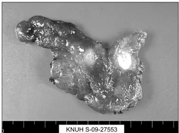
Fig. 1. Gross image of the thyroid gland showing two white solid masses. A 0.7×0.5 cm sized, white and solid nodule with infiltratigns margins located in right thyroid lobe was thy- roid papillary carcinoma. The other mass was 0.9×0.7 cm sized, well defined, bright yellow solid tumor with smooth cut surface, located just adjacent to isthmus was GCT.
Granular Cell Tumor of Thyroid Gland That Was Concomitant with Papillary Thyroid Carcinoma - A Case Report
Eun Jeong Jang, M.D., An Na Seo, M.D., Sun-Jae Lee, M.D. and Ji-Young Park, M.D. Ph.D.
Department of Pathology, Kyungpook National University Hospital, Daegu, Korea
Granular cell tumor (GCT) of the thyroid is rare and histo- genesis of the carcinoma still remains poorly understood.
Here in this study, we report a case of perithyroidal gran- ular cell tumor in a 44-year-old woman, diagnosed as me- dullary carcinoma upon the interoperative frozen diagnosis.
The tumor was comprised of white, solid mass with infiltrat- ing margin in isthmus. Microscopically, the tumor revealed abundant eosinophilic cytoplasm, elongated nucleus and eosinophilic amyloid-like materials. It was composed of dif- fuse sheets of polygonal cells with abundant eosinophilic cytoplasm and cytologically bland nucleus on permanent section. On immunohistochemical staining, S-100 and CD68 are diffusely positive. Determining the progression and the behavior of the tumor is critical for providing long-term man- agement and preventing aggressive treatment. (Korean J Endocrine Surg 2011;11:28-30)
Key Words: Granular cell tumor, Medullary carcinoma, Papillary carcinoma, Thyroid
INTRODUCTION
Due to the rare occurrence and complex pathologic nature of the carcinoma, accurate diagnosis for perithyroidal GCT is often challenged. We report a case of GCT, coincident with papillary carcinoma, just adjacent to isthmus of thyroid gland. Clinically it was presented as thyroid mass and originally diagnosed as me- dullary carcinoma on frozen section.
CASE REPORT
A 44 year-old woman with a medical history of hypertension visited local medical center due to her right neck mass which
was developed 6 months ago. She had no specific symptom and thyroid function tests were within normal range. In the subsequent ultrasonographic examination, 2 hypoechoic masses were noted in right thyroid lobe (0.6×0.6 cm) and isthmus (0.6×0.5 cm) with multiple lymph node enlargements. Clinical analysis suggested she had a metastatic thyroid malignant tumor, and fine needle aspiration (FNA) was performed to confirm the diagnosis. The FNA results indicated that her 2 thyroid masses and lymph node had papillary carcinoma and benign nodule, respectively. The as- piration cytology of lymph node revealed necrotizing lymphadeni- tis, consistent with Kikuchi lymphadenitis. The patient successively underwent right thyroid lobectomy. Specimen of the right thyroid lobe and isthmus was obtained from the surgical resection, Measuring 0.7×0.5 cm in size, white and solid nodule located in right thyroid lobe revealed typical papillary growth pattern and nu- clear features including nuclear enlargement, clearing, irregularity, pseudoinclusions, and grooves. The observed features were con- sistent with conventional papillary thyroid carcinoma.
Eun Jeong Jang, et al:Granular Cell Tumor of Thyroid Gland 29
Fig. 2. (A) The tumor cells show abundant eosinophilic cytoplasm, elongated nucleus admixed with eosinophilic collagenous materials in perithyroidal soft tissue (H&E stain, ×100). (B) Immunohistochemical staining for S-100 shows diffuse, strong cytoplasmic stain- ing (S-100 stain, ×100).
The other mass was measured 0.9×0.7 cm in size and was well defined solid tumor in bright yellow color with smooth cut surface; it was located just adjacent to isthmus. Hemorrhage or necrosis was not present on cut surface (Fig. 1). The tumor was composed of cells with abundant eosinophilic cytoplasm, elon- gated nucleus, eosinophilic amyloid-like materials. It was suspi- cious of medullary carcinoma on frozen section. On the perma- nent paraffin-embedded hematoxylin-and eosin stained section, the tumor showed sheet-like growing pattern and was composed of round, polygonal cells with eosinophilic, granular cytoplasm. It showed infiltrating margin to adjacent soft tissue and typically the granular cells encompassed small nerves. It also contained many eosinophilic collagenous materials in tumor. The nuclei were cyto- logically bland and mitosis was not found. (Fig. 2A) umor cells were positive on S-100 (Fig. 2B) and CD68 immunohistochemical staining but were negative on Cytokeratin, Thyroglobulin, Calcito- nin, CD34, HMB45 and Synatophysin.
Based on histology and immunohistochemical profile, the final diagnosis was resolved as GCT of thyroid gland.
DISCUSSION
Granular cell tumor (GCT) is a fairly common tumor of Schwann cell origin.(1-3)
Although originally considered a muscle tumor by Abrikossoff in 1926, its close association with nerve and immunohistochemical characteristics firmly identifies it as a neural lesion.(3)
GCT occurs in patients of any age and is usually located in the dermis or subcutis and less frequently in the submucosa, smooth muscle, or striated muscle.(3,4) The most common site
is head and neck lesion, especially in the tongue; but it can occur in other organs, including larynx, tracheobroncheal tree, gastro- intestinal tract, parotid gland, genitourinary tract, vulva, nerves, bile duct, and breasts.(1,3-5)
The first reported case of GCT of thyroid gland was in an 11.5-year-old girl who was receiving high dose estrogen treatment for familial tall stature since she was 9 years old.(6) After Mahoney had reported the case, 6 additional cases have been published in the English literature and 3 additional cases have been reported in the Korean literature (including this case).
(1,6-11) In all but except for a single case, 8 patients were female and the age at diagnosis ranged from 11 to 47 (average age was 28.3). Most of them were asymptomatic and the tumor was confined to thyroid.(9) One patient had weakness, anxiety, and palpitation; this patient had concomitant multifocal GCT in thyroid with papillary thyroid carcinoma.(11)
Differential diagnoses include medullary carcinoma and Hurthle cell tumor. Medullary carcinoma shows sheets or irregular islands of polygonal or plump spindle cells transversed by fibrovascular septa. Occasionally, a whirling, trabecular, pseudopapillary, ro- sette, tubular, microglandular, or cribriform pattern can be observed. The tumor cells have round to oval nuclei and nuclear pleomorphism is usually not significant. The cytoplasm is finely granular. Amyloid is found in 80∼85% and appear as pink-stain- ing amorphous materials.(12) Hurthle cell tumor of thyroid is usu- ally thickly encapsulated tumor which is composed of cells with abundant brightly eosinophilic granular cytoplasm and the cyto- plasmic outline is well defined. The cells are arranged in small follicles, trabeculae, solid sheets and the nuclei are round, with granular to coarse chromatin and distinct nucleoli. GCT shows
30 대한내분비외과학회지:제 11 권 제 1 호 2011
sheet-like growing pattern and is composed of rounded, polygonal cells with eosinophilic, granular cytoplasm that has indistinct cyto- plasmic borders. The tumor cells have eccentrically located round to oval nuclei and indistinct nucleoli. Discovering the features of encompassing small nerves help in the diagnosis for GCT.(1,13) Immunohistochemical stains are helpful in distinguishing GCT from medullary carcinoma and Hurthle cell tumor. Immunohisto- chemical profiles of medullary carcinoma shows positive staining for Calcitonin, Cytokeratine, TTF-1 and CEA. Hurthle cell tumor shows positive staining for thyroglobulin or TTF-1, Cytokeratin and S-100 protein. GCT stains uniformly positive for S-100 protein, CD68, CEA but negative for thyroglobulin or TTF-1.(1,12) Malignant granular cell tumors constitute fewer than 2% of all granular cell tumors. A history of long clinical duration and recent rapid growth has been observed. Malignant granular cell tumors show necrosis, spindling, vesicular nuclei with prominent nucleoli, increased mitotic activity (>2 mitoses/10 HPF), high nucleocyto- plasmic ratio, and pleomorphism. Excluding the malignant tumors, recurrence is rare. Of 92 cases reported by Strong et al., six recurred.(13) Hence local surgical excision is curative in nearly all cases.(3)
The malignancy for the case in this report was a very rare case of perithyroidal GCT concomitant with papillary carcinoma, appearing as medullary carcinoma on frozen section. Although perithyroidal GCT is very rare, it is important to consider some possibility of GCT when the tumor is composed of round cells with eosinophilic cytoplasm on frozen section or fine needle aspi- ration and make a differential diagnosis with malignant thyroid tumor.
REFERENCES
1) Baloch ZW, Martin S, Livolsi VA. Granular cell tumor of the thyroid:
a case report. Int J Surg Pathol 2005;13:291-4.
2) Compagno J, Hyams VJ, Ste-Marie P. Benign granular cell tumors of the larynx: a review of 36 cases with clinicopathologic data. Ann Otol Rhinol Laryngol 1975;84:308-14.
3) Sharon W. Weiss, R. Goldblum J. Benign tumors of peripheral nerves. In: William Schimitt, editors. Soft tissue tumors. 5th ed.
Philadelphia: Mosby; p.878-87.
4) Kang H, Hong E, Park M, Lee J. Granular cell tumor of the thyroid.
Korean J Pathol 1998;32:63-7.
5) Ordonez NG, Mackay B. Granular cell tumor: a review of the pathology and histogenesis. Ultrastruct Pathol 1999;23:207-22.
6) Mahoney CP, Patterson SD, Ryan J. Granular cell tumor of the thyroid gland in a girl receiving high-dose estrogen therapy. Pediatr Pathol Lab Med 1995;15:791-5.
7) Espinosa-de-Los-Monteros-Franco VA, Martinez-Madrigal F, Ortiz- Hidalgo C. Granular cell tumor (Abrikossoff tumor) of the thyroid gland. Ann Diagn Pathol 2009;13:269-71.
8) Paproski SM, Owen DA. Granular cell tumor of the thyroid. Arch Pathol Lab Med 2001;125:544-6.
9) Milias S, Hytiroglou P, Kourtis D, Papadimitriou CS. Granular cell tumour of the thyroid gland. Histopathology 2004;44:190-1.
10) Chang SM, Wei CK, Tseng CE. The cytology of a thyroid granular cell tumor. Endocr Pathol 2009;20:137-40.
11) Park J, Kang H, Park M, Cho M, Yoon J, Jaegal Y. A case of granular cell tumor of the thyroid. Korean J Head Neck Oncol 2006;22:43-6.
12) Christopher DM. Flecher. Tumors of the thyroid and parathyroid glands. In: Michael Houston, editors. Diagnostic histopathology of tumors. 3rd ed. Philadelphia: Elsevier; p.1026-46.
13) Strong EW, McDivitt RW, Brasfield RD. Granular cell myoblastoma.
Cancer 1970;25:415-22.