http://dx.doi.org/10.3988/jcn.2015.11.1.87 J Clin Neurol 2015;11(1):87-91
Introduction
Paraneoplastic limbic encephalitis (PLE) is one of the most frequent presentations of paraneoplastic encephalomyelitis syndrome. It is produced by involvement of the medial region of the temporal lobes, especially the hippocampus, which de- rive their characteristic clinical manifestations. By definition, PLE is associated with cancer, but it is not caused by a prima- ry or metastatic neoplasia in the central nervous system (CNS).
Therefore, the specific diagnosis of a PLE requires a cancer diagnosis.1
The differential diagnosis should be established, mainly to rule out infectious and autoimmune disorders (especially viral encephalitis and Whipple’s disease). However, metabolic en- cephalopathies, neurotoxic drugs, deficiency states, inflamma- tory disorders, primary or secondary CNS tumors, and neuro- degenerative disorders must be excluded.2-4 Hyperintensity in the temporal lobes, and especially in the medial region, is of- ten evident on T2-weighted and fluid attenuated inversion re- covery (FLAIR) sequences in MRI, and occasionally there is gadolinium enhancement.5 Among the many different neo- plasms known to cause PLE, small-cell lung cancer (SCLC) is the most frequently reported etiology. We report herein a case of a PLE as a debut of squamous cell carcinoma of the lung.
Paraneoplastic Limbic Encephalitis in a Male with Squamous Cell Carcinoma of the Lung
Tamara Sauri,a Àngel Izquierdo,a,b,d LLuis Ramió-Torrentà,b,c,e Àngel Sanchez-Montañez,f Joaquim Bosch-Barrera,a,b,c Rut Portaa,b,c
aDepartment of Medical Oncology, Catalan Institute of Oncology (ICO), Girona, Spain
bGirona Biomedical Research Institute (IDIBGi), Girona, Spain
cDepartment of Medical Sciences, School of Medicine, University of Girona, Girona, Spain
dCancer Registry of Girona, Girona, Spain
eDepartment of Neurology, Dr. Josep Trueta University Hospital, Girona, Spain
fDepartment of Neuroradiology, Vall d’Hebron University Hospital, Barcelona, Spain
Received July 2, 2013 Revised January 5, 2014 Accepted January 8, 2014 Correspondence Rut Porta, MD, PhD
Department of Medical Oncology, Catalan Institute of Oncology (ICO), Avinguda de França s/n,
Girona 17007, Spain Tel +34 972 225 834 Fax +34 972 21 73 54 E-mail [email protected]
BackgroundzzParaneoplastic limbic encephalitis (PLE) is a rare syndrome characterized by memory impairment, symptoms of hypothalamic dysfunction, and seizures. It commonly pre- cedes the diagnosis of cancer. Small-cell lung cancer is the neoplasm that is most frequently re- ported as the etiology underlying PLE.
Case ReportzzThis report describes a male patient who presented with neurologic symptoms consistent with anterograde amnesia, apathy, and disorientation. MRI revealed diffuse hyperin- tensities located predominantly in the medial bitemporal lobes, basal ganglia, frontal lobes, and leptomeninges on fluid attenuated inversion recovery images, suggesting PLE. Study of the pri- mary tumor revealed squamous cell carcinoma of the lung. The patient was treated with neoadju- vant chemotherapy followed by surgery and adjuvant chemoradiotherapy, which resulted in his neurologic symptoms gradually improving.
ConclusionszzPLE might be a rare debut of squamous cell carcinoma of the lung. Treatment of the primary tumor may improve the neurologic symptoms.
J Clin Neurol 2015;11(1):87-91 Key Wordszz lung cancer, squamous cell carcinoma, paraneoplastic syndrome, limbic encephalitis.
Open Access
cc This is an Open Access article distributed under the terms of the Cre- ative Commons Attribution Non-Commercial License (http://creative- commons.org/licenses/by-nc/3.0) which permits unrestricted non-com- mercial use, distribution, and reproduction in any medium, provided the ori- ginal work is properly cited.
Case Report
A 54-year-old Caucasian male who smoked 20 cigarettes per day and had an otherwise unremarkable medical history was admitted to our hospital due to subacute neurologic symp- toms. He presented with nocturnal myoclonus, sexual dys- function, mood swings, insomnia, daytime sleepiness, and occasional disorientation. His vital signs were normal at the time of admission, and a physical examination did not detect any abnormality. A neurologic examination revealed antero- grade amnesia, apathy, and a disorder index in the Mini-Men- tal State Examination of 24/30.
All blood test findings were within normal limits, and se- rology was negative for hepatitis B virus, hepatitis C virus, human immunodeficiency virus, leishmaniasis, histoplasmo- sis, syphilis, and borreliosis. The findings of immunological studies were normal for antinuclear antibodies, anti-DNA anti- body, angiotensin-converting enzyme, complement, IgA, an- tineutrophil cytoplasmic autoantibodies, anti-Ro/La antibody, and anticardiolipin antibody; a proteinogram also yielded nor- mal findings. A polymerase chain reaction performed using the cerebrospinal fluid (CSF) yielded negative findings for herpes simplex and varicella zoster virus. A cytologic analy- sis of the CSF did not reveal any malignant cells.
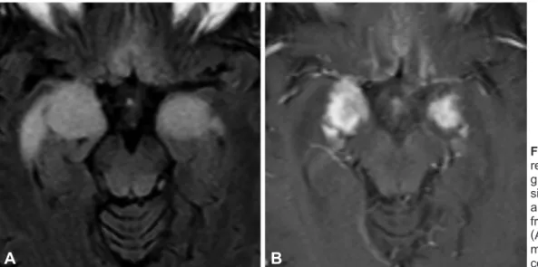
The CSF was also negative for paraneoplastic antibodies with specificity for recombinant Hu, Yo, Ri, Ta, amphiphysin, and Ma2 antigens. The electroencephalogram (EEG) was nor- mal. Cerebral CT demonstrated hypodense lesions in the me- dial bitemporal region and in the right caudate nucleus, with pseudonodular uptake in both unci. MRI demonstrated diffuse hyperintensities in the parenchyma, with predominantly me- dial bitemporal involvement extending into the basal ganglia, frontal lobes, and leptomeninges on FLAIR images, which is the most characteristic sequence, suggesting a PLE (Fig. 1).
An extension study was conducted to locate a tumor. Tes- ticular and prostate ultrasonography revealed slight changes
suggestive of chronic prostatitis and normal gonads. CT of the abdomen and chest revealed a 33-mm nodule in the right inferior lobe (RIL), suggestive of hamartoma, and lymph nodes of significant size at stations 7 and 2R. Ultrasound en- doscopy confirmed the presence of paraesophageal right hilar adenopathy, which was suggestive of malignancy. Histopa- thology of the lymph node was positive for squamous cell car- cinoma. Fibrobronchoscopy demonstrated erythematous mu- cosa in the right superior lobe. Bronchoalveolar aspiration and bronchoalveolar lavage produced negative findings. Pos- itron-emission tomography and CT (PET/CT) demonstrated a hypermetabolic lesion in the azygos-esophageal recess that have could represented a lymph-node conglomerate. Outbreaks of high metabolic activity in both hippocampal regions that were suggestive of limbic encephalitis were observed, but with no evidence of metastatic lesions.
The patient was diagnosed with squamous cell carcinoma of the lung TxN2M0 (stage IIIA). He was treated with three cycles of 75 mg/m2 cisplatin and 75 mg/m2 docetaxel every 3 weeks. A postreatment evaluation by PET/CT demonstrated a slight reduction of the mediastinal lesions. A right inferior lobectomy and a mediastinal lymphadenectomy were per- formed 3 months after completing the last cycle of chemo- therapy. A pathologic examination demonstrated that the peri- bronchial, interlobar, and subcarinal lymph nodes contained squamous cell carcinoma with extension into the soft tissues and capsular rupture. The nodule in the RIL was confirmed as a pulmonary chondroid hamartoma. The surgical margins were free of disease. Brain FLAIR imaging after the surgery re- vealed remarkable decreases in both the extension and signal intensity within the medial region of both temporal lobes. Sec- ondary mesial atrophy with temporal horns dilatation was ob- served (Fig. 2). The patient’s neurologic symptoms improved after surgery. The postoperative therapy included 4 cicles of carboplatin (area under the curve 5) and 500 mg/m2 peme- trexed administered every 3 weeks with concomitant medias-
Fig. 1. Brain fluid attenuated inversion recovery imaging performed before sur- gery showing a diffuse hyperintense signal within the medial temporal lobes and extending into the basal ganglia, frontal basal lobes, and leptomeninges (A). Avid and homogeneous enhance- ment was noted after administration of contrast medium (B).
A B
tinal radiotherapy.
After postoperative treatment, PET/CT revealed two hyper- metabolic lesions in the right pleuroesophageal recess, and MRI revealed persistence of the T2-signal intensity and me- sial bitemporal lobe atrophy. After 8 months of follow-up the patient presented with acute neurologic symptoms that re- quired admission to an intensive care unit (ICU) due to fever, upper-extremity myoclonus, and somnolence. Brain MRI and CT demonstrated a persistent T2-elongation of the mesial re- gions and no definitive gadolinium enhancement. The CSF was positive for Escherichia coli and Staphylococcus aureus.
An EEG revealed diffuse neuronal dysfunction with no focal or diffuse paroxysms suggestive of seizures. Despite treatment with broad-spectrum antibiotics, the patient had a low level
of consciousness with persistent neurologic deterioration, and died 1 month after ICU admission. The autopsy revealed per- sistent squamous cell carcinoma in the tracheal and mediasti- nal lymph nodes with extensive infiltration of the esophagus wall and a tracheoesophageal fistula. Infiltration of the right atrium, right ventricle, and renal capsule was also observed.
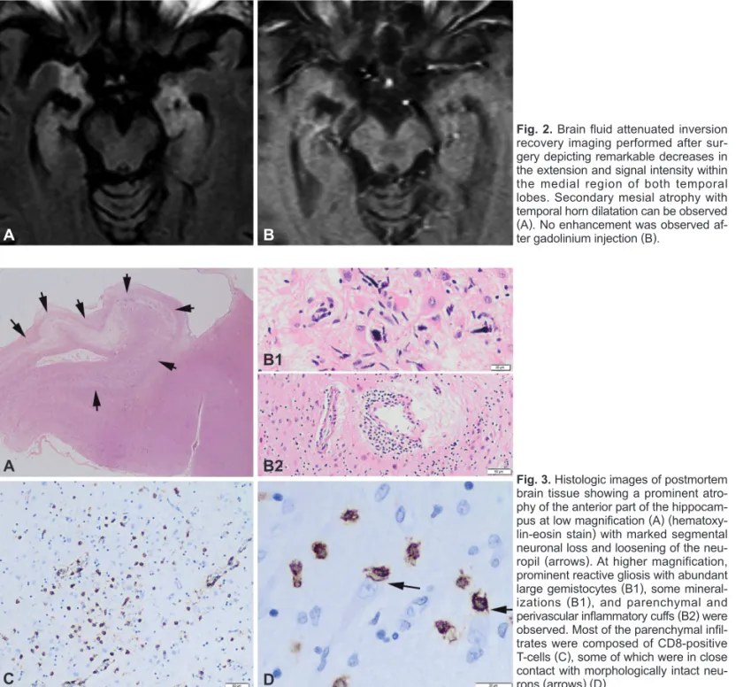
In the brain, the autopsy confirmed advanced bilateral T-cell- mediated limbic encephalitis with persistent inflammatory activity (Fig. 3) extending to the midbrain tectum.
Discussion
Paraneoplastic limbic encephalitis is a rare disease character- ized by the subacute onset of short-term memory loss, sei- A
C
B1
B2
D
Fig. 3. Histologic images of postmortem brain tissue showing a prominent atro- phy of the anterior part of the hippocam- pus at low magnification (A) (hematoxy- lin-eosin stain) with marked segmental neuronal loss and loosening of the neu- ropil (arrows). At higher magnification, prominent reactive gliosis with abundant large gemistocytes (B1), some mineral- izations (B1), and parenchymal and perivascular inflammatory cuffs (B2) were observed. Most of the parenchymal infil- trates were composed of CD8-positive T-cells (C), some of which were in close contact with morphologically intact neu- rons (arrows) (D).
Fig. 2. Brain fluid attenuated inversion recovery imaging performed after sur- gery depicting remarkable decreases in the extension and signal intensity within the medial region of both temporal lobes. Secondary mesial atrophy with temporal horn dilatation can be observed (A). No enhancement was observed af- ter gadolinium injection (B).
A B
zures, and symptoms of hypothalamic dysfunction. It com- monly precedes a diagnosis of malignancy, and it is a diagnosis of exclusion. The association between subacute encephalop- athy and a distant tumor was first described in 1960, and PLE was recognized as a separate clinical entity in 1968.6,7 PLE is most frequently associated with lung cancer (50%), of which 80% of cases are SCLC. It has also been associated with tes- ticular cancer (20%), breast cancer (8%), thymus cancer, and lymphoma (1–2%).8-11 It is not uncommon for non-SCLC (NSCLC) to present as a mixed component of SCLC, and it has been reported that up to one-third of patients with NSCLC present with neuroendocrine differentiation that could be the cause of a PLE.12 Intriguingly, our patient was affected by pure squamous cell carcinoma of the lung. To the best of our knowledge this is only the second such case to be described in the literature.13
The pathologic features of PLE are characterized by affect- ed limbic structures with microscopic perivascular lympho- cytic infiltration, neuronal cell loss, and reactive microglial proliferation. The current consensus is that paraneoplastic syn- drome is mediated by the immune system and is secondary to cytotoxic T-cells attacking neurons. Apoptotic tumor cells are captured by tissue dendritic cells and trigger a specific T- cell-mediated response.14 The clinical symptoms typically in- clude cognitive disorders (92%), with anterograde memory al- teration, confusion, and visuospatial deficiency, and psychiatric syndromes (50%) such as depression, anxiety, and hallucina- tions. One or more seizures may be observed in 58% of pa- tients, predominantly of temporal lobe origin. The neurologic signs of extralimbic damage can be observed in 42% of cases, including more diffuse brain damage, cerebellar ataxia, and peripheral neuropathy.15
MRI plays an important role in the diagnosis of limbic en- cephalitis. A hyperintensity is typically observed on T2- weighted images, although it can be better observed on FLAIR sequences and diffusion-weighted images, primarily in the temporal lobe (60–80%) and the limbic mesial cortical struc- tures (i.e., the hippocampus, amygdala, hypothalamus, and insular and cingulate cortexes).5 The CSF exhibits features of inflammation in 80% of patients, with lymphocytic pleocyto- sis, elevated protein, oligoclonal bands, or elevated IgG. PLE is by definition associated with cancer, but it is not caused by primary or metastatic neoplasia in the nervous system. There- fore, the diagnosis of limbic encephalitis requires the search for an occult malignancy, and the specific diagnosis of PLE requires a cancer diagnosis within 5 years following the onset of PLE or the presence of a well-characterized onconeuronal antibody. Major paraneoplastic antibodies are detected in 60%
of PLE cases. Anti-Hu and -ANNA-3 are predominantly as- sociated with SCLC (94%), anti-Ma2/anti-Ta with testicular
cancer, and anti-CV2/CRMP5 with lymphoma or SCLC.
However, antineuronal antibodies can be undetectable in 40%
of PLE cases, as observed in our patient, and their absence does not preclude the diagnosis.1,16,17 In fact, PLE with posi- tive onconeuronal antigens typically improves after treatment of the primary tumor because the mechanism has an immune component.2
The differential diagnosis of PLE in a patient with cancer may be difficult and includes many other cancer-related com- plications, such brain metastases, toxic and metabolic en- cephalopathies, infections (especially herpes simplex enceph- alitis), and neurotoxic adverse effects of chemotherapy or other drugs. In addition, the diagnosis requires the exclusion of de- ficiency states such as Wernicke-Korsakoff syndrome, inflam- matory disorders (i.e., acute disseminated encephalomyelitis), primary CNS malignancies (e.g., lymphoma), and neurode- generative disorders (e.g., Creutzfeldt-Jakob disease).3,4
In conclusion, this is only the second report in the literature to describe PLE associated with squamous cell carcinoma of the lung. Our case presented with neurologic features and MRI alterations consistent with PLE with no detectable on- coneuronal antibodies. The neurologic symptoms in our pa- tient improved after surgery to remove the primary tumor;
however, autopsy confirmed the persistence of bilateral lim- bic encephalitis.
Conflicts of Interest
The authors have no financial conflicts of interest.
Acknowledgements
Histological images were kindly provided by the Neurological Tissue Bank of the Biobanc-Hospital Clinic-Institut d’Investigacions Biomèdiques August Pi i Sunyer IDIBAPS (Dr. Ellen Gelpi).
The authors thank to Dr. Francesc Graus Ribas from the Onconeurolo- gy Unit of the Clinic University Hospital of Barcelona for his generous advices. This manuscript version has been kindly reviewed by the Eng- lish Teacher Katie Linder.
REFERENCES
1. Graus F, Delattre JY, Antoine JC, Dalmau J, Giometto B, Grisold W, et al. Recommended diagnostic criteria for paraneoplastic neurological syndromes. J Neurol Neurosurg Psychiatry 2004;75:1135-1140.
2. Bataller L, Kleopa KA, Wu GF, Rossi JE, Rosenfeld MR, Dalmau J.
Autoimmune limbic encephalitis in 39 patients: immunophenotypes and outcomes. J Neurol Neurosurg Psychiatry 2007;78:381-385.
3. Dropcho EJ. Update on paraneoplastic syndromes. Curr Opin Neurol 2005;18:331-336.
4. Dirr LY, Elster AD, Donofrio PD, Smith M. Evolution of brain MRI abnormalities in limbic encephalitis. Neurology 1990;40:1304-1306.
5. Lawn ND, Westmoreland BF, Kiely MJ, Lennon VA, Vernino S. Clini- cal, magnetic resonance imaging, and electroencephalographic find- ings in paraneoplastic limbic encephalitis. Mayo Clin Proc 2003;78:
1363-1368.
6. Brierley JB, Corsellis JA, Hierons R, Nevin S. Subacute encephalitis of later adult life. Mainly affecting the limbic areas. Brain 1960;83:
357-368.
7. Corsellis JA, Goldberg GJ, Norton AR. “Limbic encephalitis” and its association with carcinoma. Brain 1968;91:481-496.
8. Burton GV, Bullard DE, Walther PJ, Burger PC. Paraneoplastic limbic encephalopathy with testicular carcinoma. A reversible neurologic syndrome. Cancer 1988;62:2248-2251.
9. Dögel D, Beuing O, Koenigsmann M, Diete S. [Paraneoplastic limbic encephalitis resulting from non-Hodgkin-lymphoma: two case re- ports]. Fortschr Neurol Psychiatr 2008;76:41-46.
10. Alamowitch S, Graus F, Uchuya M, Reñé R, Bescansa E, Delattre JY. Limbic encephalitis and small cell lung cancer. Clinical and immu- nological features. Brain 1997;120(Pt 6):923-928.
11. Rajappa S, Digumarti R, Immaneni SR, Parage M. Primary renal lym- phoma presenting with paraneoplastic limbic encephalitis. J Clin On- col 2007;25:3783-3785.
12. Howe MC, Chapman A, Kerr K, Dougal M, Anderson H, Hasleton PS.
Neuroendocrine differentiation in non-small cell lung cancer and its
relation to prognosis and therapy. Histopathology 2005;46:195-201.
13. Dabbeche C, Guyon D, Loubes-Lacroix F, Manelfe C. [Paraneoplastic limbic encephalitis associated with epidermoid lung carcinoma]. J Neuroradiol 2005;32:278-280.
14. Voltz R. Paraneoplastic neurological syndromes: an update on diagno- sis, pathogenesis, and therapy. Lancet Neurol 2002;1:294-305.
15. Gultekin SH, Rosenfeld MR, Voltz R, Eichen J, Posner JB, Dalmau J.
Paraneoplastic limbic encephalitis: neurological symptoms, immuno- logical findings and tumour association in 50 patients. Brain 2000;
123(Pt 7):1481-1494.
16. Schüller M, Jenne D, Voltz R. The human PNMA family: novel neu- ronal proteins implicated in paraneoplastic neurological disease. J Neuroimmunol 2005;169:172-176.
17. Darnell RB, Posner JB. Paraneoplastic syndromes involving the ner- vous system. N Engl J Med 2003;349:1543-1554.