J http://dx.doi.org/10.13065/jksdh.2014.14.04.601 Original Article
Copyrightⓒ2014 by Journal of Korean Society of Dental Hygiene
This is an open-access article distributed under the terms of the Creative Commons Attribution Non-Commercial License (http://creativecommons.org/
licenses/by-nc/3.0/), which permits unrestricted non-commercial use, distribution, and reproduction in medium, provided the original work is properly cited.
JKSDH is available at http://www.jksdh.or.kr/ pISSN 2287-1705 / eISSN 2288-2294
Pseudomonas aeruginosa
exotoxin A(PEA)가 사람혀 편평암종세포에서 나타나는 세포자멸사 작용
최별보라⋅김규천1
동서대학교 치위생학과⋅1부산대학교 치의학전문대학원 구강해부학교실
Apoptotic effect of
Pseudomonas aeruginosaexotoxin A in human tongue squamous cell carcinoma(SCC) 25 cells
Byul Bo-ra Choi⋅Gyoo-Cheon Kim1
Department of Dental Hygiene, Dongseo University⋅1Department of Oral Anatomy, School of Dentistry, Pusan National University
Received : 17 April, 2014 Revised : 25 July, 2014 Accepted : 11 August, 2014
ABSTRACT
Objectives : The purpose of the study is to examine the apoptotic effects of Pseudomonas aeruginosa exotoxin A(PEA) in squamous cell carcinoma(SCC) 25 cells.
Methods : Cell growth reduction and apoptosis induced by PEA were confirmed by WST-1 assay, Hoechst 33258 staining, flow cytometry analysis, and Western blot assay.
Corresponding Author Gyoo-Cheon Kim
Department of Oral Anatomy School of Dentistry
Pusan National University Yangsan 626-870, Korea.
Tel : + 82-51-510-8243 + 82-10-5251-8214 Fax : + 82-51-510-8241 E-mail : [email protected]
Results : The PEA treatment decreased the cell viability in a dose and time dependent manner:
control; 100±0e(p<0.01), 0.1875 nM; 87±4.36d(p<0.01), 0.375 nM; 82±0.58d(p<0.01), 0.75 nM;
72±1.67c(p<0.01), 1.5 nM; 51±1.53bc(p<0.01), 7.5 nM; 31±1.20ab(p<0.01), 15 nM; 26±0.67a(p<0.01), control; 100±0a(p<0.05), 24 h; 51±1.53b(p<0.05), 48 h; 16±0.5c(p<0.05), 72 h; 12±1.67d%(p<0.05).
The PEA was observed on SCC 25 cells with the half maximal inhibitory concentration(IC50) value of 1.5 nM at 24 hours. The PEA treated SCC 25 cells demonstrated several types of apoptotic indications, such as nuclear condensation, the increase of sub G1, and the cleavage of PARP-1 and DFF 45.
Conclusions : PEA showed anti-cancer activity against SCC 25 cells via apoptosis. PEA may potentially contribute to human oral cancer treatment.
Key Words : apoptosis, Pseudomonas aeruginosa exotoxin A, squamous cell carcinoma 색인 : 녹농균 외독소 A, 세포자멸사, 편평상피암
서론
두경부암은 구강편평상피세포암으로 거의 대부분을 차지 하고 있으며 발병 시 5년 동안 생존율이 50%에 불과하다1,2). 또한 수술 시 광범위한 얼굴의 대부분을 절제하므로 치유율 도 낮고 좋지 못한 예후를 가지고 있다. 현재 치료법으로
외과 수술요법, 방사선 치료법, 화학 요법 또는 광 역학 치료 요법 등이 쓰이고 있으나 정상조직에서 부작용을 유발하거나 암 조직에 대한 저항성을 가지는 등 완벽한 치료법이 없는 실정이다.
그러는 중에 암 치료법으로 저항성을 저해시키고 항암효과 를 증대시키는 박테리아를 발견하게 되었다. Coley3)에 따르
부터 elongation factor 2(EF-2)에 ADP ribosyl moiety의 전이 를 촉매한다. 이 EF2에 ADP-ribosylation은 단백질 합성의 기 능을 방해시켜 결국 숙주 세포를 사멸케 한다7). 최근 Behdani8) 등에 따르면 PEA를 나노 물질과 중합하여 처리했 을 시 암세포의 성장을 억제한다는 보고가 있었고 Jenkins9)등 의 연구에서 알레르기 반응을 유발시키는 비만세포에서 cas- pase-3과 8을 통한 세포자멸사가 유도된다는 보고도 있었다.
암 치료법의 기전으로 흔히 세포 계획사로 불리는 세포자 멸사, 자가 포식, 혈관 생성 저해, 전이 및 침습 억제를 들
수 있다10,11). 그 중 세포자멸사는 암세포에 있어서 증식을
억제하거나 스스로 사멸하게 하는 기전으로 암 치료에 있어 서 매우 중요한 지표라 할 수 있다.
세포자멸사는 주위의 정상세포의 파괴 없이 제거시키는 능 동적 기전이며, 발생할 시 나타나는 특성으로는 핵의 응축, DNA의 단편화, 세포의 부피 감소 그리고 원형질 막의 수포형 성 등이 있다12). 세포자멸사는 외인성 경로와 내인성 경로로 크게 두 가지의 세포 죽음 기전으로 나뉘어져 있다. 그 중 하나는 외인성 경로로 TNF와 Fas와 같은 세포 외부의 death receptor를 통해 caspase-8또는 caspase-9의 활성화를 통해 유 도된다13). 나머지 하나는 내인성 경로로 mitochondria path- way와 관련된 밀접한 관계가 있다14). 내인성 경로는 PARP-1 과 DFF 45와 같은 단백질이 감소하거나 분절이 되어 최종적 으로 caspase-3의 활성화를 통해 유도된다15).
국내외의 여러 연구동향을 분석한 결과 몇몇 박테리아는 암세포 치료에 매우 효과적인 물질을 가지고 있는 것으로 보고된다.
본 연구에서는 Psuedomonas aeruginsosa가 분비하는 세포 밖 분비물에서 항암효과가 있는 PEA를 이용하여, 사람구강편 평상피암종세포(SCC 25 세포)에 처리하여 암세포 성장억제 에 미치는 효과와 그 기전을 제시하고자 한다.
2. 세포배양
사람혀편평암종세포인 SCC 25 세포는 American Type Culture Collection(Rockville, MD, USA)에서 구입하였고, DMEM/F12 1:1 배지(Gibco, Gaithersburg, MD, USA)에 10%
fetal bovine serum(FBS)와 항생제(100 μg/ml pen- icillin/streptomycin, 4 mM L-glutamine)를 첨가한 다음 37℃
의 5% CO2 humidified air incubator에서 배양되었다.
3. PEA 처리 후 나타나는 세포 생존율 측정
세포 생존율은 WST-1(Roche Applied Science, Mannheim, Germany) 방법으로 사용되었다16). 96 well plate에서 5x10³으 로 분주한 뒤 24시간 배양하였다. 24시간 뒤 세포의 상층 배지를 제거한 다음, PEA를 농도별(0.1875-15 nM)로 24, 48, 72시간동안 반응시켰다. 그런 다음 각 시간에 맞추어 배지를 제거하고 phosphate buffered saline(PBS)로 세척하였다.
WST-1 용액을 배지와 1:9로 희석한 다음 100 µl씩 2시간 동안 배양기에서 반응을 시켰다. 이후, ELISA reader (Sunrise Remote Control, Tecan, Austria)로 450 nm 파장에서 흡광도 를 측정한 다음 그래프로 환산하여 나타내었다. 다음의 결과 는 3회 반복하여 실험하였다.
4. PEA 처리 후 나타나는 세포자멸사 실험 4.1. Hoechst 33258 염색 후 형광 현미경 촬영
세포를 100 mm dish에 1x10⁶로 분주한 뒤 24시간 배양 시킨 후 PEA를 1.5 nM의 농도로 처리하였다. 24시간 뒤 dish의 배지를 제거하고 세포의 핵을 염색시키는 Hoechst 33258 (Sigma, St. Louis, MO, USA)를 4 µg/mL의 농도로 처리 한 후 37℃ 에서 10분 동안 처리하였다. 염색된 세포는 PBS로 세척한 뒤 수거하여 형광 현미경(Axioskop, Carl Zeiss, Germany)을 통해 관찰하였다.
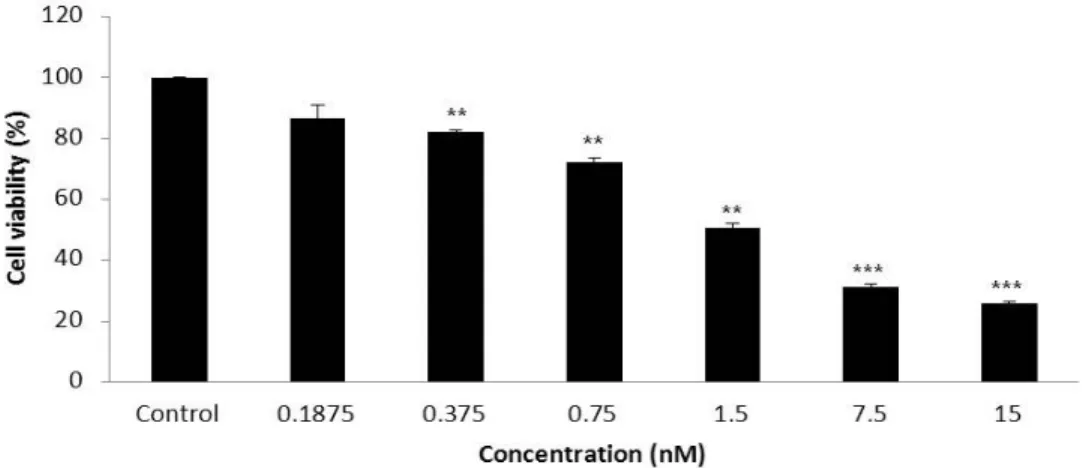
Fig. 1. PEA showed cell proliferation in a dose-dependent manner by WST-1 assay on SCC 25 cells(**p<0.01, ***p<0.001)
4.2. Propidium iodide(PI) 염색 후 유세포 분석기 측정
세포를 100 mm dish에 1x10⁶로 분주한 뒤 24시간 배양시킨 후 PEA를 1.5 nM의 농도로 처리하였다. 처리된 세포들은 각 각 12, 24, 48시간 뒤 dish의 배지를 제거하고 PBS로 세척한 뒤 70% 에탄올에 고정하였다. 24시간 후 고정된 세포들은 PBS로 세척한 후 RNase A를 100 µg/ml이 되도록 넣어준 뒤 4℃ 에서 30분 동안 처리하였다. 그런 다음 PI(Sigma, St.
Louis, MO, USA)를 100 µg/ml 의 농도로 처리 한 후 4℃
에서 5분 동안 염색하였다. 염색된 세포는 수거하여 유세포 분석기용 tube에 담아 유세포 분석기 CYTOMICS FC500 (Beckman Coulter, FL, CA, USA)를 통해 FL 3파장에서 관찰하 였다.
4.3. Western blot 분석
세포를 100 mm dish에 24시간 배양한 다음, PEA를 1.5 nM 의 농도로 처리하였다. 6시간에서 48시간 뒤 dish의 배지를 제거하고 4℃에서 세포를 모은 다음 PBS로 세척하였다. 이후 14,000 rpm에 10분 동안 원심분리 한 다음 다시 차가운 PBS로 세척했다. 적당량의 lysis buffer(10 mM Tris/HCl, pH 7.2, 1%
Tripton X-100, 150 nM NaCl, 5 mM EDTA, protease inhibitor cocktail)를 첨가해 4℃에서 1시간 동안 반응시킨 후, 14000 rpm에 30분간 원심 분리해 상층액에 있는 총 단백질을 분리 했다. 각 단백질의 농도는 Bio-Rad 단백질 정량시약(Bio-Rad laboratories Hercules, CA)을 사용해 정량한 다음 5X sample buffer를 섞어 sample을 만들었다. 동량의 sample을 7.5-15%
polyacrylamide SDS gel을 이용해 전기영동으로 분리 한 후, PVDF membrane으로 전이시켰다. 전이시킨 PVDF mem- brane을 5%의 skim milk를 처리해 2시간 동안 blocking을
하고, 1차 항체(Santa Cruz Biotechnology, Santa Cruz, CA, USA)를 4℃에서 overnight 시켰다. 다음 날 TBS-T(20 mM Tris, 150 mM NaCl, 0.1% Tween 20)로 세척을 하고 2차 항체 를 희석하여 2시간 동안 반응시켰다. 그런 다음 여러 번 다시 TBS-T로 세척하고 ECL Western blotting reagent을 적용시킨 다음 특정단백질의 발현 양을 Alpha Imager HP(Alpha Innotech, Santa Clara, CA, USA)를 통해 분석하였다.
5. 자료 분석
수집된 실험 자료는 통계분석용 소프트웨어인 IBM SPSS Statistics 21(SPSS Inc., NY, USA)을 통해 결과를 산출하였다.
대조군과 실험군의 통계 처리는 비모수 검정인 Kruskal-Wallis test를 사용하였고 사후검정은 Tukey test us- ing ranks를 사용하였다. 실험 결과는 *p<0.05, **p<0.01, 그리 고 ***p<0.001을 통계학적으로 유의성이 있는 것으로 판정하 였다.
연구결과
1. PEA에 의한 SCC 25 세포 성장 억제 유도
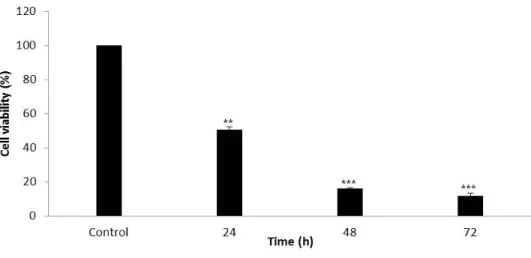
SCC 25 세포에서 PEA가 생존율에 미치는 영향을 보기 위해 서 WST-1 분석을 했다. 먼저 SCC 25 세포에 PEA를 각각의 농도(0.1875-15 nM)로 처리한 후 24시간이 지났을 때, 대조군 은 100±0e, 각 농도별로 87±4.36d, 82±0.58d, 72±1.67c, 51±1.53bc, 31±1.20ab, 26±0.67a%로 농도의존적인 감소를 보 였으며(p<0.01)<Fig. 1>, 1.5 nM를 IC 50의 농도로 잡고 시간 별로 확인을 했을 때는 대조군은 100±0a, 각 농도별로 51±1.53b, 16±0.5c, 12±1.67d%로 시간의존적인 감소형태를
Fig. 3. PEA induces apoptosis by hoechst 33258 stain using fluorescence microscope on SCC 25 cells
Fig. 2. PEA showed cell proliferation in time-dependent manner by WST-1 assay on SCC 25 cells(**p<0.01, ***p<0.001)
보였다(p<0.05)<Fig. 2>.
2. PEA에 의한 SCC 25 세포의 세포자멸사 유도 확인
PEA를 SCC 25세포에 처리 후, 세포의 형태학적인 모양 변 화를 확인하기 위해 Hoechst 33258 염색을 하였다.
<Fig. 3>의 결과, SCC 25세포의 대조군은 핵 모양이 정상적 인 형태인 둥근 모양인 반면, PEA를 24시간 처리했을 때 핵의 모양은 대조군에 비해 줄어들고 응축된 모양을 관찰 할 수 있었다. SCC 25세포의 핵의 형태학적 모양을 관찰했을 때, PEA를 처리했을 시 세포자연사로 유도되었음을 확인할 수 있었다.
세포자멸사와 관련된 sub-G1과 단백질양의 변화를 알아보 기 위해 유세포 분석과 Western blot분석을 하였다.
<Fig. 4>의 실험결과를 보면 세포자연사의 지표인 sub-G1 이 대조군에 비해 PEA를 처리 후, 24시간부터 계속 증가하는 것을 확인할 수 있었다(대조군: 9.7%, 24시간: 18.7%, 48시간 28.5%).
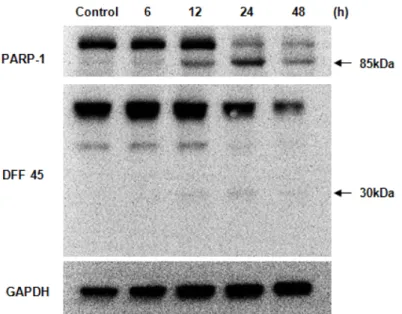
세포자멸사 신호전달에서 대표적인 caspase-3의 기질로 알 려진 PARP-1과 DFF 45의 결과를 Western blot 분석법으로 확인하였다. 실험결과 PEA를 처리를 했을 때, 세포의 DNA를 회복시키는 기능을 하는 PARP-1는 116 kDa은 줄어들고 85 kDa은 증가된 양상을 보였고 caspase를 활성화시키는 CAD 를 억제하고 있는 DFF 45(ICAD)는 45 kDa과 40 kDa은 줄어
Fig. 5. PEA induces apoptosis by PARP-1 and DFF 45 using western blot on SCC 25 cells
Fig. 4. PEA induces apoptosis by PI stain using flow cytometry on SCC 25 cells
들고 30 kDa에서 증가된 양상을 보아 시간이 지남에 따라 각각의 기질들이 쪼개짐으로써 세포자연사가 유도되었음을 확인할 수 있었다<Fig. 5>.
총괄 및 고안
아주 오래 전부터 박테리아의 독소를 이용한 암 치료는 계 속 연구되고 시행되어 왔다. Koo 등17)에 의해 탄저균으로부 터 발생되는 lethal factor을 이용한 in vivo실험에서 누드 마우 스의 사람 흑색종 조직 크기가 줄어들었다는 보고가 있었고 Bhattacharjee 등18)의 의해서도 Verotoxin 1이 대장암세포에 서 세포주기 중 S기를 정지시켜 높은 세포 독성을 나타낸다는 보고도 있었다. Punj 등19)은 Pseudomonas aeruginosa에서 발
생하는 azurin을 실험한 결과 유방암에서 세포자멸사를 유도 한다고 보고하였다. 또한 실험에 사용된 PEA는 Chang와 Kwon7)에 의해 자궁암세포에서 세포 자멸사를 통한 세포 죽 음을 유도한다는 사실도 보고되었다. PEA는 66 kDa의 613개 의 아미노산의 단백질로 구성되어 있으며 Pseudomonas aer- uginosa로부터 분비되는 세포 밖 분비물에서 발생되어 나온 독소이다7).
본 연구에서는 구강암으로 알려진 SCC 25세포에서 PEA에 의한 세포 성장에 미치는 효과와 이와 관련하여 세포자멸사 유발 여부를 조사하였다. 이를 위해 암세포의 증식 억제효과 를 알아보기 위해 WST-1의 분석을 한 결과 PEA를 처리 시 농도 의존적으로 감소하였으며 또한 시간 의존적으로 급격히 감소된 것을 관찰할 수 있었다. 이 결과로 볼 때, PEA가 구강 암의 한 종류로 알려진 사람혀편평암종세포의 생존율을 농
를 염색하는 형광 물질인 PI(propidium iodide)로 나타나는 분석법이다. 세포 자멸사가 일어날 시 나타나는 특성으로 핵 의 쪼개짐이 발생하게 되는데 이를 sub-G1이라 한다. 즉, 다 시 말해 이 sub-G1을 세포 주기 내 전체 DNA에서 차지하는 apoptotic cell을 %로 환산해 나타내는 것이다. 세포 자멸사의 유발 정도를 나타내는 sub-G1의 빈도를 측정한 결과, 대조군 에서는 9.7%에 불과했지만 PEA를 처리 후 24시간이 지났을 때는 18.7%, 48시간에는 28.5%로 현저히 증가되는 것을 확인 하였다. 이는 암세포의 증식억제와 더불어 세포자멸사와 밀 접한 관련을 보이는 것으로 관찰된다.
PARP-1은 DNA의 수복, 염색질 기능 그리고 유전자 전사에 있어서 매우 중요한 역할을 한다20). 하지만 세포자멸사 과정 에서 caspase-3가 활성이 되면 핵 단백질인 PARP-1이 분해가 일어나면서 단편화 현상이 관찰되고 이는 DNA의 수복 과정 이 억제되어진다21). 또한 세포자멸사가 일어날 시 나타나는 현상 중 하나인 DNA 단편화가 있는데 여기와 관련되어 중요 한 역할을 하는 인자가 DFF이다22). DFF는 두 가지의 인자로 구성이 되어 있는데 그 중 하나는 caspase-activated DNase인 DFF40이고 다른 하나는 inhibitor of caspase-activated DNase 인 DFF 45이다23). 그 중 DFF 45는 세포자멸사가 유발되면 활성화된 caspase에 의하여 단편화가 일어나게 된다22).
단백질에서의 세포자멸사 효과를 알아보기 위해 Western blot분석법을 시행하였다. 그 결과 PARP-1에서 12시간부터 기질이 단편화되기 시작하면서 48시간에는 거의 대부분이 단편화 현상을 보이는 것을 확인하였다. DFF 45에서는 PARP-1와 마찬가지로 12시간부터 단편화가 시작되어 24시간 에 최대로 단편화 현상을 보이고 48시간에는 거의 대부분 단백질 발현 감소의 형태를 보였다.
이상의 결과를 보면 PEA는 구강암세포인 SCC 25에서 세포 증식 억제 및 핵 형태 변화를 보였으며 이는 세포자멸사와 밀접한 관련이 있으며 특히 caspase-3의 기질인 PARP-1과 DFF 45의 단편화를 유도했기 때문에 보다 확실한 세포자멸사 의 기전을 통해 이루어진 것으로 보인다. 이는 PEA가 구강암
SCC 25 세포에서 세포자멸사 효과에 대한 분자생물학적 기전 을 검증하기 위해 실험을 수행하였다. 본 연구의 실험을 위하 여 구강암 세포 중 하나인 SCC 25 세포를 사용하였고 WST-1 분석법, Hoechst 33258 염색법, 유세포 분석기, Western blot 분석을 포함한 실험방법을 시행하여 다음과 같은 결론을 얻 었다.
1. SCC 25세포에 PEA를 0.1875-12 nM의 농도로 처리한 후 24시간이 지났을 때, 대조군은 100±0e, 각 농도별로 87±4.36d, 82±0.58d, 72±1.67c, 51±1.53bc, 31±1.20ab, 26±0.67a%로 농도의존적인 감소를 보였으며(p<0.01), 1.5 nM를 IC 50의 농도로 잡고 시간별로 확인을 했을 때는 대조군은 100±0a, 각 농도별로 51±1.53b, 16±0.5c, 12±1.67d%로 시간의존적인 감소를 보였다(p<0.05).
2. 24시간 동안 1.5 nM의 PEA를 처리한 SCC 25 세포의에서 의 핵 모양을 관찰하기 위해 Hoechst 33258를 염색을 한 후 형광현미경으로 분석한 결과, 1.5 nM 농도의 PEA 를 처리한 군이 처리하지 않은 군보다 세포의 수는 적어 지고 세포자멸사 형태인 핵 쪼개짐이 나타났다.
3. PI를 염색 시킨 후 유세포 분석기를 통한 sub-G1의 정량 적 측정을 분석한 결과 대조군은 9.7%, 1.5 nM의 PEA를 24시간 처리한 군은 18.7%, 그리고 48시간 처리한 군은 28.5%로 증가되는 양상을 확인하였다.
4. Western blot 분석법으로 PARP-1과 DFF 45의 단백질 변화를 확인한 결과, 1.5 nM 농도의 PEA를 처리한 군에 서 시간이 지남에 따라 각각의 기질들이 쪼개진 것을 확인하였다.
이상의 연구 결과, PEA는 구강암 세포 사멸 효과가 뛰어나 며 그 기전은 세포자멸사에 의한 것으로 관찰된다. 이로써 PEA에 대한 향후 지속적인 연구를 통해 구강암 치료 향상에 기여할 것으로 사료된다.
References
1. Lee SS, Lee SK. Molecular cloning of novel genes from subtracted cDNA library or oral squamous cell carcinoma. Korean J Oral Maxillofac Pathol 2001; 25(3): 225-41.
2. Sasahira T, Kirita T, Kuniyasu H. Update of molecular pathobiology in oral cancer: a review. Int J Clin Oncol 2014 Epub ahead of print.
3. Coley WB. The treatment of malignant tumors by repeated inoculations of erysipelas. With a report of ten original cases. Am J Med Sci 1893; 105: 487-11.
4. Lundin JI, Checkoway H. Endotoxin and cancer. Environ Health Perspect 2009; 117(9): 1344-50. http://dx.doi.org/10.1289/ehp.0800439.
5. Fillon S, Lang F, Jendrossek V. Pseudomonas aeruginosa triggered apoptosis of human epithelial cells depends on the temperature during infection. Cell Physiol Biochem 2002; 12(4): 207-14.
6. Sharma AK, FitzGerald D. Pseudomonas exotoxin kills drosophila S2 cells via apoptosis. Toxicon 2010; 56(6): 1025-34.
http://dx.doi.org/10.1016/j.toxicon. 2010.07.007.
7. Chang JH, Kwon HY. Expression of 14-3-3delta, cdc2 and cyclin B proteins related to exotoxin A-induced apoptosis in HeLa S3 cells. Int Immunopharmacol 2007; 7(9): 1185-91.
8. Behdani M, Zeinali S, Karimipour M, Khanahmad H, Schoonooghe S, Aslemarz A, Seyed N, Moazami-Godarzi R, Baniahmad F, Habibi-Anbouhi M, Hassanzadeh-Ghassabeh G, Muyldermans S.
Development of VEGFR2-specific Nanobody Pseudomonas exotoxin A conjugated to provide efficient inhibition of tumor cell growth. N Biotechnol. 2013; 30(2): 205-9. http://dx.doi.org/
10.1016/j.nbt.2012.09.002.
9. Jenkins CE, Swiatoniowski A, Issekutz AC, Lin TJ. Pseudomonas aeruginosa exotoxin A induces human mast cell apoptosis by a caspase-8 and –3-dependent mechanism. J Biol Chem. 2004;
279(35): 37201-7.
10. Lim JH, Lee HJ, Lee EO, Lee HJ, Kwon HY Shim BS, et al.
Apoptotic effect of vitisin A from vitis amurensis against MES-SA uterine cancer cells. Korean J Oriental Physiology &
Pathology 2008; 22(2): 290-5.
11. Yang HS, Kim JY, Lee JH, Lee BW, Park KH, Shim KH, et al.
Celastrol isolated from tripterygium regelii induces apoptosis through both caspase-dependent and -independent pathways in human breast cancer cells. Food Chem Toxicol 2011; 49(2):
527-32. http://dx.doi.org/10.1016/j.fct.2010.11.044.
12. Verma M, Singh SK, Bhushan S, Sharma VK, Datt P, Kapahi BK, et al. In vitro cytotoxic potential of polyalthia longifolia on human cancer cell lines and induction of apoptosis through mitochondrial-dependent pathway in HL-60 cells. Chem Biol Interact 2008; 171(1): 45-56.
13. Chen L, Gong MW, Peng ZF, Zhou T, Ying MG, Zheng QH, et al. The marine fungal metabolite, dicitrinone B, induces A375 cell apoptosis through the ROS-related caspase pathway. Mar Drugs 2014; 12(4): 1939-58. http://dx.doi.org/10.3390/md12041939.
14. Thangam R, Senthilkumar D, Suresh V, Sathuvan M, Sivasubramanian S, Pazhanichamy K, et al. Induction of ROS-dependent mitochondria mediated intrinsic apoptosis in MDA-MB-231 cells by glycoprotein from codium decorticatum. Agric Food Chem 2014. Epub ahead of print.
15. Cheng TC, Lu JF, Wang JS, Lin LJ, Kuo HI, Chen BH.
Antiproliferation effect and apoptosis mechanism of prostate cancer cell PC-3 by flavonoids and saponins prepared from Gynostemma pentaphyllum. J Agric Food Chem 2011; 59(20):
11319-29. http://dx.doi.org/10.1021/jf2018758.
16. Olszewska P, Szymański J, Mikiciuk-Olasik E, Szymański P.
New cyclopentaquinoline derivatives with fluorobenzoic acid induce G1 arrest and apoptosis in human lung adenocarcinoma cells. Eur J Pharmacol 2014; 15(729): 30-6. http://dx.doi.org/
10.1016/j.ejphar.2014.02.003.
17. Koo HM, VanBrocklin M, McWilliams MJ, Leppla SH, Duesbery NS, Vande Woude GF. Apoptosis and melanogenesis in human melanoma cells induced by anthrax lethal factor inactivation of mitogen-activated protein kinase kinase. Proc Natl Acad Sci U S A 2002; 99(5): 3052-7.
18. Bhattacharjee RN, Park KS, Uematsu S, Okada K, Hoshino K, Takeda K, et al. Escherichia coli verotoxin 1 mediates apoptosis in human HCT116 colon cancer cells by inducing overexpression of the GADD family of genes and S phase arrest. FEBS Lett 2005; 579(29): 6604-10.
19. Punj V, Bhattacharyya S, Saint-Dic D, Vasu C, Cunningham EA, Graves J, et al. Bacterial cupredoxin azurin as an inducer of apoptosis and regression in human breast cancer. Oncogene 2004; 23(13): 2367-78.
20. Yuan K, Sun Y, Zhou T, McDonald J, Chen Y. PARP-1 regulates resistance of pancreatic cancer to TRAIL therapy. Clin Cancer Res 2013; 19(17): 4750-9. http://dx.doi.org/10.1158/1078-0432.
21. Sodhi RK, Singh N, Jaggi AS. Poly(ADP-ribose) polymerase-1 (PARP-1) and its therapeutic implications. Vascul Pharmacol 2010;
53(3-4): 77-87. http://dx.doi.org/ 10.1016/j.vph.2010.06.003.
22. Boulares AH, Zoltoski AJ, Sherif ZA, Yakovlev A, Smulson ME.
Roles of DNA fragmentation factor and poly(ADP-ribose) polymerase-1 in sensitization of fibroblasts to tumor necrosis factor-induced apoptosis. Biochem Biophys Res Commun 2002;
290(2): 796-801.
23. Rao PV, Jayaraj R, Bhaskar AS, Kumar O, Bhattacharya R, Saxena P, et al. Mechanism of ricin-induced apoptosis in human cervical cancer cells. Biochem Pharmacol 2005; 69(5):
855-65.