CASE REPORT
급격한 간기능 저하를 보인 급성 문맥혈전증이 동반된 간경변증 환자의 항응고제 사용 효과
조훈길, 김유리
1, 조은영
원광대학교 의과대학 내과학교실, 영상의학교실1
The Effect of Anticoagulant in Patients with Cirrhosis Associated with Acute Portal Vein Thrombosis
Hoon Gil Jo, Youe Ree Kim1 and Eun Young Cho
Departments of Internal Medicine and Radiology1, Wonkwang University School of Medicine, Iksan, Korea
The treatment of portal vein thrombosis (PVT) in patients with liver cirrhosis (LC) has been controversial, and it is generally case- and institution-dependent. The occurrence of acute or extensive PVT is critical and requires urgent treatment because it is usually accompanied by symptoms, particularly when total occlusion occurs, causing acute decompensation of liver disease. Even in severe cases, drug selection and treatment duration are determined based on each institution’s experience. Therefore, consistent guidelines for the treatment of patients with LC with PVT are required. Recently, a patient with acute occlusive PVT with LC who showed signs of acute decompensation was treated by administering low molecular weight heparin as anticoagulant therapy. After anticoagulant treatment, the portal vein was almost completely recanalized, and the deteriorated liver function improved. In addition, the patient recovered well and showed no recurrence of PVT for more than a year. Thus, the most recent knowledge regarding the treatment of nonmalignant PVT in LC was reviewed along with a case report. (Korean J Gastroenterol 2021;78:177-182)
Key Words: Liver cirrhosis; Portal vein; Venous thrombosis; Anticoagulants
Received April 2, 2021. Revised May 26, 2021. Accepted May 26, 2021.
CC This is an open access article distributed under the terms of the Creative Commons Attribution Non-Commercial License (http://creativecommons.org/licenses/
by-nc/4.0) which permits unrestricted non-commercial use, distribution, and reproduction in any medium, provided the original work is properly cited.
Copyright © 2021. Korean Society of Gastroenterology.
교신저자: 조은영, 54538, 익산시 익산대로 460, 원광대학교 의과대학 내과학교실
Correspondence to: Eun Young Cho, Department of Internal Medicine, Wonkwang University School of Medicine, 460 Iksan-daero, Iksan 54538, Korea. Tel:
+82-63-859-2566, Fax: +82-63-850-2025, E-mail: [email protected], ORCID: http://orcid.org/0000-0003-2604-918X Financial support: None. Conflict of interest: None.
서 론
문맥혈전증(portal vein thrombosis)은 간경변증 환자에서 비교적 흔히 발생되는 합병증으로, 특히 진행성 간경변증에서 더 흔하게 관찰된다. 최근 문맥혈전증에 대한 적극적인 항응고 요법이 환자의 생존율을 향상시킨다는 보고들이 증가하고 있으 나,1,2 여전히 출혈 위험성이 높은 진행성 간경변증 환자에서 문맥혈전증에 대한 치료가 꼭 필요한지에 대해서는 이견이 있 다. 전통적인 항응고제인 와파린의 단점을 개선한 직접작용 경구항응고제(direct acting oral anticoagulants, DOAC)가
개발되어 사용되면서 항응고제 관련 출혈 위험성이 현저히 줄어 들었다는 보고들이 있으나, Child-Turcotte-Pugh B (CTP B) 등급 이상 진행된 간경변증 환자에서는 아직 안전성이 확인되어 있지 않다.3저자들은 급성 문맥혈전증 발생과 함께 기존 간경변 증이 급격히 악화된 환자에게 저분자량 헤파린(low molecular weight heparin, LMWH)인 enoxaparin을 이용한 항응고 치료 후 간문맥의 혈류가 회복되고 간기능 악화의 증상과 징후가 호전된 예를 경험하였다. 이에 현재까지의 비악성(non-malig- nant) 문맥혈전증의 치료에 대한 문헌고찰과 함께 이를 보고하 고자 한다.
A
A BB
C
C DD
Fig. 1. Pre-treatment axial view of abdominal computed tomography (CT) images. (A, B) At admission, the axial portal phase CT image showed an occluded right and main portal vein (arrow of A, right portal vein; arrowhead of A, main portal vein). In addition, the precontrast CT image showed a newly developed hyperdense thrombosis in the right portal vein compared to the main portal vein (arrow of B, 58 HU indicated acute thrombus; arrowhead of B, 27 HU indicated chronic thrombus). (C, D) Seven days before admission, axial portal phase CT image showed partial thrombosis in the right portal vein (arrow of C). This thrombosis showed low density in the precontrast image (arrow of D, 27 HU). HU, housefield unit.
증 례
57세 여자가 찢어지는 듯한 흉골 하 및 명치 부위 통증으 로 응급실을 방문하였다. 활력징후는 혈압 90/60 mmHg, 맥 박 71회/분, 호흡수 16회/분, 체온 36.3℃였다. 통증은 일주 일 전부터 시작되었으며 물이나 음식을 먹으면 증상이 악화 되어 1-2분간 지속되다 호전되는 양상을 반복하였고, 일주일 전 같은 증상으로 응급실에 왔다 호전되어 퇴실하였다. 20년 전 타 병원에서 만성 C형간염으로 인터페론을 투여한 후 완 치 판정 받은 기왕력이 있으며, 15년 전 간경변증으로 진단 받았고, 당시 복수가 있어 이뇨제를 복용하였다. 최근까지 3차 례 식도정맥류출혈로 치료한 병력이 있었다. 당뇨병이 있어
투약 중이었고, 음주와 흡연은 하지 않았다. 내원 당시 혈압 저하로 승압제를 투여하였고 2병일째부터 발생한 38℃ 이상 의 고열로 ceftriaxone을 투여하였다. 내원 6개월 전 간기능은 total bilirubin 1.48 mg/dL, albumin 3.0 g/dL, PT (INR) 1.23, AST 57 U/L, ALT 23 U/L였으나, 내원 당시 total bi- lirubin 6.27 mg/dL, albumin 2.3 g/dL, AST 128 U/L, ALT 48 U/L, ALP 270 U/L, GGT 180 U/L, PT (INR) 1.71 로 악화된 소견을 확인할 수 있었다. 그 외 혈액 검사에서 혈 색소 15.1 g/dL, 백혈구 17,880/mm3 (중성구 14,100/mm3), 혈소판 42,000/mm3였고, BUN 49.0 mg/dL, 크레아티닌 0.93 mg/dL, 나트륨 123 mEq/L, 칼륨 4.1 mEq/L, 암모니아 35 μmol/L, CRP 8.05 mg/dL, HbA1c 10.3%, anti-HCV (+),
A
B
C D E
Fig. 2. Post-treatment ultrasonography (US) and axial view of abdominal computed tomography (CT) images. (A) After 2 weeks of anticoagulant therapy, the US image showed no flow signal at the right proximal portal vein using super microvascular imaging (SMI) (arrow). (B) After 3 weeks of anticoagulant therapy, the US image showed a restored portal vein flow signal on SMI (arrow). (C) After 1 month of anticoagulant therapy, the axial portal phase CT image showed recanalization of the right portal vein (arrow), but remaining preexisting chronic thrombus.
(D) At 6 months after the end of anticoagulant therapy, the axial portal phase CT image showed improvements of partial thrombosis (arrow), recanalized left portal vein (dotted arrow), and decreased ascites. (E) At 6 months after the end of anticoagulation therapy, axial portal phase CT image showed cavernous transformation (arrow).
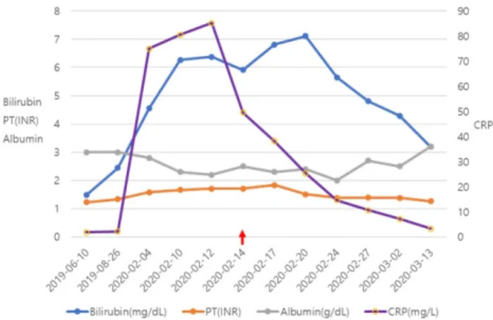
Fig. 3. Changes in the laboratory findings during anticoagulant therapy. LMWH (enoxaparin) was administered for 1 month, starting with HD 4 (2020-02-14) (red arrow). LMWH, low molecular weight heparin; PT, prothrombin time; INR, international normalized ratio;
HD, hospital day; CRP, C-reactive protein.
HCV RNA (-), AFP 1.79 ng/mL, PIVKA II 16 mAU/mL로 염증 지표가 높았다. 혈액배양 검사에서 B군 사슬알균(group B
streptococcus
,Streptococcus agalactiae
)이 2회 이상 배양되어 의미 있는 병원체로 판단하였고 ceftriaxone에 감 수성이 있었다. 통증의 원인을 찾기 위해 시행한 복부 CT에 서 문맥기에 주문맥과 양측 문맥에 혈전증이 진행되어 완전 히 폐색된 상태였고(Fig. 1A), 복벽에 뿌옇게 증가된 음영과 복강 내 소량의 복수가 관찰되어 문맥의 완전 폐색에 의한 전신 울혈이 확인되었다. 또한 조영 전 복부 CT에서 새로 진 행된 문맥혈전은 고밀도(hyperdense, 58 housefield unit [HU])로 관찰되었다(Fig. 1B). 한편 일주일 전 실시한 복부 CT에서는 주문맥 및 왼쪽 문맥 가지의 부분 문맥혈전증(Fig.1C)이 확인되었고, 이 부분은 조영 전 CT에서 저밀도(low density, 27 HU)로 관찰되었다(Fig. 1D). 따라서 급격하게 진행된 완전 폐색을 동반한 급성의 광범위한 문맥혈전증 에 의한 증상들로 판단하고 enoxaparin을 4주간 투여하였 다. Enoxaparin 투여 15일 후 실시한 복부 초음파의 미세 혈관 영상(super microvascular imaging; Aplio i800 with a convex broadband probe, Canon Medical Systems Corporation, Otawara, Japan)에서는 우측 문맥의 근위부 에 흐름신호(flow signal)가 관찰되지 않고 원위부 일부에서 약한 흐름신호만 관찰되었으나(Fig. 2A), enoxaparin 투여 3 주 후 실시한 복부 초음파에서는 문맥의 흐름신호가 근위부 까지 복원되었음을 확인할 수 있었다(Fig. 2B). 치료 1개월 후 실시한 복부 CT (Fig. 2C)에서 우측 문맥의 혈류가 회복 되었음을 확인할 수 있었으나 일부 만성 혈전이 남아있었다.
환자는 증상과 혈액 검사 소견(Fig. 3)이 호전되어 퇴원하였 고, 이후 외래를 통해 증상의 악화 없이 이뇨제를 서서히 감
량할 수 있었다. 퇴원 6개월 후 실시한 복부 CT (Fig. 2D, E) 에서 복수의 유의미한 감소와 함께 우측 문맥의 부분 혈전의 일부가 호전되었음과 해면 변형(cavenous transformation) (Fig. 2E) 및 좌측 문맥의 혈류가 회복되었음을 확인할 수 있 었고, 치료 후 1년 이상 증상과 혈액 검사 소견의 악화 없이 외래 추적 검사 중이다.
고 찰
문맥혈전증은 원인 질환에 따라 악성과 비악성, 간경변성 과 비간경변성으로 나눌 수 있고 상태에 따라 급성과 만성, 폐색성과 비폐색성으로 나눌 수 있다.3 비악성 문맥혈전증의 가장 흔한 원인은 간경변증으로 약 1-25%에서 문맥혈전증이 발생한다고 알려져 있다.4,5 간경변증 환자에서 발생하는 문맥 혈전증은 간내 섬유화의 진행으로 인한 간 구조의 찌그러짐으 로 인해 감소된 문맥 흐름 속도가 중요한 기여 인자이며,6 간 질환이 진행할수록 문맥혈전증의 발생 빈도는 증가한다.
문맥혈전증의 증상은 급성으로 발생하였는지 또는 만성으 로 생긴 것인지에 따라 달라지며, 폐색의 정도에 따라 증상의 중증도가 다를 수 있다.7급성 문맥혈전증의 경우는 복통, 열, 복수의 악화 등의 증상을 보일 수 있고, 완전 폐색이 되면 증 상이 더 심하게 나타나서 급성 간부전의 증상과 징후들을 보 일 수 있다. 만성 또는 부분 폐색된 문맥혈전증의 경우 대개 무증상이며 복부 영상 검사에서 우연히 발견되는 경우가 많 다. 만성 문맥혈전증은 지속되면 간 문맥 주위에 많은 측부혈 관을 형성하는 해면 변형을 유도하게 된다. 또, 부분 문맥혈전 증의 경우는 혈전의 범위가 증가되거나 완전히 폐색된 상태로 진행할 수 있어서 이에 대한 면밀한 확인이 필요하다. 본 증례 도 급성으로 문맥혈전의 범위가 증가되면서 완전 폐색으로 진 행되어 복통과 열 등의 증상으로 내원하였고, 복수와 전신부 종의 악화, bilirubin의 상승, PT의 연장 등의 간부전의 징후 를 보였다.
문맥혈전증의 진단은 영상 검사로 한다. 문맥혈전증에 대 한 평가 시 가장 먼저 종양 혈전(tumor thrombus)인지 양성 혈전(bland thrombus, nonmalignant thrombus)인지 구분 해야 한다. 종양 혈전은 종양에 대한 치료로 호전되지만, 양성 혈전인 경우에는 항응고요법을 할 것인지 결정해야 한다. 문 맥혈전 발생의 급/만성 여부를 구분하는 것은 단기로 실시한 영상 검사를 비교할 수 없으면 어려울 수 있는데, 대개 조영 전 CT에서 고 감쇠(high attenuation)를 보이면 급성으로 생 긴 혈전일 수 있고, 낮은 감쇠를 보이면 만성 혈전일 수 있다.
이러한 차이는 급성기에는 혈전 내부의 헤모글로빈의 단백질 분율이 증가하면서 혈전의 감쇠 계수가 증가하고, 시간이 갈 수록 간동맥으로의 혈액 공급이 증가하고 지방 변성이 진행되
어 혈전의 감쇠 계수가 감소되기 때문이다.8 환자는 우선 복부 CT에서 간내 종괴가 없어 양성 혈전으로 확인할 수 있었다.
또, 조영 전 복부 CT에서 고 감쇠의 문맥혈전을 보였고, 입원 일주일 전 복부 CT에 비해 유의미한 문맥혈전의 진행이 확인 되어 급성 양성 혈전증으로 진단할 수 있었다. 한편 본 증례의 경우 내원 당시 감염을 시사하는 소견과 혈액배양 검사에서 균주가 동정이 되어 급성 혈전증이 세균 감염으로 인해 발생 하였을 가능성을 생각해 볼 수 있다. 정맥염(pylephlebitis)으 로 간문맥혈전증이 진행되는 경우는 영상 검사에서 문맥벽의 비후나 조영증강 및 문맥 주위 지방 침윤으로 인한 저 음영 소견 등을 보일 수 있다.9그러나 본 증례에서는 문맥벽의 변 화나 주변 간 실질의 변화가 관찰되지 않아 정맥염으로 진행 된 간문맥혈전증보다는 감염으로 급격히 악화된 간기능으로 생긴 급성 간문맥혈전증으로 볼 수 있지만 이 둘을 명확하게 구분하는 것은 어려울 수 있다.
문맥혈전의 치료는 위험과 이익을 고려해야 한다. 간경변 증 환자에서 생긴 부분 문맥혈전에 대해 항응고요법을 하지 않았을 때의 임상 경과에 대한 소수의 연구가 있는데, 20-27개 월 관찰 중 30-50%에서 문맥혈전이 진행하고, 이는 간부전으 로의 진행 및 사망률의 증가와 연관되어 있다고 보고하였
다.7,10,11 따라서 항응고요법이 필요할 수 있겠으나 응고장애
를 지닌 간경변증 환자에서 항응고요법은 치료 중 출혈 위험 을 증가시킬 수 있어 주의가 필요하다. 2016년 유럽 간학회 가이드라인에서는 간경변증 환자에서 문맥혈전증의 치료를 시행하기 전에 위장관 출혈, 특히 정맥류의 출혈에 대한 예방 을 위한 베타 차단제의 사용 또는 밴드결찰술이 필요하며, 완 전한 재개통 및 재발의 방지를 위해 적어도 6개월 이상 항응 고 치료를 유지할 것을 권고하고 있지만, 각각의 사례별로 각 기관에 자체적인 치료 알고리즘에 따라 치료하도록 권고하고 있다.12 문맥혈전증에 대한 항응고 치료제로는 Xa인자의 간접 억제제인 LMWH, 비타민 K를 길항하는 와파린, 그리고 Xa인 자의 직접 억제제인 DOAC이 있다. 전통적인 항응고제인 와 파린에 대한 연구로 Chung 등13은 비악성 문맥혈전증 환자 에서 와파린 투여군과 대조군을 비교하는 연구를 진행하여 치 료군에서 높은 문맥 재개통률을 확인하였고 대조군과 비교해 출혈 합병증이 높지 않음을 보고하였다. 그러나 와파린 투여 시 그 용량이 적절한 치료 목표(INR 2-3)에 있는지 주기적인 혈액 검사를 통해 모니터링 해야 하고, 특히 간경변증 환자에 서는 이러한 치료 목표가 아직 불분명하여 사용이 제한적일 수 있다.14 또 대부분의 연구에서 와파린은 단독 치료보다는 LMWH 치료 후 가교 치료로 사용하는 경우가 많았다.15 다음 으로 LMWH는 진행된 간경변에서도 비교적 안전하게 사용 할 수 있고, 주기적인 혈액 검사를 통한 추적이 불필요하지 않다는 장점이 있으나, 피하주사를 해야 하므로 장기간 투여
가 어렵고 입원이 필요할 수 있다. 최근 출시된 DOAC은 이 러한 단점을 보완하여 경구로 투여하고 혈액 검사 확인이 필 요 없다고 되어 있으나, 대개의 연구가 간경변증 환자를 배제 하고 진행되어서 간경변증 환자에서의 효과와 안전성에 대한 자료가 부족하다.16,17 대상성 간경변증을 대상으로 DOAC과 와파린을 비교한 연구에서는 DOAC이 효능과 안전성면에서 우수하였고, DOAC과 LMWH를 비교한 연구에서는 비슷한 효능과 안전성을 보였다.18,19 DOAC은 현재까지는 CTP A의 간경변증에서는 비교적 안전하게 사용할 수 있고, 비대상성 간경변증인 CTP B와 CTP C는 사용에 주의를 요하거나 사용 하지 않는 것이 유리하다고 하겠다.3 따라서 항응고요법으로 대상성 간경변증에서는 DOAC이나 LMWH를 사용할 수 있 겠고, 비대상성 간경변증에서는 LMWH를 사용해볼 수 있겠 다. 본 증례도 입원 당시 CTP C (11점)로 비대상성 간경변증 에 해당하였고, PT (INR) 1.7 이상 지연되어 항응고 치료와 연관된 출혈의 가능성이 높았다. 그러나 급격히 악화된 간질 환의 원인이 급성 폐색성 문맥혈전증으로의 진행으로 판단하 여 LMWH인 enoxaparin을 투여하였고, 투여 1개월 동안 출 혈은 없었다.
문맥혈전증에 대한 항응고요법 시 막힌 문맥의 혈류가 회 복되는 데 기여하는 주요한 인자는 진단과 치료 시작까지의 간격으로 6개월 이내의 시간 간격인 경우가 성공률이 높았 다.20 따라서 문맥혈전의 치료에서는 조기 진단과 치료가 치료 성공에 가장 중요하다고 하겠다. 본 증례는 기저 간경변증이 있었고 급격히 진행된 문맥혈전증으로 인해 만성 간질환의 급 성 악화가 발생한 증례로 빠른 진단과 치료로 막힌 문맥의 혈류를 회복시킬 수 있었고 간부전의 징후들도 일부 호전시킬 수 있었다.
저자들은 문맥혈전증이 급격히 진행하여 생긴 간부전을 동 반한 진행성 간경변증 환자에서 LMWH를 이용한 항응고 치 료로, 성공적으로 간문맥의 혈류를 회복시키고 간기능을 호전 시킨 증례를 경험하였다. 비대상성 간경변증에서는 LMWH로 항응고 치료를 하는 것이 선택 가능한 치료이며, DOAC이 간 경변증 환자의 문맥혈전증을 치료하는 주요 치료제가 되기 위 해서는 진행성 간경변증에서 안전성에 대한 추가 연구가 필요 하다.
REFERENCES
1. Pettinari I, Vukotic R, Stefanescu H, et al. Clinical impact and safe- ty of anticoagulants for portal vein thrombosis in cirrhosis. Am J Gastroenterol 2019;114:258-266.
2. Noronha Ferreira C, Reis D, Cortez-Pinto H, et al. Anticoagulation in cirrhosis and portal vein thrombosis is safe and improves prog- nosis in advanced cirrhosis. Dig Dis Sci 2019;64:2671-2683.
3. Weinberg EM, Palecki J, Reddy KR. Direct-acting oral anti- coagulants (DOACs) in cirrhosis and cirrhosis-associated portal vein thrombosis. Semin Liver Dis 2019;39:195-208.
4. Nery F, Chevret S, Condat B, et al. Causes and consequences of portal vein thrombosis in 1,243 patients with cirrhosis: results of a longitudinal study. Hepatology 2015;61:660-667.
5. Okuda K, Ohnishi K, Kimura K, et al. Incidence of portal vein thrombosis in liver cirrhosis. An angiographic study in 708 patients. Gastroenterology 1985;89:279-286.
6. Zocco MA, Di Stasio E, De Cristofaro R, et al. Thrombotic risk fac- tors in patients with liver cirrhosis: correlation with MELD scoring system and portal vein thrombosis development. J Hepatol 2009;51:682-689.
7. Sarin SK, Philips CA, Kamath PS, et al. Toward a comprehensive new classification of portal vein thrombosis in patients with cirrhosis. Gastroenterology 2016;151:574-577.e3.
8. Qi X, Han G, He C, et al. CT features of non-malignant portal vein thrombosis: a pictorial review. Clin Res Hepatol Gastroenterol 2012;36:561-568.
9. Balthazar EJ, Gollapudi P. Septic thrombophlebitis of the mesen- teric and portal veins: CT imaging. J Comput Assist Tomogr 2000;
24:755-760.
10. Girleanu I, Stanciu C, Cojocariu C, Boiculese L, Singeap AM, Trifan A. Natural course of nonmalignant partial portal vein thrombosis in cirrhotic patients. Saudi J Gastroenterol 2014;20:288-292.
11. Luca A, Caruso S, Milazzo M, et al. Natural course of extrahepatic nonmalignant partial portal vein thrombosis in patients with cirrhosis. Radiology 2012;265:124-132.
12. European Association for the Study of the Liver. EASL clinical practice guidelines: vascular diseases of the liver. J Hepatol
2016;64:179-202.
13. Chung JW, Kim GH, Lee JH, et al. Safety, efficacy, and response predictors of anticoagulation for the treatment of nonmalignant portal-vein thrombosis in patients with cirrhosis: a propensity score matching analysis. Clin Mol Hepatol 2014;20:384-391.
14. Tripodi A, Anstee QM, Sogaard KK, Primignani M, Valla DC.
Hypercoagulability in cirrhosis: causes and consequences. J Thromb Haemost 2011;9:1713-1723.
15. Delgado MG, Seijo S, Yepes I, et al. Efficacy and safety of anti- coagulation on patients with cirrhosis and portal vein thrombosis.
Clin Gastroenterol Hepatol 2012;10:776-783.
16. Weitz JI, Lensing AWA, Prins MH, et al. Rivaroxaban or aspirin for extended treatment of venous thromboembolism. N Engl J Med 2017;376:1211-1222.
17. Bruins Slot KM, Berge E. Factor Xa inhibitors versus vitamin K an- tagonists for preventing cerebral or systemic embolism in pa- tients with atrial fibrillation. Cochrane Database Syst Rev 2018;
3:CD008980.
18. Hanafy AS, Abd-Elsalam S, Dawoud MM. Randomized controlled trial of rivaroxaban versus warfarin in the management of acute non-neoplastic portal vein thrombosis. Vascul Pharmacol 2019;
113:86-91.
19. Nagaoki Y, Aikata H, Daijyo K, et al. Efficacy and safety of edox- aban for treatment of portal vein thrombosis following danapa- roid sodium in patients with liver cirrhosis. Hepatol Res 2018;
48:51-58.
20. Rodriguez-Castro KI, Vitale A, Fadin M, et al. A prediction model for successful anticoagulation in cirrhotic portal vein thrombosis.
Eur J Gastroenterol Hepatol 2019;31:34-42.