Functional Electric Stimulation-assisted Biofeedback Therapy System for Chronic Hemiplegic Upper Extremity Function
Yeung Ki Kim1, Jun Chan Song2, Jae Won Choi3, Jang Hwan Kim4, Yoon Tae Hwang5
1Leaders Rehabilitation Center, 2Department of Physical Therapy, Daegu Health College, 3Department of Physical Therapy, Kyungbuk College,
4Department of Prosthetics and Orthotics, Hanseo University, 5Department of Physical Therapy, Gangneung Yeongdong College
Purpose: Rehabilitative devices are used to enhance sensorimotor training protocols, for improvement of motor function in the hemiplegic limb of patients who have suffered a stroke. Sensorimotor integration feedback systems, included with these devices, are very good therapeutic frameworks. We applied this approach using electrical stimulation in stroke patients and examined whether a functional electric stimulation-assisted biofeedback therapy system could improve function of the upper extremity in chronic hemiplegia.
Methods: A prototype biofeedback system was used by six subjects to perform a set of tasks with their affected upper extremity during a 30-minute session for 20 consecutive working days. When needed for a grasping or releasing movement of objects, the functional electrical stimulation (FES) stimulated the wrist and finger flexor or extensor and assisted the patients in grasping or releasing the objects. Kinematic data provided by the biofeedback system were acquired. In addition, clinical performance scales and activity of daily living skills were evaluated before and after application of a prototype biofeedback system.
Results: Our findings revealed statistically significant gradual improvement in patients with stroke, in terms of kinematic and clinical performance during the treatment sessions, in terms of manual function test and the Purdue pegboard. However, no significant difference of the motor activity log was found.
Conclusion: Hemiplegic upper extremity function of a small group of patients with chronic hemiparesis was improved through two weeks of training using the FES-assisted biofeedback system. Further research into the use of biofeedback systems for long-term clinical improvement will be needed.
Keywords: Electric stimulation, Biofeedback, Hemiplegia, Rehabilitation
I. Introduction
Stroke is the most common serious neurologic disorder in the United States, comprising half of all patients admitted to hospital for a neurologic disease. Although decreased mortality has been contributed by improved medical management of the complications, most of survivors have significant functional handicap.1 In general the option for management of hemiplegia has been manual exercise
therapy, orthotic fitting, functional substitution. Functional electrical stimulation (FES) was reported that effective on reduce of spasticity, prevention of joint contracture, motor recovery and functional improvement.2,3 When functional electric stimulation assisted with exercise applied to hemiplegic upper extremity function was improved.4
A number of recent reports using sensorimotor integration training protocols have shown the improvement of neurologic impairment and functional deficit in the exercised limb of patients with stroke, compared with control individuals. In particular, studies using the training with robotic devices indicated that that sensorimotor integration, use of a feedback system, and active participation of patients leads to a better motor outcome compared to controls.5-8 Received November 27, 2012 Accepted December 11, 2012
Corresponding author Jun Chan Song, [email protected]
Copyright © 2012 by The Korean Society of Physical Therapy
This is an Open Access article distributed under the terms of the Creative Commons Attribution Non-Commercial License (http://creativecommons.org/licenses/by-nc/3.0/) which permits unrestricted non-commercial use, distribution, and reproduction in any medium, provided the original work is properly cited.
The Journal of Korean Society of Physical Therapy Original Article
In this study, we aimed to develop functional electric stimulation-assisted biofeedback therapy system combined with exercise; the focus was on sensorimotor integration, feedback mechanism for an exercise pattern, and active participation of patients in the exercise program. In addition, we also had a purpose of investigating the effectiveness of the FES-assisted biofeedback therapy system on upper extremity hemiplegic function.
II. Materials and Methods
1. Subjects
We studied six patients (mean age 46.0±16.9 years) with a history of stroke. Three patients were right hand dominant and the others were left. They had a diagnosis of a single cerebrovascular accident (CVA), and were more than one- year post-CVA (mean duration post-stroke 17.5±8.8 months) (Table 1). Each of patients was deserved as their own controls.
The patients were chosen according to the following criteria:
1) having a history of stroke having occurred more than a year before the study, 2) able to move only their arm, but unable to flex or extend their hand voluntarily, 3) no interval change of the clinical performance of the hemiplegic upper extremity over the last two months. The following exclusion criteria were also used: 1) unable to flex or extend impaired hand by electrical stimulation, 2) serious cognitive impairment (Mini- Mental State Examination score<16), aphasia, and visual hemi-neglect, 3) other serious medical conditions (e.g., have metallic device in heart. Six subjects were enrolled in the study. All patients gave written informed consent prior to the study, and approved by our hospital’s Ethics Committee.
2. Apparatus
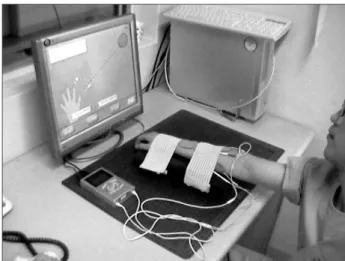
The system was designed for clinical neurological applications. The therapeutic system consisted of a sliding board, the FES stimulator and a computer system including monitor (Figure 1). The sliding board with 4 solid castors was attached to the patient’s forearm and enabled the patient to move voluntarily his or her hemiplegic arm above the desk with little resistance. The FES stimulator was attached to the wrist and finger flexor and extensor muscle groups of the patients. When needed for a grasping or releasing movement of objects, the FES stimulated the wrist and finger flexor or extensor and assisted the patients in grasping or releasing the objects.
The training was achieved by a set of video-game that required a grasping, moving and releasing of objects displayed on the screen of the monitor with a hand figure also displayed on the screen. The hand figure on the screen moved in accordance with the sliding board on the desk;
therefore, patients felt that they had moved the hand figure displayed on the screen by moving the sliding board. Patients were required to move their arm (displayed on the screen as the hand figure), attached to the sliding board, to the place where the objects were displayed on the screen. When the hand figure on the screen was located at the objects (e.g., apple on tree), the FES stimulated the wrist and finger flexor and assisted with the grasping movement without volitional efforts. Then the patients were required to move the objects
Figure 1. Diagram of functional electric stimulation-assisted bio- feedback therapy system.
Table 1. Clinical characteristics of subjects Subject
no.
Age
(year) Sex Duration
(month)* Diagnosis
1 2 3 4 5 6
54 18 47 43 70 44
Male Male Female Male Female Male
13 15 12 13 17 35
Intracranial hemorrhage Intracranial hemorrhage Intracranial hemorrhage Cerebral infarct Cerebral infarct Intracranial hemorrhage
*Months of post-stroke.
to another target position displayed on the screen (e.g., basket on floor). When the hand figure holding the objects moved to the target position (e.g., basket on floor), the FES stimulated the wrist and finger extensor group and assisted with the releasing movement of the patients without volitional efforts.
After the training session the computer system analyzed the average time for the movements between two target positions and the total number of objects transferred during the whole session.
The instrument used in our research was the M511 developed by Cybermedic (Iksan, Korea). Surface electrodes are 3×3 cm sized. The optimal current intensity of the FES system was adjusted just prior to the main training session.
Rising time was 1 second; duration of impulse time was 4 seconds; repose between impulse time was 5 seconds;
stimulating frequency was 50 Hz; amplitude and width of pulse were individually adjusted to get appropriate motor reaction without pain.
This system relies on the sensorimotor integration principle using the sliding board and FES system. An additional training effect was visual motor training, using the video-game and feedback exercise protocol, by the operating system of the video-game.
3. Experimental procedure
Subjects received treatment on their hemiplegic upper extremity for 30 minutes daily for 20 consecutive workdays.
The treatment session consisted of two tasks, which composed of reaching, grasping, moving (e.g., pushing, pulling) and releasing an object displayed on the computer screen. One task was a figure matching skill that consisted of a certain type of figure (e.g., circle, square, triangle, star) and a hole that matched with the shape of the figure. Patients were required to grasp one figure and move it to the hole that matched with the figure. The other task was picking an apple from an apple tree and moving it to a collecting box. Because the total number was one of assessments, subjects repeated each task their best.
Outcomes were assessed by next three divisions:
kinematic measures, clinical performance scales and daily living activity skills. Kinematic measures were obtained from
the computer system. These measures recorded were the averaged time taken to reach, grasp, move and release an object and the total number of objects that were transferred.
Clinical performance scales included a manual function test (MFT) and the Purdue pegboard test. The functional independence measure (FIM) was used to assess the patients’
ability to execute activities of daily living. A motor activity log (MAL)9 has been used to measure the activities of daily living of subjects. The assessments were carried out three times:
pretreatment, two weeks after the start of treatment, and post-treatment.
All data were analyzed in comparison differences between repeated tests using the one-way ANOVA with repeated measurement. All statistical analysis was conducted with PASW 18.0 (IBM Co., Armonk, NY, USA), and p<0.05 was set up as the criterion for statistical significance.
III. Results
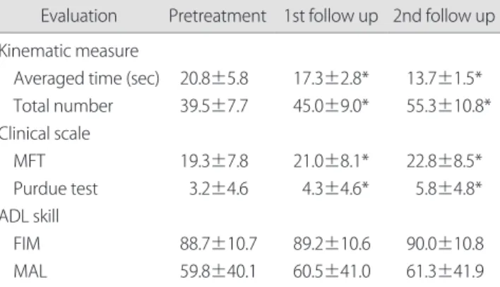
The changes in kinematic measures, clinical performance scales and daily living activity skills were averaged over all patients. Table 2 shows changes of averaged data over all patients.
The data showed a gradual improvement in subject’s kinematic performance with continued use of the bio- feedback system. Statistical analysis of the kinematic
Table 2. Changes in kinematic measures, clinical performance scales and activity of daily living skills averaged over all patients
Evaluation Pretreatment 1st follow up 2nd follow up Kinematic measure
Averaged time (sec) Total number Clinical scale MFT Purdue test ADL skill FIM MAL
20.8±5.8 39.5±7.7
19.3±7.8 3.2±4.6
88.7±10.7 59.8±40.1
17.3±2.8*
45.0±9.0*
21.0±8.1*
4.3±4.6*
89.2±10.6 60.5±41.0
13.7±1.5*
55.3±10.8*
22.8±8.5*
5.8±4.8*
90.0±10.8 61.3±41.9 Values are presented as mean±standard deviation.
MFT: manual function test, ADL: activity of daily living, FIM: functional independent measure, MAL: motor activity log.
*p<0.05.
measures showed that improvement of subjects’ kinematic performance was statistically significant (p<0.05).
The data also demonstrated a gradual improvement in the subjects’ clinical performance with continued use of the biofeedback system. Statistical analysis of the MFT and the Purdue pegboard test showed that the improvement of the subject’s performance was statistically significant (p<0.05).
However, the ANOVA of both the FIM and MAL did not show statistically significant effects of the treatment.
IV. Discussion
Our device is focused on the goal directed, task-specific activity by fulfilling the mission which is operated by executing mission. We intended our system could augment the patient’s ability to move their hemiplegic arm voluntarily by sliding board and facilitate the hand function by FES.
We designed the activity pattern according to visuomotor relationship principle by matching the real movement of paretic arm with visual movement of hand on the screen. In addition, we can succeed the feedback mechanism of upper extremity movement by reaching, holding, and releasing activity during executing mission. Because the mission was designed to enjoy playing the game, the system could provoke the subject’s motivation and make the subjects to participate the exercise program actively. The system was easy to operate to both therapist and participants.
We designed the exercise sequence as the natural movement pattern of normal upper extremity; placing the hand - holding the material - relocating the hand - releasing the material. The exercise pattern of our system was based on functional activity because those sequences of motor execution were very similar to many activities of daily living. In addition, many hemiplegic patients have difficulties on control of their hemiparetic upper extremity, it is very important to divide the whole mission into many subcomponents easy to handle. As the result, the system was very friendly to most of participant to operate.
The result of this study indicated that a functional electric stimulation-assisted biofeedback therapy system improved kinematic and clinical performance of the upper extremity
in stroke patients with chronic hemiplegia of the upper extremity. All subjects included in the study were chronic hemiplegia (time interval between start of intervention and onset was more than a year) those improvements is believed to be sent genuine in function of chronic hemiplegia of the upper extremity. There is some possibility that our exercise program using a functional electric stimulation-assisted biofeedback therapy system can augment the potential weakness in chronic hemiplegia rather than neurologic recovery.
Although our exercise program was focused on functional activities, the scores of activity of daily living (ADL) failed to show a statistical significant improvement. This may have been due to the fact that the most of participant have difficulties on fine hand movement already prior to participate the intervention. The main improvements in arm and forearm function and less degree of improvement of hand function could not elevate the score of ADL. Another possibility is that the degree of improvement was not large enough to make a significant difference in ADL scores.
Because of the absence of a control group and low number of study subjects, we cannot completely rule out the possibility that additional traditional exercise therapy, of a similar duration, would be as effective as the biofeedback system. However, the main goal of our study was to develop the FES assisted biofeedback system and evaluate the possibility of clinical use of our biofeedback system. In addition, our study did not evaluate a long term effect of the biofeedback system. In the future, experiments evaluating the long-term cumulative effect of the biofeedback system, in chronic hemiplegic patients, require a larger sample of subjects with a blinded and controlled study design.
Forced-use or constraint-induced strategies have exploited the natural context of the patient environment by requiring increased use of the paralyzed limb.10 These efforts were consistent with experiments where an additional sensorimotor training, for the paralyzed limb, was employed to improve outcome.11-13 Animals with focal cortical injury exposed to an enriched or challenging sensorimotor environment have been shown to exhibit greater anatomic responses.14-16 These findings suggest the important principle
of use it or loose it. We believe our system concentrates on the sensorimotor integration principle using a sliding board and FES system. An added effect is visual motor training using video-game and a feedback exercise protocol with the operating system of the video-games. Our findings suggest that our system, with its focus on sensorimotor integration, visual motor relationships, feedback exercise therapy as well as the active participation of patients in the exercise program, can improve upper extremity chronic hemiplegia in post stroke patients.
References
1. Joel AD. Physical medicine and rehabilitation 4th edition.
Philadelphia, Lippincott, Williams & Wilkins, 2004.
2. Kwon YH, Kwon JW, Park SY et al. Cortical activation by trans- cranial direct current stimulation and functional electrical stimulation in normal subjects: 2 case studies. J Korean Soc Phys Ther. 2011;23:77-82.
3. Malhotra S, Rosewilliam S, Hermens H et al. A randomized con- trolled trial of surface neuromuscular electrical stimulation applied early after acute stroke: effects on wrist pain, spasticity and contractures. Clin Rehabil. 2012. [Epub ahead of print]
4. Randall LB. Physical medicine and rehabilitation. Philadelphia, WB Saunders Co., 1996.
5. Gritsenko V, Prochazka A. A functional electric stimulation-assisted exercise therapy system for hemiplegic hand function. Arch Phys Med Rehabil. 2004;85(6):881-5.
6. Fasoli SE, Krebs HI, Stein J et al. Effects of robotic therapy on
motor impairment and recovery in chronic stroke. Arch Phys Med Rehabil. 2003;84(4):477-82.
7. Lum PS, Burgar CG, Shor PC et al. Robot-assisted movement training compared with conventional therapy techniques for the rehabilitation of upper-limb motor function after stroke. Arch Phys Med Rehabil. 2002;83(7):952-9.
8. Volpe BT, Krebs HI, Hogan N. Is robot-aided sensorimotor training in stroke rehabilitation a realistic option? Curr Opin Neurol. 2001;
14(6):745-52.
9. Taub E, Miller NE, Novack TA et al. Technique to improve chronic motor deficit after stroke. Arch Phys Med Rehabil. 1993;74(4):347- 54.
10. Ryu IT, Hwang BY, Kim JH et al. The effects of constraint- induced movement therapy on improvement of hand function in hemiplegic side. J Korean Soc Phys Ther. 2009;21:9-14.
11. Kwakkel G, Wagenaar RC, Twisk JW et al. Intensity of leg and arm training after primary middle-cerebral-artery stroke: a randomised trial. Lancet. 1999;354(9174):191-6.
12. Kwakkel G, Wagenaar RC, Koelman TW et al. Effects of intensity of rehabilitation after stroke. A research synthesis. Stroke.
1997;28(8):1550-6.
13. Nudo RJ, Wise BM, SiFuentes F et al. Neural substrates for the effects of rehabilitative training on motor recovery after ischemic infarct. Science. 1996;272:1791-4.
14. Jones TA, Schallert T. Use-dependent growth of pyramidal neurons after neocortical damage. J Neurosci. 1994;14(4):2140-52.
15. Kozlowski DA, James DC, Schallert T. Use-dependent exaggeration of neuronal injury after unilateral sensorimotor cortex lesions. J Neurosci. 1996;16(15):4776-86.
16. Kozlowski DA, Schallert T. Relationship between dendritic pruning and behavioral recovery following sensorimotor cortex lesions.
Behav Brain Res. 1998;97:89-98.