Detection of Herpes Simplex Virus, Varicella Zoster Virus, Helicobacter Pylori and Candida in Saliva of Patients with
Recurrent Aphthous Ulceration
Hur Woong, D.D.S.,M.S.D.,Ph.D., Chang-Lyuk Yoon, D.D.S.,M.S.D.,Ph.D., Jong-Mo Ahn, D.D.S.,M.S.D.,Ph.D..
Department of Oral Medicine, Institute of Forensic Odontology, College of Dentistry, Chosun University
To examine whether HSV, VZV, H. pylori and Candida that are known to be microorganisms causing ulcerative disease in oral cavity and have the relatively high contigiousness are detected in saliva of patients with RAU and related to the development with RAU, PCR and culture were performed on the saliva of 29 patients with RAU and 29 control subjects who visited the Department of Oral Medicine, Dental Hospital, Chosun University.
The results were obtained as follows;
1. HSV DNA was detected in 41.4% patients with RAU, and 55.2% control subjects, however, a significant difference between the two groups was not detected, (P>0.05), and VZV DNA was not detected in both groups.
2. H. pylori DNA was detected in 27.6% patients with RAU, and 48.3% control subjects, however, a significant difference between the two groups was not detected (P>0.05).
3. Candida was cultured in 13.8% patients with RAU, and 6.9% control subjects, however, a significant difference between the two groups was not detected (P>0.05).
This results suggest that HSV, VZV, H. pylori and Candida can not be regarded to play a direct role in the development of RAU. Thus it is considered that in future, on a larger sample, also, it has to be examined whether other microorganisms acts as a trigger factor of the development of RAU.
Key word : Recurrent aphthous ulcer, HSV, VZV, H. pylori, Candida
Corresponding author : Assist. Prof. Jong-Mo Ahn Department of Oral Medicine, Institute of Forensic Odontology, College of Dentistry, Chosun University, 421, Seosuk-Dong, Dong-Gu, Gwang-Ju, 501-825, Korea.
E-mail : [email protected] received: 2005-06-15
accepted: 2005-07-30
* This study was supported in part by research funds from Chosun University, 2004.
Ⅰ. INTRODUCTION
Recurrent aphthous ulceration (RAU) that is referred to as "canker Sore" is a soft tissue lesion which painful ulcer occurs repeatedly in the buccal and labial mucosa, the tongue and the floor of the mouth etc., and it is detected in approximately 20%
of adults
1-3). RAU is classified as minor aphthous
ulceration, major aphthous ulceration and
herpetiform aphthous ulceration. And it is generally
cured within 7~10 days, however, severe lesions
may last for over 6 weeks
4).
Although the etiology of RAU has not been elucidated yet, genetic factors, hormone change, microbiological factors, trauma, nutrient deficiency, stress, allergy, etc. have been suggested to be possible etiologic factor. Among such etiologic factors, the study on microbiological factors has been performed by numerous investigators. Brice et al.
2)investigated Herpes Simplex Virus (HSV) 1 and 2, Varicella Zoster Virus (VZV), Epstein-Bar Virus (EBV), Cytomegalovirus, etc. in the oral mucosa and peripheral blood cell of patients with RAU, and Safronova et al.
5)performed the study that separates bacteria and fungus in the oral mucosa of children with RAU. In addition, the investigation about the correlation of RAU and Helicobacter pylori (H. pylori) that is known as the important pathogen occuring gastrointestinal ulcer was performed by Shimoyama et al.
6)and Briek et al.
7)However, in such studies and other studies,
2, 8-11)it was investigated by using samples obtained from the smear of the lesion and biopsy, or sample obtained from peripheral blood, but the microbiological cause of RAU has not been elucidated yet.
Since RAU is an oral mucosa disease, it is influenced by various oral environmental factors.
Particularly, it is thought to be closely associated the saliva that has the digestive function mediated by amylase, the lubricative and protective function for oral mucosa, anti-bacterial function by lysozyme, lactoferrin, and lactoperoxidase, anti-viral function by secretory IgA and the function of soft tissue healing, etc.
12)In various oral
Clinical characteristic Mean of age (yr) Gender
Patients with RAU
(n = 29) 38.2 13/29 (44.8%) Male
16/29 (55.2%) Female Control subjects
(n = 29) 27.6 8/29 (27.6%) Male
21/29 (72.4%) Female n : Number of individuals
Table 1. Clinical characteristics of patients with RAU and control subjects
diseases and systemic diseases, the composition of saliva changes, its diagnostic value thus is very high. However, the studies about microbiological factors in the saliva of patients with RAU are not abundant.
In this study, different from previous studies performed about microbiological factors in the lesion and the blood of patients by numerous investigators, using the saliva that mediates a great effect on the oral environment and has the protective function of oral mucosa, it was examined whether HSV, VZV, H. pylori, and Candida that have been shown to be able to induce ulcerative disease in oral cavity and have the relatively high contagiousness are detected in the saliva of patients with RAU, and related to the development of RAU.
Ⅱ. MATERIALS AND METHODS
1. Materials
Among patients with RAU visited the Department of Oral Medicine, Dental Hospital, Chosun University, 29 individuals who had the history of more than 2 years were selected as the patients group, and 29 healthy individuals whose oral condition was clinically good and without the history of RAU were selected as the control group.
The age range of patients group was from 9 to 71
years (average age: 38.2 years), 13 males and 16
females. The age range of the control group was
from 23 to 39 years (average age: 27.6 years), and
8 males and 21 females (Table 1).
2. Methods
1) The sampling of unstimulated whole saliva The study subjects were asked to rinse the oral cavity with water and subsequently waited for 10 minutes without stimulation to allow the secretion of the saliva. The secreted saliva was collected in a 1.5 ml microcentrifuge tube, and stored frozen at -20℃ prior to experiments.
2) DNA extraction
From the collected saliva, DNA was extracted as follows by using the AccuPrep
ⓇGenomic DNA Extraction Kit (Bioneer, Daegeon, Korea). First, 200
㎕ of phosphate buffered saline (PBS), 20 ㎕ proteinase K and 200 ㎕ binding buffer were added to a 1.5 ㎖ microcentrifuge tube containing the saliva, and mixed. They were heated at 60℃ for 10 minutes and 100 ㎕ isopropanol was added to the tube, and vortexing for 5 seconds. The mixed solution was transferred to a prepared binding column tube, centrifuged at 8,000 rpm for 1 minute, and the filtered solution was discarded. 500 ㎕ of W1 buffer was added to the column, centrifuged at 8,000 rpm for 1 minute, the filtered solution was discarded, 500 ㎕ of W2 buffer was added, centrifuged at 8,000 rpm for 1 minute, the filtered solution was discarded, and finally, centrifuged one more time at 13,000 rpm for 1 minute, and thus ethanol remained in the column was removed completely. The binding column tube was transferred to a new 1.5 ㎖ microcentrifuge tube, 200 ㎕ of EL buffer was added to the binding column tube, reacted for approximately 1 minute at room temperature, centrifuged at 8,000 rpm, and the filtered solution was used for PCR.
The concentration of genomic DNA was determined at 260 nm and 280 nm with Ultraspec 2000 (Pharmacia biotech, Cambridge, England).
Purity of DNA were determined by evaluating the A260/A280 ratio.
3) The amplification by Polymerase chain reaction (PCR)
A. HSV DNA
PCR of HSV DNA was performed by using the RealArt
TMHSV 1/2 GEL PCR Kit (Artus, Hamburg, Germany). 45 ㎕ of the master mix contained in the kit was mixed with 5㎕ of the template DNA extracted from the saliva in the oral cavity, and it was used for PCR. The PCR was performed under conditions of 94℃ for 5 minutes, 1 cycle; 94℃ for 30 seconds, 60℃ for 30 seconds, 72
℃ for 45 seconds, 5 cycles; 94℃ for 30 seconds, 58
℃ for 30 seconds, 72℃ for 45 seconds, 35 cycles; 72
℃ for 10 minutes, 1 cycle.
Besides, while performing PCR, the negative control and positive control contained in the RealArt
TMHSV 1/2 GEL PCR Kit (Artus, Hamburg, Germany) were amplified every time, and thus the possibility of contamination and false positive was ruled out. All PCR procedures were performed by the MiniCycler
TM(MJ Research, Massachusetts, U.S.A).
B. VZV DNA
PCR of VZV DNA was performed by using RealArt
TMVZV GEL PCR Kits (Artus, Hamburg, Germany). 45 ㎕ of the master mix contained in the kit was mixed with 5 ㎕ of the template DNA extracted from the saliva in the oral cavity, and used for PCR.
The PCR was performed under conditions of 94℃
for 5 minutes, 1 cycle; 94℃ for 30 seconds, 55℃ for 30 seconds, 72℃ for 45 seconds, 10 cycles; 94℃ for 30seconds, 53℃ for 30 seconds, 72℃ for 45 seconds, 10 cycles; 94℃ for 30 seconds, 50℃ for 30 seconds, 72℃ for 45 seconds, 10 cycles; 72℃ for 10 minutes, 1 cycle.
Besides, while performing PCR, the negative
control and positive control contained in the
RealArt
TMVZV GEL PCR Kit (Artus, Hamburg,
Germany) were amplified every time, and thus the
possibility of contamination and false positive was
ruled out. All PCR procedures were performed by
the MiniCycler
TM(MJ Research, Massachusetts,
U.S.A).
C. H. pylori DNA
For the first PCR of H. pylori genomic DNA, to the AccuPower PCR Premix (Bioneer, Daegeon, Korea) containing 10 mM Tris-HCl (pH 9.0), 40 mM KCl, 1.5 mM MgCl
2, 250 ㎛dNTP, and 1 U Tag DNA Polymerase, 5 ㎕ of the template DNA and 2
㎕ of each Primer (10 pmole/㎕) were added, and the final volume was adjusted to 20 ㎕ with 8-mop.
The PCR was performed under conditions of 94℃
for 5 minutes, 1 cycle; 94℃ for 30 seconds, 55℃ for 30 seconds, 72℃ for 50 seconds, 30 cycles; 72℃ for 5 minutes, 1 cycle.
As the mixture for the second PCR of H. pylori genomic DNA, to the AccuPower PCR Premix (Bioneer, Daegeon, Korea) containing 10 mM Tris-HCl (pH 9.0), 40 mM KCl, 1.5 mM MgCl
2, 250
㎛ dNTP, and 1 U Tag DNA Polymerase, 2 ㎕ of the first PCR product and 2 ul of each Primer (10 pmole/㎕) were added, and the final volume was adjusted to 20 ㎕ with 8-mop. The condition of second PCR was the same of first PCR.
Besides, while performing PCR, the negative control and the positive control were amplified every time, and thus the possibility of contamination and false positive was ruled out. All PCR procedures were performed by the MiniCycler
TM(MJ Research, Massachusetts, U.S.A).
4) Electrophoresis
To 1.5% agarose gel containing Ethidium bromide (0.5 ㎍/㎖), 9 ㎕ of the PCR product, 1 ㎕
Group HSV VZV H. pylori Candida
Patients
(n=29) 12 (41.4%)
*0 (0%) 8(27.6%)
*4 (13.8%)
*Control
(n=29) 16 (55.2%) 0 (0%) 14 (48.3%) 2 (6.9%)
n : Number of individuals
* : Statistical Significance (P>0.05)
Table 2. The Detection rate of HSV, VZV, H. pylori and Candida in saliva of patients with RAU and control subjects
of 10 X DNA loading buffer (20% Ficoll 400; 0.1 M EDTA pH 8.0; 1% SDS; 0.25% Bromphenol Blue;
0.25% Xylene cyanol) were mixed and electrophoresis was performed, and subsequently, by using a UV Transilluminator, the size of PCR products was analyzed. As the Marker, 100 bp DNA Ladder (Bioneer, Daegeon, Korea) was used.
5) Candida culture test
The saliva in the oral cavity was obtained by a sterilized cotton swab, smeared widely on the CHROMagar
Ⓡ Candida(CHROMagar, Paris, France), and incubated for 2~3 days in a 37℃ incubator (KUKJE Scien, Seoul, Korea).
6) Statistical analysis
For the statistical analysis of the patients group with RAU and the control group, Pearson's χ
2test was performed using the SPSS windows 10.0 program.
Ⅲ. RESULTS
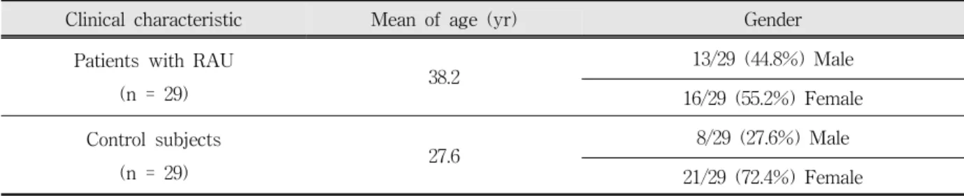
1. Comparison of the expression of HSV and VZV DNA
The result of the electrophoresis on 1.5% agarose gel showed that the 148bp size HSV DNA detected in 12 patients with RAU (41.4%), and 16 control subjects (55.2%), however, a significant difference between the two groups was not detected(P>
0.05)(Fig. 1 and Table 2).
1 2 3 4 5 6 7 8
400bp 148pb
Fig. 1. Result of PCR amplification products to HSV DNA analyzed on 1.5% agarose gel electrophoresis
Lane 1 : 100bp DNA Ladder Lane 2 : Negative control Lane 3 : Positive control Lane 4 : Saliva sample[Negative]
Lane 5, 6, 7, 8 : Saliva sample[Positive]
1 2 3 4 5 6 7 8 9 10 11 12
400bp 224pb
Fig. 2. Result of PCR amplification products to VZV DNA analyzed on 1.5% agarose gel electrophoresis
Lane 1 : 100bp DNA Ladder Lane 2 : Negative control Lane 3 : Positive control
Lane 4-12 : Saliva sample[Negative]
And the 224bp size VZV DNA band was not detected in both groups (Fig. 2 and Table 2).
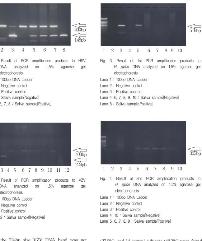
2. Comparison of the expression of H. pylori DNA
The result of the electrophoresis on 1.5% agarose gel of the first and second PCR products to H.
pylori DNA showed that 8 patients with RAU
1 2 3 4 5 6 7 8 9 10
516bp
Fig. 3. Result of 1st PCR amplification products to H. pylori DNA analyzed on 1.5% agarose gel electrophoresis
Lane 1 : 100bp DNA Ladder Lane 2 : Negative control Lane 3 : Positive control
Lane 4, 6, 7, 8, 9, 10 : Saliva sample[Negative]
Lane 5 : Saliva sample[Positive]
1 2 3 4 5 6 7 8 9 10
325bp
Fig. 4. Result of 2nd PCR amplification products to H. pylori DNA analyzed on 1.5% agarose gel electrophoresis
Lane 1 : 100bp DNA Ladder Lane 2 : Negative control Lane 3 : Positive control
Lane 4, 10 : Saliva sample[Negative]
Lane 5, 6, 7, 8, 9 : Saliva sample[Positive]
(27.6%), and 14 control subjects (48.3%) were found to be positive, however, a significant difference between the two groups was not detected (P>0.05) (Fig. 3 and 4, Table 2).
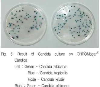
3. Comparison of the result of Candida culture
The result of the culture on CHROMagar
ⓇFig. 5. Result of Candida culture on CHROMagar
ⓇCandida
Left : Green - Candida albicans Blue - Candida tropicalis Rose - Candida krusei Right : Green - Candida albicans
Candida (CHROMagar, Paris, France) showed that 4 patients with RAU (13.8%) and 2 control subjects (6.9%) were found to be positive, however, a significant difference between the two groups was not detected (P>0.05) (Fig. 5, Table 2).
Ⅳ. DISCUSSION
The nutritional and physiological environment of the oral cavity is suitable to the proliferation of microorganism, and thus numerous microorganisms form the normal flora that presents constantly in the oral cavity. If the balance of normal flora is disrupted by a certain cause, it is possible that microorganism in the oral cavity causes a specific oral disease. Particularly, the saliva that its composition and action may be different among individuals controls the colonies of microorganism, affects the affinity of microorganism to the tooth surface or the mucosal surface, and it mediates a great effect as a pathophysiologic variable of microorganisms in the oral cavity. Hence, numerous molecular biological and microbiological studies on the saliva have been carried out recently.
As the study methods to find the etiology of RAU, immunological, microbiological and molecular biological studies have been performed, and primarily, the studies have been performed using
cells in the peripheral blood, the biopsy tissue of lesions, and the saliva. Among them, the saliva is known to be the ideal source for the extraction of genomic DNA, and the method using the saliva is inexpensive, rapid, reliable, and safe. In addition, in comparison with other methods that require to collect the blood or biopsy, its advantages are that it does not harm the subjects and it is not contagious.
In this study, therefore, to find the etiology of RAU, microbiological analysis in the saliva was performed by molecular biological methods.
Ulceration by viral infection that is frequently discovered by dentists is similar to the clinical pattern of RAU, and thus its diagnosis and treatment may be difficult
13). Particularly, in patients with AIDS or HIV positive individuals, herpetic lesions and RAU are oral symptoms developed generally, therefore, as the infectious etiologic factor of RAU, the studies on the relation with virus has been carried out actively
14, 15). In regard to the study on the HSV and VZV that can infect the oral cavity frequently, Pederson
16, 17)have reported that the elevation of lymphocyte toxicity was not the characteristic of autoimmune response, but it is immune response against infected cells by virus, and they claimed that the reactivation of HSV and VZV was the cause of the enhancement of lymphocyte toxicity, and Studd et al.
18)have reported that in patients with Béhçet’s syndrome or RAU, in comparison with healthy individuals, HSV-1 DNA fragment in circulating monocytes and anti-HSV-1 antibodies in serum were significantly higher.
However, Galliani et al.
19)have reported that food, the allergy to dental materials, and immune response to virus are not the etiologic factor of RAU, Lee et al.
20)extracted HSV DNA in the saliva of the patient with Béhçet’s syndrome but they could not detect a significance of the presence HSV DNA and ulcers in the oral cavity, Brice et al.
2)have reported that the detection of herpes virus DNA in RAU patients was not a direct cause of RAU but it appeared because of normal viral shedding.
In this study, HSV DNA was found to be positive
in saliva of 12 patients with RAU (41.4%) and 16 control subjects (55.2%), however, a significance was not detected, and VZV DNA was not detected in both groups. Thus, it is considered that these two viruses did not mediate a direct effect on the development of RAU.
The studies that characterized the association of RAU and bacteria are very limited, Riggio et al.
10)isolated Streptococcus oralis from RAU lesions, Donatsky et al.
21)suggested the association of RAU and α-hemolytic Streptococcus, coagulase-negative Staphylococcus and Neisseria through bacteria culture, and Narikawa et al.
22)have reported that in the oral lesion of the patients with Béhçet’s disease, Streptococcus sanguis was isolated at a high frequency. However, these bacteria are not accepted as the etiologic factor of RAU presently.
On the other hand, the association of RAU with H.
pylori that is known to be a major pathogen in the development of gastrointestinal ulcers has been suggested recently
6, 7, 23-26). This is due to that H.
pylori detected in the oral cavity and the stomach area same strain with identical nucleic acid sequence, and the histological pattern of the ulcer in the oral cavity is similar to gastric ulcer
27). In addition, although H. pylori resides in the human stomach primarily and plays an important role in gastric-duodenal diseases, H. pylori has the ability to colonize in the saliva and dental plaques, it thus may be associated with the development of oral diseases
28).
Reviewing the related studies on RAU and H.
pylori, Birek et al.
7)have reported that RAU and H.
pylori are associated since the positive reaction of H. pylori in RAU lesions was detected in 32 cases of 39 patients with RAU (71.8%), and Richter et al.
29)have reported that H. pylori infection may be related to the development of RAU since H. pylori was isolated from the saliva of patients with RAU.
However, Fritscher et al.
30)have reported that H.
pylori infection and the development of RAU were not related based on the result of the isolation of H.
pylori from dental plaques and RAU lesions in 105 children and adults and Iamaroon et al.
31)have
claimed based on the result of the analysis of the samples from the dorsum of tongue and ulcerous lesions in 20 cases of patients with RAU by nested PCR that H. pylori could not play a role in the pathogenesis of RAU.
In this study, H. pylori was detected in saliva of 8 patients with RAU (27.6%), and 14 control subjects (48.3%), however, it was not statistically significant, and furthermore, it was isolated more in control group. Therefore, H. pylori was speculated to be a normal flora in the oral cavity.
The major microorganism in development of Candidiasis in the oral cavity, Candida albicans is a normal flora, and it co-habits with numerous microorganisms in the oral cavity. In the yeast phase, the toxicity of Candia albicans is relatively low, hence, it becomes pathogens upon the change of environment suitable to the overgrowth and the infiltration to tissues (diabetes, long-term administration of antibiotics, the reduction of the salivary secretion, etc.). Atrophic candidiasis such as antibiotic candidiasis, denture stomatitis and angular cheilitis is ulcerative lesions, and their appearance may be similar to RAU. Although the report on the association of RAU and Candida was difficult to find, Safronova et al.
5)have reported that in quantitatively and qualitatively analyzing of microorganism in the oral cavity of children with RAU and healthy children, the expression of Candida between the two groups was not different, and Oudshoorn et al.
32)have reported that RAU was not associated with Candida infection.
In this study, Candida was cultured in saliva of 4 patients with RAU (13.8%) and 2 control subjects (6.9%), however, a significance was not detected.
Summarizing the above results, HSV, VZV, H.
pylori and Candida could not be a direct etiologic
factor in the development of RAU. Therefore, it is
considered that in future, in the cases of patients
with RAU inducing ulceration and the damage of
the mucosa, it is required to examine whether two
or more than one microorganisms function as a
triggering factor may serve as an abnormal local
immune response.
Ⅴ. CONCLUSIONS
To examine whether HSV, VZV, H. pylori and Candida that are known to be microorganisms causing ulcerative disease in oral cavity and have the relatively high contigiousness are detected in saliva of patients with RAU and related to the development with RAU, PCR and culture were performed on the saliva of 29 patients with RAU and 29 control subjects who visited the Department of Oral Medicine, Dental Hospital, Chosun University.
The results were obtained as follows;
1. HSV DNA was detected in 41.4% patients with RAU, and 55.2% control subjects, however, a significant difference between the two groups was not detected, (P>0.05), and VZV DNA was not detected in both groups.
2. H. pylori DNA was detected in 27.6% patients with RAU, and 48.3% control subjects, however, a significant difference between the two groups was not detected (P>0.05).
3. Candida was cultured in 13.8% patients with RAU, and 6.9% control subjects, however, a significant difference between the two groups was not detected (P>0.05).
This results suggest that HSV, VZV, H. pylori and Candida can not be regarded to play a direct role in the development of RAU. Thus it is considered that in future, on a larger sample, also, it has to be examined whether other microor- ganisms acts as a trigger factor of the development of RAU.
REFERENCES
1. Park SB, Kim BG, Bae JS. Detection of viruses and changes of protein of saliva in patients with recurrent aphthous ulcer. J Kor Acad Oral Med 1999;24(2):
125-135.
2. Brice SL, Cook D, Leahy H, Huff JC, Weston WL.
Examination of the oral mucosa and peripheral blood cells of patients with recurrent aphthous ulceration
for human herpesvirus DNA. Oral Surg Oral Med Oral Pathol Oral Radiol Endod 2000;89(2):193-198.
3. Embil JA, Stephens RG, Manuel FR. Prevalence of recurrent herpes labialis and aphthous ulcers among young adults on six continents. J Can Med Assoc 1975;113627-630.
4. Regezi JA, Sciubba J. Ulceration conditions. In Oral pathology Clinical-pathologic correlations edited by Regezi JA, Sciubba J, Philadelphia, 1999, WB Saunders, 46-53.
5. Safronova LA, Poltavs'kyi OM, Tsaruk'janova IH et al. Oral cavity microbial coenobia in healthy children and childre with chronic recurrent aphthous stomatitis. Mikrobiol Z 2003;65(6):49-58.
6. Shimoyama T, Horie N, Kato T, Kaneko T, Komiyama K. Helicobacter pylori in oral ulcerations.
J Oral Sci 2000;42(4):225-229.
7. Briek C, Grandhi R, McNeil K et al. Detection of Helicobacter pylori in oral aphthous ulcers. J Oral Pathol Med 2000;29(10):523-525.
8. Di Luca D, Mirandola P, Ravaioli T et al. Human herpesvirus 6 and 7 in salivary glands and shedding in saliva of healthy and human immunodeficiency virus positive individuals. J Med Virol 1995;45(4):462-468.
9. Ghodratnama F, Riggio MP, Wray D. Search for human herpesvirus 6, human cytomegalovirus and varicella zoster virus DNA in recurrent aphthous stomatitis tissue. J Oral Pathol Med 1997;26(4):
192-197.
10. Riggio MP, Lennon A, Ghodratnama F, Wray D. Lack of association between streptococcus oralis and recurrent aphthous stomatitis. J Oral Pathol Med 2000;29(1):26-32.
11. Hoover CI, Olson JA, Greenspan JS. Humoral responses and cross-reactivity to viridans strepto- cocci in recurrent aphthous ulceration. J Dent Res 1986;65(8):1101-1104.
12. Mandel ID. The function of saliva. J Dent Res 1987;66:623-627.
13. Sciubba JJ. Herpes simplex and aphthous ulceration : presentation, diagnosis and management an update.
Gen Dent 2003;51(6):510-516.
14. Gileva O, Sazhina H, Gileva E, Efimov A, Scully C.
Spectrum of oral manifestation of HIV/AIDS in the perm region (Russia) and identification of self-induced ulceronegotic lingual lesions. Med Oral 2004;9(3):212-215.
15. Ramos-Gomez F. Dental corsiderations for the
paediatric AIDS/HIV patient. Oral Dis 2002;2(8):
49-54.
16. Pedersen A. Are recurrent oral aphthous ulcers of viral etiology? Med Hypotheses 1991;36(3):206-210.
17. Pedersen A, Hornsleth A. Recurrent aphthous ulceration: a possible clinical manifestation of reactivation of varicella zoster or cytomegalovirus infection. J Oral Pathol Med 1993;22(2):64-68.
18. Studd M, McCance DJ, Lehner T. Detection of HSV-1 DNA in patients with Béhçet’s syndrome and in patients with recurrent oral ulcers by the polymerase chain reaction. J Med Microbiol 1991;34:39-43.
19. Galliani EA, Infantolino D, Tarantello M, Cipriani R, De Lazzari F. Recurrent aphthous stomatitis: which role for viruses, food, and dental materials? Ann Ital Med Int 1998;13(3):152-156.
20. Lee S, Bang D, Cho YH, Lee ES, Sohn S. Polymerase chain reaction reveals herpes simplex virus DNA in saliva of patients with Béhçet’s disease. Arch Dermatol Res 1996;288:179-183.
21. Donatsky O, Justensen T, Lind K, Vestergaard BF.
Microorganism in recurrent aphthous ulcerations.
Scand J Dent Med 1977;85(6):426-433.
22. Narikawa S, Suzuki Y, Takahashi M et al.
Streptococcus oralis previously identified as uncommon 'streptococcus sanguis' in Béhçet’s disease. Arch Oral Biol 1995;40(8):685-690.
23. Vidoria JM, Kalopothakis E, Silva JdeF, Gomez RS.
Helicobacter pylori DNA in recurrent aphthous stomatitis. J Oral Pathol Med 2003;32(4):219-223.
24. Goodwin CS, Mendall MM, Northfield TC.
Helicobacter pylori infection. Lancet 1999;349:
265-269.
국문요약
재발성 아프타성 궤양 환자의 타액에서 Herpes Simplex Virus, Varicella Zoster Virus, Helicobacter pylori 그리고 Candida 검출