Degradation and Conversion of Blood Group Antigens in Saliva
Sang-Wook Lim, D.D.S.
1, Hee-Kyung Park, D.D.S.,M.S.D.,Ph.D.
2, Seung-Eun Jung, B.S.,M.S.
1, Hong-Seop Kho, D.D.S.,M.S.D.,Ph.D.
1, Young-Ku Kim, D.D.S.,M.S.D.,Ph.D.
1Department of Oral Medicine & Oral Diagnosis, College of Dentistry, Seoul National University1 Section of Forensic Dentistry, Department of Forensic Medicine, National Institute of Scientific Investigation2
Mucin glycoproteins are the primary carriers of the oligosaccharide moieties that constitute the blood group substances in human saliva. The aim of this study was to determine whether or not the conversion of either the A or B blood group antigens to the H antigen can occur during the degradation process of stored saliva samples. Forty subjects (20 subjects in each A and B blood group) identified as secretors were enrolled in this study. Fresh whole saliva samples and their clarified supernatants were stored at room temperature for 1 week. The conversion of the blood group antigens was detected by SDS-PAGE and immunoblotting. Among the subjects showing the conversion in whole saliva, glandular saliva samples were obtained from 8 subjects (4 subjects in each A and B blood group). Submandibular-sublingual saliva (SMSL) and a mixture of SMSL and parotid saliva (PS) were stored at room temperature for 1 week. The conversion of the blood group antigens was detected by the same method.
The obtained results were as follows:
1. In the clarified samples of whole saliva, the A antigen was detected as being either intact (5%) or degraded molecules (95%) after the 1 week period. Conversion of the A antigen to the H antigen was detected in 5 subjects (25%). In the unclarified samples, the A antigen was either detected as degraded molecules (90%) or was not detected (10%).
Conversion of the antigen had occurred in 4 subjects (20%).
2. In the clarified samples of whole saliva, the B antigen was detected as intact (20%) or as degraded molecules (65%) or was not detected (15%) after the 1 week period. Conversion of the B antigen to the H antigen was detected in 7 subjects (35%). In the unclarified samples, the B antigen was detected as intact (5%) or as degraded molecules (65%), or was not detected (30%). Conversion of the antigen was observed in 2 subjects (10%).
3. In the glandular saliva samples, only one of the four subjects displayed an antigenic conversion from the A to H antigen or from the B to H antigen. The conversion had occurred in both the SMSL samples and the SMSL and PS mixture. No degradation of the antigens was detected in the other three samples of the A or B blood groups, nor was there any conversion.
The results demonstrated that conversion of the blood group antigens could occur in saliva, and suggested that the enzymes responsible for the conversion are present in saliva. Further studies on the origin and activity of the specific glycosidases in saliva as well as quantitative measurements of the antigenic conversion will be needed.
Key words : Saliva, Blood group antigens, Conversion
Corresponding author: Prof. Young-Ku Kim
Department of Oral Medicine and Oral Diagnosis College of Dentistry, Seoul National University Yunkeun-Dong 28, Chongro-Ku, Seoul 110-749, Korea E-mail: [email protected]
received: 2004-11-06
revised: 2004-12-20 / accepted: 2005-01-12
Ⅰ. INTRODUCTION
About 80% of individuals, so-called secretors, have the ability to secrete blood group substances in their secretions, such as saliva, tears and gastric secretions
1). In the saliva, the carbohydrate moieties of the salivary glycoproteins carry the blood group antigens, which are expressed in the salivary glands and secreted in the saliva
1-3). Although there are genetic differences in the glycosylation and expression pattern of the blood group antigens between ethnic groups
4), there is no doubt that the high-molecular-weight salivary mucin (MG1) is the primary carrier of the oligosaccharide moieties that constitute the ABH, Lea and Leb blood group substances in human saliva
2,5,6). The carbohydrate moieties of the salivary glycoproteins play important roles in the interaction between the salivary glycoproteins and oral microorganisms
7). Therefore, it is possible that the expression of the ABH blood group antigens in the saliva might alter the specific interactions between the microorganisms and their salivary glycoprotein receptors, influence the ability of the salivary glycoproteins to mediate the bacterial adherence/clearance, and then intervene in the development and prevention of oral infectious diseases
2,8,9).
It is well known that some exoglycosidases modify the A or B blood group antigens. The hydrolysis of the terminal N-acetyl-α-D-galacto- samine by N-acetyl-α-D-galactosaminidase (EC 3.2.1.49) converts the A antigen to the H antigen, and similarly, hydrolysis of the terminal α -D-galactose residue by α-D-galactosidase (EC 3.2.1.22) converts the B antigen to the H antigen
10,11). Human feces contain enzymes produced by the enteric bacteria that degrade the blood group antigens of the gut mucin glycoproteins
12,13). The bacterial, plant and recombinant glycosidases degrade the erythrocytes surface glycoconjugates without significantly affecting the viability of the cells
14-16). The enzymatically converted group O RBCs from the group B RBCs can be safely transfused to
otherwise incompatible blood group A and O patients without fatal transfusion reactions
17). A previous study showed that some changes occur in the ABH blood group antigens in the saliva samples as a result of the progressive degradation of the salivary components carrying the blood group antigens by the enzymes that originate from the bacteria and serum
5). However, there is no information as to whether the conversion of the blood type can occur in blood group antigens in the saliva. The aim of this study was to investigate whether or not conversion of the blood group antigens in the saliva can occur during the degradation process.
Ⅱ. MATERIALS AND METHODS 1. Subjects
Forty healthy subjects, dental students and staffs in the College of Dentistry, Seoul National University were enrolled in the study. There were 20 A blood type and 20 B blood type subjects (10 men and 10 women in each group). They were found to be blood group secretors from previous experiments
6). Their ages ranged from 22 to 37 years (mean age, 24.7 ± 3.8 years). The subjects had no history of serious illness and had not taken antibiotics in the 3 months before the study.
2. Collection of unstimulated whole saliva
Unstimulated whole saliva was collected into a
chilled sterile centrifuge tube for approximately 10
min by a standard, reproducible method described
elsewhere
5). Half of the sample (about 2 ml) was
clarified by centrifugation at 3,500 xg for 10 min to
remove the cellular debris and the other half was
not clarified. The unclarified saliva samples and the
clarified supernatants were then used immediately
for the experiments. The remainder of the samples
were stored at room temperature (about 25℃), and
after 1 week, the same experiments were performed
to determine if there were any changes in the ABH
blood group antigens in the saliva samples. The clarified whole saliva sample of the same subject stored at -20℃ for 1 week was used as a control.
3. Electrophoresis and immunoblotting
Saliva samples were subjected to 10% sodium dodecyl sulfate-polyacrylamide gel electrophoresis (SDS-PAGE) and then immunoblotted to Immobilon -P membranes (polyvinylidene difluoride [PVDF]
membranes; Millipore Corp., Bedford, MA) as previously described
5). The membranes were blocked with Tris-buffered saline (TBS: 10 mM of Tris-HCl, 154 mM of NaCl, pH 7.5) containing 3%
bovine serum albumin (BSA) for more than 1 h. The membranes were then incubated with a 1:100 dilution of the monoclonal mouse anti-human blood group antigens A, B and H (DAKO Corp., Carpinteria, CA) as the primary antibodies for 3 h at room temperature. After being washed 3 times with TBS containing 0.01% BSA for 5 min each, the blots were incubated with a 1:1,000 dilution of horseradish peroxidase conjugate rabbit anti-mouse IgG (DAKO Corp.) as the secondary antibody for 2 h. The same washing cycle with TBS containing 0.01% BSA was then repeated, and the blots were visualized with a solution of 6 mg of DAB (diaminobenzidine tetrahydrochloride, DAKO Corp.) in 10 ml of 50 mM Tris-HCl buffer (pH 7.6) by adding 100 μl of 3% H
2O
2. The saliva samples of blood type A subjects were analyzed with anti-A and H antibodies and those of blood type B were analyzed with anti-B and H antibodies.
4. Examination of blood group antigen conversion in glandular saliva
Among the subjects displaying the conversion of the blood group antigens in the whole saliva samples, 4 subjects in each of the A and B blood groups participated in this experiment. Parotid saliva (PS) was collected using a modified Lashley cup placed directly over the Stensen's duct orifice
18). Submandibular-sublingual saliva (SMSL)
was collected using a custom-made mouthpiece of rubber base impression material, as described by Block and Brottman
19). Salivary flow was stimulated by applying 2% citric acid onto the lateral border of the tongue every 30 sec during the collection period (generally 5 min). Stimulated glandular saliva collected during the first 2 min was discarded. Each saliva sample was collected into a chilled sterile tube and centrifuged at 3,500 xg for 10 min to remove any cellular debris. The resulting supernatants from the SMSL and the mixture of supernatants from the PS and SMSL (1:1) were immediately subjected to electrophoresis and immunoblotting. The same samples were stored at room temperature for 1 week, after which the same experiments were then performed to determine if there were any changes in the ABH blood group antigens in the glandular saliva samples. The clarified SMSL sample of the same subject stored at -20℃ for 1 week was used as a control sample.
Ⅲ. RESULTS
1. Degradation and conversion of the AB blood group antigens in the whole saliva samples
In the clarified samples, the A antigen was
detected as being either intact (5%) or degraded
molecules (95%) after the 1 week period. Conversion
of the A antigen to the H antigen was detected in
5 subjects (25%). In the unclarified samples, the A
antigen was either detected as degraded molecules
(90%) or was not detected (10%). Conversion of the
antigen had occurred in 4 subjects (20%) (Table 1,
Fig. 1). The B antigen was detected as intact (20%)
or as degraded molecules (65%) or was not detected
(15%) in the clarified samples after the 1 week
period. Conversion of the B antigen to the H antigen
was detected in 7 subjects (35%). In the unclarified
samples, the B antigen was detected as intact (5%)
or as degraded molecules (65%), or was not detected
(30%). Conversion of the antigen was observed in
2 subjects (10%) (Table 2, Fig. 2).
Fig. 1. SDS-PAGE(10%)/Western blotting of the whole saliva samples from the subjects of the blood type A secretor. Immunoblotting of the electrophoretically separated saliva samples with monoclonal mouse antibodies to the A (left panel) and H (right panel) antigens as the primary antibodies. Lane 1, clarified unstimulated whole saliva (UWS) of a subject (SHL) stored at room temperature (RT) for 1 week; Lane 2, unclarified UWS of (SHL) stored at RT for 1 week; Lane 3, clarified UWS of another subject (JMC) stored at RT for 1 week; Lane 4, unclarified UWS of (JMC) stored at RT for 1 week; Lane 5, clarified UWS of (SHL) preserved at -20℃ for 1 week; Lane 6, clarified UWS of (JMC) preserved at -20℃ for 1 week.
In the left panel, the A antigen was not detected (lane 2) or was detected as degraded molecules (lane 1, 3, and 4) compared to the samples preserved at -20℃ (lane 5 and 6). In the right panel, the H antigen was detected with increased intensity in the clarified sample (lane 1) of (SHL) and the clarified and unclarified samples (lane 3 and 4) of (JMC) as compared to the samples stored at -20℃ (lane 5 and 6). The arrow indicates the interface between the stacking and separating gel.
Fig. 2. SDS-PAGE(10%)/Western blotting of the whole saliva samples from the subjects of the blood type B secretor. Immuno- blotting of the electrophoretically separated saliva samples with mono- clonal mouse antibodies to B (left panel) and H (right panel) antigens as the primary antibodies. Lane 1, clarified unstimulated whole saliva (UWS) of a subject (SKJ) stored at room temperature (RT) for 1 week; Lane 2, unclarified UWS of (SKJ) stored at RT for 1 week; Lane 3, clarified UWS of another subject (KWL) stored at RT for 1 week; Lane 4, unclarified UWS of (KWL) stored at RT for 1 week; Lane 5, clarified UWS of (SKJ) preserved at -20
℃ for 1 week; Lane 6, clarified UWS of (KWL) preserved at -20℃ for 1 week. In the left panel, the B antigen was detected as an intact molecule (lane 1) or was detected as degraded molecules (lane 2, 3, and 4) compared to the samples preserved at -20℃ (lane 5 and 6). In the right panel, the H antigen was detected with increased intensity in the clarified sample (lane 1) in (SKJ) and the clarified and unclarified samples (lane 3 and 4) of (KWL) compared to the samples stored at -20℃ (lane 5 and 6). The arrow indicates the interface between the stacking and separating gel.
Type of unstimulated whole saliva Results of immunoblotting Number of subjects
Anti-A + / Anti-H + 0
Anti-A + / Anti-H - 1
Anti-A (+) / Anti-H + 5
Clarified Anti-A (+) / Anti-H - 14
Anti-A - / Anti-H + 0
Anti-A - / Anti-H - 0
Anti-A + / Anti-H + 0
Anti-A + / Anti-H - 0
Unclarified Anti-A (+) / Anti-H + 4
Anti-A (+) / Anti-H - 14
Anti-A - / Anti-H + 0
Anti-A - / Anti-H - 2
Anti-A +: positive response as an intact molecule by monoclonal mouse anti-human blood group antigen A as the primary antibody; Anti-A (+): positive response as the degraded state; Anti-A -: negative response; Anti-H +:
positive response by monoclonal mouse anti-human blood group antigen H as primary antibody; Anti-H -: negative response
Table 1. Summary of the immunoblotting experiments using the whole saliva samples stored at room temperature for 1 week in the blood type A subjects
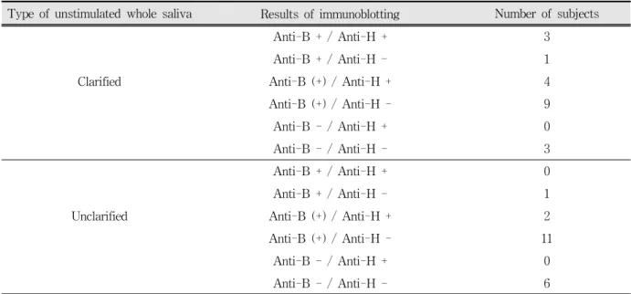
Type of unstimulated whole saliva Results of immunoblotting Number of subjects
Anti-B + / Anti-H + 3
Anti-B + / Anti-H - 1
Clarified Anti-B (+) / Anti-H + 4
Anti-B (+) / Anti-H - 9
Anti-B - / Anti-H + 0
Anti-B - / Anti-H - 3
Anti-B + / Anti-H + 0
Anti-B + / Anti-H - 1
Unclarified Anti-B (+) / Anti-H + 2
Anti-B (+) / Anti-H - 11
Anti-B - / Anti-H + 0
Anti-B - / Anti-H - 6
Anti-B +: positive response as an intact molecule by monoclonal mouse anti-human blood group antigen B as the primary antibody; Anti-B (+): positive response as the degraded state; Anti-B -: negative response; Anti-H +:
positive response by monoclonal mouse anti-human blood group antigen H as primary antibody; Anti-H -:
negative response
Table 2. Summary of the immunoblotting experiments using the whole saliva samples stored at room temperature for 1 week in the blood type B subjects
Fig. 3. SDS-PAGE(10%)/Western blotting of the glandular saliva samples from subjects of the blood type A and B secretors.
Immunoblotting of the electrophoretically separated saliva samples with mono- clonal mouse antibody to the H antigen as the primary antibody. Lane 1, clarified submandibular-sublingual saliva (SMSL) of a subject (HSK, blood group A secretor) stored at room temperature (RT) for 1 week; Lane 2, mixture (1:1) of clarified SMSL and parotid saliva (PS) of (HSK) stored at RT for 1 week; Lane 3, clarified SMSL of (HSK) preserved at -20℃ for 1 week; Lane 4, clarified SMSL of another subject (SHC, blood group B secretor) stored at RT for 1 week; Lane 5, mixture (1:1) of clarified SMSL and PS of (SHC) stored at RT for 1 week;
Lane 6, clarified SMSL of (SHC) preserved at -20℃ for 1 week. The greater affinity of the antibody to the H antigen was detected in the SMSL as well as in the SMSL and PS mixture stored at RT compared to the SMSL preserved at -20℃. The arrow indicates the interface between the stacking and separating gel.
2. Degradation and conversion of the AB blood group antigens in the glandular saliva samples
Of the four subjects, only one displayed an antigenic conversion from the A to H antigen or from the B to H antigen. The conversion had occurred in both the SMSL samples and the SMSL and PS mixture (Fig. 3). No degradation of the antigens was detected in the other three samples of the A or B blood groups, nor was there any conversion.
Ⅳ. DISCUSSION
The conversion of the A or B blood group antigens to the H antigen occurred in both the clarified and unclarified whole saliva samples. The fact that the conversion occurred in the clarified saliva suggested that the enzymes are secreted and presented in the saliva. Although there is no information on the origin of the enzymes responsible for the conversion of the antigens in the saliva, oral bacteria or serum components such as leukocytes are the most likely source. Blood group-degrading enzymes in human feces were shown to originate from enteric bacteria. A subpopulation of the human fecal microbiota is unique in specializing in the hydrolysis of the complex carbohydrates from the mucin glycopro- teins and glycosphingolipids. Detailed information on the glycosidase specificities produced by the various strains of enteric bacteria has been reported
20-22).
Even though the conversion of the blood group
antigens occurred in the clarified glandular saliva,
the possibility of secreting blood group-degrading
enzymes from salivary glands appears to be low
because the conversion occurred in only one subject
from each A or B blood group, and the degradation
did not occur in the other subjects. Contamination
from bacterial or host products during the collection
of the PS or SMSL could have happened. The design
of collection device covering a wide area of the
mouth floor may allow some contamination during the collection, especially in the case of SMSL.
A clarified saliva sample from the same subject preserved at -20℃ was used as the control, because the blood group antigens have been detected as intact molecules in saliva samples preserved at -20℃ for at least 6 months
5). The 7 day-stored samples were used in the experiments because preliminary experiments showed the conversion of the antigen in the whole saliva samples occurring between 3 to 10 days at room temperature. If samples stored during various periods were used instead, the conversion might be detected in a larger number of samples showing degradation.
The conversion of the blood group antigens in the saliva has meaningful significance in biological aspect. The mucin glycoproteins are recognized as components of the tooth and mucosal pellicles
23,24)and their carbohydrate moieties can function as receptors for pathogenic oral microorganisms
25). Therefore, the conversion of the blood group antigens might affect the pathogenesis of oral diseases.
V. CONCLUSIONS
The aim of this study was to determine whether or not the conversion of either the A or B blood group antigens to the H antigen can occur during the degradation process of stored saliva samples.
Forty subjects (20 subjects in each A and B blood group) identified as secretors were included in this study. Fresh whole saliva samples and their clarified supernatants were stored at room temperature for 1 week. The conversion of the blood group antigens was detected by SDS-PAGE and immunoblotting. Among the subjects showing the conversion in whole saliva, glandular saliva samples were obtained from 8 subjects (4 subjects in each A and B blood group).
Submandibular-sublingual saliva (SMSL) and a mixture of SMSL and parotid saliva (PS) were stored at room temperature for 1 week. The
conversion of the blood group antigens was detected by the same method.
The obtained results were as follows:
1. In the clarified samples of whole saliva, the A antigen was detected as being either intact (5%) or degraded molecules (95%) after the 1 week period. Conversion of the A antigen to the H antigen was detected in 5 subjects (25%). In the unclarified samples, the A antigen was either detected as degraded molecules (90%) or was not detected (10%). Conversion of the antigen had occurred in 4 subjects (20%).
2. In the clarified samples of whole saliva, the B antigen was detected as intact (20%) or as degraded molecules (65%) or was not detected (15%) after the 1 week period. Conversion of the B antigen to the H antigen was detected in 7 subjects (35%). In the unclarified samples, the B antigen was detected as intact (5%) or as degraded molecules (65%), or was not detected (30%). Conversion of the antigen was observed in 2 subjects (10%).
3. In the glandular saliva samples, only one of the four subjects displayed an antigenic conversion from the A to H antigen or from the B to H antigen. The conversion had occurred in both the SMSL samples and the SMSL and PS mixture. No degradation of the antigens was detected in the other three samples of the A or B blood groups, nor was there any conversion.
The results demonstrated that conversion of the
blood group antigens could occur in saliva, and
suggested that the enzymes responsible for the
conversion are present in saliva. However, the
methods used in these experiments provide only
qualitative and limited information. Future
experiments on the detection of the specific
glycosidase activities in the saliva as well as
quantitative measurements of the antigenic
conversion will be needed to properly explain this
phenomenon.
REFERENCES
1. Hamper K, Caselitz J, Seifert G , Seitz R, Poschmann A. The occurrence of blood group substa nces (A, B, H, Le-a, Le-b) in salivary glands and salivary gland tumors. An immunohistochemical investigation. J Oral Pathol 1986;15:334-338.
2. Prakobphol A, Leffler H, Fisher S. The high-molecular-weight human mucin is the primary salivary carrier of ABH, Lea, and Leb blood group antigens. Crit Rev Oral Biol Med 1993;4:325-333.
3. Greenwell P. Blood group antigens: molecules seeking a function? Glycoconj J 1997;14:159-173.
4. Tanegashima A, Nishi K, Fukunaga T, Rand S, Brinkmann B. Ethnic differences in the expression of blood group antigens in the salivary gland secretory cells from German and Japanese non-secretor individuals. Glycoconj J 1996;13:537-545.
5. Kim W, Kim YK, Chung SC, Lee SW, Kho HS.
Detection of ABH blood group antigens in the saliva of Koreans and their stability according to storage of saliva samples. Forensic Sci Int 2002;129:58-63.
6. Shin ES, Chung SC, Kim YK, Lee SW, Kho HS. The Relationship between Oral Candida carriage and the secretor status of blood group antigens in saliva. Oral Surg Oral Med Oral Pathol Oral Radiol. Endod 2003;96:48-53.
7. Levine MJ, Reddy MS, Tabak LA, Loomis RE, Bergey EJ, Jones PC, Cohen RE, Stinson MW, Al-Hashimi I. Structural aspects of salivary glycoproteins. J Dent Res 1987;66:436-441.
8. Burford-Mason AP, Weber JCP, Willoughby JMT.
Oral carriage of Candida albicans, ABO blood group and secretor status in healthy subjects. J Med Vet Mycol 1988;26:49-56.
9. Lamey P-J, Darwazeh AMG, Muirhead J, Rennie JS, Samaranayake LP, MacFarlane TW. Chronic hyperplastic candidosis and secretor status. J Oral Pathol Med 1991;20:64-67.
10. Levy G, Aminoff D. Purification and properties of α -N-acetylgalactosaminidase from Clostridium perfringens. J Biol Chem 1980;225:11737-11742.
11. Yatziv S, Flowers H. Action of α-D-galactosidase on glycoprotein from human B-erythrocytes. Biochem Biophys Res Commun 1971;45:514-518.
12. Hoskins LC. Bacterial degradation of gastrointestinal mucins. II. Bacterial origin of fecal ABH(O) blood group antigen-destroying enzymes. Gastroenterology 1968;54:218-224.
13. Hoskins LC, Zamcheck N. Bacterial degradation of gastrointestinal mucins. I. Comparison of mucin constituents in the stools of germ-free and conventional rats. Gastroenterology 1968;54:210-217.
14. Falk P, Hoskins LC, Lindstedt R, Svanborg C, Larson G. Deantigenation of human erythrocytes by bacterial glycosidases-evidence for the noninvolvement of medium- sized glycosphingolipids in the Dolichos biflorus lectin hemagglutination. Arch Biochem Biophys 1991;290:312-319.
15. Hobbs L, Mitra M, Phillips R, Haibach H, Smith D.
Deantigenation of human type B erythrocytes with Glycine max alpha-D-galactosidase. Biomed Pharmacother 1995;49:244-250.
16. Vosnidou NC, Johnson SA, Mitra MM, Wells DC, Li CQ, Evans ML, Harmata MA, Walker JC, Smith DS.
Seroconversion of type B to O erythrocytes using recombinant Glycine max α-D-galactosidase.
Biochem Mol Biol Int 1998;46:175-186.
17. Kruskall MS, AuBuchon JP, Anthony KY, Herschel L, Pickard C, Biehl R, Horowitz M, Brambilla DJ, Popovsky MA. Transfusion to blood group A and O patients of group B RBCs that have been enzymatically converted to group O. Transfusion 2000;40:1290-1298.
18. Lashley KS. Reflex secretion of the human parotid gland. J Exp Psychol 1916;1:461-465.
19. Block PL, Brottman S. A method of submaxillary saliva collection without cannulization. N Y State Dent J 1962;28:116-118.
20. Hoskins LC, Agustines M, McKee WB, Boulding ET, Kriaris M, Niedermeyer G. Mucin degradation in human colon ecosystems. Isolation and properties of fecal strains that degrade ABH blood group antigens and oligosaccharides from mucin glycoproteins. J Clin Invest 1985;75:944-953.
21. Larson G, Falk P, Hoskins LC. Degradation of human intestinal glycosphingolipids by extracellular glycosi- dases from mucin-degrading bacteria of the human fecal flora. J Biol Chem 1988;263:10790-10798.
22. Falk P, Hoskins LC, Larson G. Bacteria of the human intestinal microbiota produce glycosidases specific for lacto-series glycosphingolipids. J Biochem (Tokyo) 1990;108:466-474.
23. Al-Hashimi I, Levine MJ. Characterization of in vivo salivary-derived enamel pellicle. Arch Oral Biol 1989;34:289-295.
24. Bradway SD, Bergey EJ, Jones PC, Levine MJ. Oral mucosal pellicle: Adsorption and transpeptidation of
salivary components to buccal epithelial cells.
Biochem J 1989;261:887-896.
25. Schenkels LCPM, Gururaja TL, Levine MJ. Salivary mucins: their role in oral mucosal barrier function and
국문요약
혈액형 항원의 분해와 변환에 관한 연구
서울대학교 치과대학 구강내과진단학교실1, 국립과학수사연구소 법의학과 법치의학실2
임상욱
1․박희경
2․정승은
1․고홍섭
1․김영구
1Mucin 당단백질은 인체 타액 성분 중 혈액형 항원을 표지하는 주요 성분으로 알려져 있다. 본 연구의 목적은 보관중인 타액 검체에서 일어나는 분해과정 중에 A형 혹은 B형 혈액형 항원이 H형 항원으로 변환이 일어나는 지를 조사하는데 있다.
A형과 B형 각각 20명씩 총 40명의 분비자로부터 채취한 전타액과 상층액을 실온에서 1주일간 보관한 다음, SDS-PAGE와 immunoblotting법을 이용하여 혈액형 항원의 변환 여부를 조사하였다. 전타액에서 변환을 보인 연구대상 중에서 A형과 B형 각각 4명씩 총 8명에서 이하선 타액과 악하선-설하선 타액을 채취하였고, 악하선-설하선 타액과 악하선-설하선 타액 및 이하 선 타액의 혼합액을 실온에서 1주일간 보관하였으며, 같은 방법으로 혈액형 항원의 변환 여부를 조사하여 다음과 같은 결론 을 얻었다.
1. 전타액 상층액을 1주일 보관한 검체의 경우, A형 항원이 분해되지 않은 경우가 5%, 분해된 경우가 95%이었고, A형 항원의 H형 항원으로의 변환이 5명(25%)의 연구대상에서 관찰되었다. 원심분리하지 않은 전타액 검체에서는 90%의 경우에서 A형 항원이 분해된 형태로 관찰되었고 10%에서는 관찰되지 않았으며, 혈액형 항원의 변환은 4명(20%)의 연구대상에서 관찰되었다.
2. 전타액 상층액을 1주일 보관한 검체의 경우, B형 항원이 분해되지 않은 경우가 20%, 분해된 경우가 65%이었고 15%에서는 관찰되지 않았으며, B형 항원의 H형 항원으로의 변환이 7명(35%)의 연구대상에서 관찰되었다. 원심분리하지 않은 전타액 검체에서는 5%의 경우 B형 항원이 분해되지 않은 형태로 관찰되었고, 65%의 경우에서 분해된 형태로 관찰되었으며 30%
에서는 관찰되지 않았다. 이 경우 혈액형 항원의 변환은 2명(10%)의 연구대상에서 관찰되었다.
3. 개별 타액선 타액검체의 경우, 4명중 각각 1명에서 A형 항원 혹은 B형 항원에서 H형 항원으로의 변환이 관찰되었다. 이 경우 혈액형 항원의 변환은 악하선-설하선 타액과 악하선-설하선 타액 및 이하선 타액의 혼합액 모두에서 일어났다. 그외 3명의 검체에서는 항원의 분해나 변환이 관찰되지 않았다.
이상의 결과는 타액에서 혈액형 항원의 변환이 일어남을 보여주는 것으로 타액에 이러한 변환을 가능하게 하는 효소가 존재함을 암시해 주었다. 그러므로 향후 혈액형 항원 변환의 정량적 연구와 함께 타액에 존재 가능성이 있는 특정 당분해효소 의 근원이나 활성에 대한 추가 연구가 필요하다.
주제어 : 타액, 혈액형 항원, 변환
drug delivery, in Rathbone MJ (Ed). Oral mucosal drug delivery: New York 1996, Marcel Dekker Inc., 191-220.