http://dx.doi.org/10.5090/kjtcs.2012.45.3.161 ISSN: 2233-601X (Print) ISSN: 2093-6516 (Online)
Departments of 1Thoracic and Cardiovascular Surgery and 2Surgery, Seoul National University Hospital, Seoul National University College of Medicine
†This article was presented at the 42nd Autumn Academic Meeting of the Korean Society for Thoracic and Cardiovascular Surgery.
Received: September 14, 2011, Revised: October 25, 2011, Accepted: November 11, 2011
Corresponding author: Ki-Bong Kim, Department of Thoracic and Cardiovascular Surgery, Seoul National University Hospital, Seoul National University College of Medicine, 101 Daehak-ro, Jongno-gu, Seoul 110-744, Korea
(Tel) 82-2-2072-3482 (Fax) 82-2-747-5245 (E-mail) [email protected]
C The Korean Society for Thoracic and Cardiovascular Surgery. 2012. All right reserved.
CC This is an open access article distributed under the terms of the Creative Commons Attribution Non-Commercial License (http://creative- commons.org/licenses/by-nc/3.0) which permits unrestricted non-commercial use, distribution, and reproduction in any medium, provided the original work is properly cited.
Subxiphoid Incisional Hernia Development after Coronary Artery Bypass Grafting
Hye-seon Kim, M.D.1, Ki-Bong Kim, M.D., Ph.D.1, Ho Young Hwang, M.D., Ph.D.1, Hyung Woo Chang, M.D.1, Kyu-Joo Park, M.D.2
Background: Median sternotomy can weaken the upper abdominal wall and result in subxiphoid incisional hernia.
We evaluated risk factors associated with the development of subxiphoid incisional hernias after coronary artery by- pass grafting (CABG). Materials and Methods: Of 1,656 isolated CABGs performed between January 2001 and July 2010, 1,599 patients who were completely followed up were analyzed. The mean follow-up duration was 49.5±34.3 months. Subxiphoid incisional hernia requiring surgical repair developed in 13 patients (0.8%). The hernia was diagnosed 16.3±10.3 months postoperatively, and hernia repair was performed 25.0±26.1 months after the ini- tial operation. Risk factors associated with the development of subxiphoid incisional hernia were analyzed with the Cox proportional hazard model. Results: Five-year freedom from the hernia was 99.0%. Univariate analysis re- vealed that female sex (p=0.019), height (p=0.019), body surface area (p=0.046), redo operation (p=0.012), off-pump CABG (p=0.049), a postoperative wound problem (p=0.041), postoperative bleeding (p=0.046), and low car- diac output syndrome (p<0.001) were risk factors for the development of the hernia. Multivariable analysis showed that female sex (p=0.01) and low cardiac output syndrome (p<0.001) were associated with subxiphoid hernia formation. Conclusion: Female sex and postoperative low cardiac output syndrome were risk factors of subxiphoid hernia. Therefore, special attention is needed for patients with high-risk factors.
Key words: 1. Hernia
2. Coronary artery bypass 3. Sternotomy
INTRODUCTION
A median sternotomy that extends toward the epigastric area can weaken the upper abdominal wall and result in the development of subxiphoid incisional hernia (Fig. 1). Report- ed incidence of subxiphoid incisional hernia has ranged from 1% to 4.2% [1,2]. The subxiphoid hernia is known for its re- pair complexities and high recurrence rate because the sub-
xiphoid area is a complex structure consisting of boney struc- tures, the rectus abdominis muscles, linea alba, and the dia- phragm [1-5]. Few studies have reported on subxiphoid inci- sional hernia while numerous studies that focused on abdomi- nal incisional hernias have been reported [6-8]. We evaluated the risk factors associated with the development of sub- xiphoid incisional hernia in patients who underwent coronary artery bypass grafting (CABG).
Table 1. Patient characteristics
Variables All patients (n=1,599)
Study patients (n=13) Sex (male:female)
Age (yr)
Mean BMI (kg/m2) Smoking
Hypertension DM Dyslipidemia Chronic renal failure COPD
LV dysfunction (LVEF <35%)
1,136:463 63.6±9.2 24.8±2.9 759 (47.5) 1,086 (67.9) 745 (46.6) 397 (24.8) 141 (8.8) 12 (0.8) 152 (9.5)
5:8 65±9 25.0±2.5 7 (53.9) 6 (46.2) 5 (38.5) 3 (23.1) 1 (7.7)
0 2 (15.4) Values are presented as mean±standard deviation or number (%).
BMI, body mass index; DM, diabetes mellitus; COPD, chronic obstructive pulmonary disease; LV, left ventricle; LVEF, left ventricular ejection fraction.
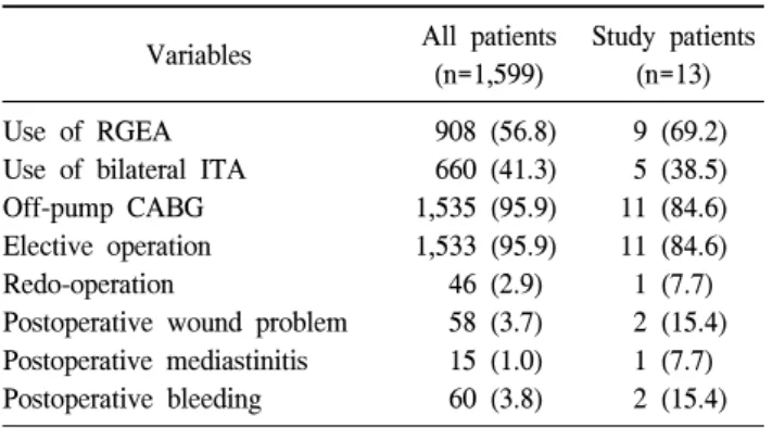
Table 2. Operative and postoperative data
Variables All patients
(n=1,599)
Study patients (n=13) Use of RGEA
Use of bilateral ITA Off-pump CABG Elective operation Redo-operation
Postoperative wound problem Postoperative mediastinitis Postoperative bleeding
908 (56.8) 660 (41.3) 1,535 (95.9) 1,533 (95.9) 46 (2.9) 58 (3.7) 15 (1.0) 60 (3.8)
9 (69.2) 5 (38.5) 11 (84.6) 11 (84.6) 1 (7.7) 2 (15.4)
1 (7.7) 2 (15.4) Values are presented as number (%).
RGEA, right gastroepiploic artery; ITA, internal thoracic artery;
CABG, coronary artery bypass graft surgery.
Fig. 1. Subxiphoid incisional hernia on computed tomography (CT) ima- ge. There is a fascial defect that re- sulted in the hernia at the subxiph- oid area on the CT scan (arrow).
MATERIALS AND METHODS 1) Patient characteristics
Between January 2000 and July 2010, 1,656 patients un- derwent isolated CABG in our institution. Among them, 1,599 patients (male:female=1,136:463) who were followed up completely were enrolled in the present study. The mean age was 63.6±9.2 years and mean body mass index (BMI) was 24.8±2.9 kg/m2. During the study period, the fascial lay- er was closed by a continuous suture (1-0 Vicryl; Ethicon, Somerville, NJ, USA) and was reinforced by additional multi- ple 1-0 silk interrupted sutures.
Patients showed various comorbidities, such as hyperten- sion (1,086, 67.9%), diabetes mellitus (745, 46.6%), and dys- lipidemia (397, 24.8%) (Table 1). Forty-six patients (2.9%)
had a history of prior cardiac surgery. Postoperative wound problems developed in 58 patients (3.63%), including post- operative mediastinitis in 15 patients (0.94%). Sixty patients (3.75%) underwent reoperation for bleeding control due to postoperative bleeding (Table 2).
2) Clinical follow-up
Patients underwent regular postoperative follow-up through the outpatient clinic at 3-month or 4-month intervals. Routine physical examination including surgical wound observation was performed at the outpatient clinic. The mean follow-up duration was 49.5±34.3 months.
3) Statistical analysis
Statistical analysis was performed using the SPSS ver. 19.0 (SPSS Inc., Chicago, IL, USA). Patient demographic and clin- ical data that was considered as having an association with the development of subxiphoid hernia were analyzed using
Fig. 2. Kaplan-Meier curve of freedom from subxiphoid incisional hernia. Five- and ten-year freedom from subxiphoid incisional her- nias was 99.0% and 99.0%, respectively. All subxiphoid incisional hernias developed within 5 years after surgery.
the Cox proportional hazard model. Preoperative risk factors such as sex, age, BMI, smoking, hypertension, diabetes melli- tus, chronic renal failure, dyslipidemia, chronic renal failure, preoperative left ventricular dysfunction (ejection fraction
<30%), chronic obstructive pulmonary disease, and prior sur- gical history were included in the analysis. Operation-related factors such as use of the right gastroepiploic artery (RGEA) graft, use of the bilateral internal thoracic artery graft, use of cardiopulmonary bypass, and emergent or urgent surgery were incorporated. Presence of wound problems including media- stinitis, reoperation for bleeding control, and postoperative low cardiac output syndrome (LCOS) were also included in the analysis.
Low cardiac output syndrome was defined as the need for a postoperative intraaortic balloon pump or inotropic support for longer than 30 minutes at the intensive care unit to main- tain the systolic blood pressure (>90 mmHg) and the cardiac index (>2.2 L/min/m2) [9].
Multivariable analysis was performed with the univariate variables whose p-values were less than 0.1. A p-value of less than 0.05 was considered to be statistically significant.
RESULTS
A subxiphoid incisional hernia requiring surgical repair de- veloped in 13 patients (0.81%). It was diagnosed 16.3±10.3 months postoperatively, and hernia repair was performed 25.0±26.1 months after the initial operation. Once patients were diagnosed with an incisional hernia, they were referred to a general surgeon. Operations for the hernia were elec- tively performed after examination by a general surgeon. All 13 patients underwent hernia repair using polyprolene mesh such as Marlex mesh (Phillips, Bartlesville, OK, USA) or Prolene mesh (Ethicon, Somerville, NJ, USA). None of the patients suffered from recurrence of subxiphoid hernia.
Five-year and ten-year freedom from subxiphoid incisional hernia were 99.0% and 99.0%, respectively. In all 13 cases, the hernia had developed within five years after the surgery (Fig. 2).
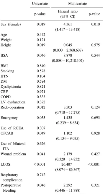
Univariate analyses revealed that female sex (p=0.019), height (p=0.019), body surface area (p=0.046), redo operation (p=0.012), off-pump CABG (p=0.049), a postoperative wound
problem (p=0.041), postoperative bleeding reoperation (p=
0.046), and LCOS (p<0.001) were related to the develop- ment of subxiphoid incisional hernia (Table 3). On multi- variable analysis, female sex (relative risk, 4.361; p=0.01) and postoperative LCOS (relative risk, 26.407; p<0.001) in- creased the risk of subxiphoid hernia.
DISCUSSION
The present study produced two findings: subxiphoid inci- sional hernia requiring surgical repair developed in 0.8% of patients who underwent isolated CABG, and female sex and postoperative LCOS were associated with the development of subxiphoid incisional hernia.
Subxiphoid incisional hernia is usually associated with me- dian sternotomy [3]. Subxiphoid incisional hernias have been known as difficult lesions to repair and have shown a high recurrence rate owing to the anatomical complexity of the subxiphoid area [2-5]. Incidences of subxiphoid incisional hernias associated with median sternotomy have been reported as between 1% and 4.2%, and a majority of the hernias have been reported to have developed within 2 to 3 years after surgery [2-5]. As complex cardiac surgical procedures asso- ciated with prolonged operation time were reported as risk factors of sternal wound infection [10], we limited the study population to only include patients who underwent isolated
Table 3. Analysis of risk factors for the development of sub- xiphoid incisional hernia using the Cox proportional hazard model
Univariate Multivariate
p-value Hazard ratio
(95% CI) p-value Sex (female)
Age Weight Height
BSA
BMI Smoking HTN DM Dyslipidemia CRF COPD LV dysfunction Redo-operation
Emergency
Use of RGEA OPCAB
Use of bilateral ITA
Wound problem
LCOS
Respiratory complication Postoperative
bleeding
0.019
0.442 0.121 0.019
0.046
0.840 0.578 0.104 0.584 0.821 0.971 0.833 0.372 0.012
0.055
0.307 0.049
0.626
0.041
<0.001
0.742
0.046
4.361 (1.417−13.418)
0.045 (0.000−2,368.607)
8.878 (0.008−10,218.102)
3.503 (0.710−17.275)
1.435 (0.239−8.634)
1.102 (0.134−9.035)
2.179 (0.320−14.852)
26.407 (8.074−86.367)
2.292 (0.446−11.788)
0.010
0.575
0.544
0.124
0.693
0.928
0.427
<0.001
0.321
CI, confidence interval; BSA, body surface area; BMI, body mass index; HTN, hypertension; DM, diabetes mellitus; CRF, chronic renal failure; COPD, chronic obstructive pulmonary dis- ease; LV, left ventricle; RGEA, right gastroepiploic artery;
OPCAB, off-pump coronary artery bypass; ITA, internal thora- cic artery; LCOS, low cardiac output syndrome.
CABG. Patients who underwent valve surgery and CABG/valve surgery were excluded in the analysis to mini- mize the effect of procedure type and operation time. In the present study, subxiphoid incisional hernia requiring surgical
repair developed in 0.8% of patients who underwent isolated CABG, and all subxiphoid hernias were detected within 5 postoperative years.
A previous study showed that obesity (BMI >30 kg/m2) increased the risk of sternal wound complications and sub- xiphoid hernia [3]. In the present study, we did not find the BMI to be a risk factor because the BMI of all the patients including the patients with subxiphoid hernia were 24.8±2.9 kg/m2 and 25.0±2.5 kg/m2, respectively. We investigated the risk factors for the development of incisional hernia after car- diac surgery among the common risk factors for sternal wound infection, such as old age, female sex, obesity, dia- betes, chronic obstructive pulmonary disease, bilateral use of the internal thoracic artery, LCOS, and re-exploration for bleeding [10-13]. Female sex has been reported to be an in- dependent risk factor for CABG and the higher morbidity in female patients was suggested to be associated with the high- er proportion of women with diabetes and obesity who under- go this procedure [14-17]. Low cardiac output syndrome was also a risk factor of deep sternal infection [12,13]. As sub- xiphoid incisional hernia is a problem for wound healing, the negative effects of postoperative LCOS on wound the healing process may increase the risk of subxiphoid incisional hernia.
In the present study, female sex and postoperative LCOS were identified as risk factors for incisional hernia.
The development of subxiphoid incisional hernia was ex- plained by fascial dehiscence, which develops from a weak- ness in the soft tissue structures as well as from distraction mediated by the xiphoid region’s osseous geometry [18].
Subxiphoid incisional hernia is obviously related to median sternotomy. However, variables commonly considered to be risk factors for wound complications were not demonstrated to be significant in this study. The use of RGEA, which re- quired a long incision extended toward the abdomen area and the peritoneal opening, was also shown to be an insignificant factor for the development of the hernia. Although previous studies have reported high recurrence rates after the repair of subxiphoid incisional hernia (24% to 44%) [3,19,20], no re- currence was observed in this study. Currently, we are apply- ing multiple interrupted sutures at the fascia layer of the sub- xiphoid area to prevent development of incisional hernia.
There are limitations to the present study. First, it was a
retrospective study in a single institution. Second, the analysis was performed in a limited sample population who underwent isolated CABG. Therefore, it is difficult to generalize this re- sult to patients who have undergone other cardiac surgical procedures. Third, we were not able to perform physical ex- aminations for all the patients to detect subxiphoid incisional hernia. Thus, the incidence of development of subxiphoid in- cisional hernia might be underestimated.
CONCLUSION
In this study, female sex and postoperative LCOS showed an increased risk of subxiphoid incisional hernia. It is recom- mended that the surgeon pay attention when performing wound closures for high-risk patients and consider the possi- bility of subxiphoid hernia development during the follow-up period.
REFERENCES
1. Losanoff JE, Basson MD, Laker S, Weiner M, Webber JD, Gruber SA. Subxiphoid incisional hernias after median ster- notomy. Hernia 2007;11:473-9.
2. Davidson BR, Bailey JS. Incisional herniae following me- dian sternotomy incisions: their incidence and aetiology. Br J Surg 1986;73:995-6.
3. Mackey RA, Brody FJ, Berber E, Chand B, Henderson JM.
Subxiphoid incisional hernias after median sternotomy. J Am Coll Surg 2005;201:71-6.
4. Cohen MJ, Starling JR. Repair of subxiphoid incisional her- nias with Marlex mesh after median sternotomy. Arch Surg 1985;120:1270-1.
5. Landau O, Raziel A, Matz A, Kyzer S, Haruzi I. Laparo- scopic repair of poststernotomy subxiphoid epigastric hernia.
Surg Endosc 2001;15:1313-4.
6. Penttinen R, Gronroos JM. Mesh repair of common abdomi- nal hernias: a review on experimental and clinical studies.
Hernia 2008;12:337-44.
7. Franz MG. The biology of hernia formation. Surg Clin
North Am 2008;88:1-15, vii.
8. Yahchouchy-Chouillard E, Aura T, Picone O, Etienne JC, Fingerhut A. Incisional hernias. I. Related risk factors. Dig Surg 2003;20:3-9.
9. Rao V, Ivanov J, Weisel RD, Ikonomidis JS, Christakis GT, David TE. Predictors of low cardiac output syndrome after coronary artery bypass. J Thorac Cardiovasc Surg 1996;112:
38-51.
10. Filsoufi F, Castillo JG, Rahmanian PB, et al. Epidemiology of deep sternal wound infection in cardiac surgery. J Cardi- othorac Vasc Anesth 2009;23:488-94.
11. Tang GH, Maganti M, Weisel RD, Borger MA. Prevention and management of deep sternal wound infection. Semin Thorac Cardiovasc Surg 2004;16:62-9.
12. Careaga Reyna G, Aguirre Baca GG, Medina Concebida LE, Borrayo Sanchez G, Prado Villegas G, Arguero Sanchez R.
Risk factors for mediastinitis and sternal dehiscence after cardiac surgery. Rev Esp Cardiol 2006;59:130-5.
13. Risk factors for deep sternal wound infection after sternot- omy: a prospective, multicenter study. J Thorac Cardiovasc Surg 1996;111:1200-7.
14. Roques F, Nashef SA, Michel P, et al. Risk factors and out- come in European cardiac surgery: analysis of the Euro- SCORE multinational database of 19030 patients. Eur J Cardiothorac Surg 1999;15:816-22.
15. Koch CG, Khandwala F, Nussmeier N, Blackstone EH.
Gender profiling in coronary artery bypass grafting. J Thorac Cardiovasc Surg 2003;126:2044-51.
16. Zitser-Gurevich Y, Simchen E, Galai N, Mandel M; ISCAB Consortium. Effect of perioperative complications on excess mortality among women after coronary artery bypass: the Israeli Coronary Artery Bypass Graft Study (ISCAB). J Thorac Cardiovasc Surg 2002;123:517-24.
17. Tokmakoglu H. Operative and early results of coronary ar- tery bypass grafting in female patients in different body mass indexes. J Cardiothorac Surg 2010;5:119.
18. Askar OM. Surgical anatomy of the aponeurotic expansions of the anterior abdominal wall. Ann R Coll Surg Engl 1977;59:313-21.
19. Davidson BR, Bailey JS. Repair of incisional hernia after median sternotomy. Thorax 1987;42:549-50.
20. Read RC, Yoder G. Recent trends in the management of in- cisional herniation. Arch Surg 1989;124:485-8.