ISSN: 2233-601X (Print) ISSN: 2093-6516 (Online)
− 401 −
Received: October 16, 2015, Revised: November 9, 2015, Accepted: November 10, 2015, Published online: October 5, 2016
Corresponding author: Joon Bum Kim, Department of Thoracic and Cardiovascular Surgery, Asan Medical Center, University of Ulsan College of Medicine, 88 Olympic-ro 43-gil, Songpa-gu, Seoul 05505, Korea
(Tel) 82-2-3010-5416 (Fax) 82-2-3010-6966 (E-mail) [email protected]
© The Korean Society for Thoracic and Cardiovascular Surgery. 2016. All right reserved.
This is an open access article distributed under the terms of the Creative Commons Attribution Non-Commercial License (http://creativecommons.org/
licenses/by-nc/4.0) which permits unrestricted non-commercial use, distribution, and reproduction in any medium, provided the original work is properly cited.
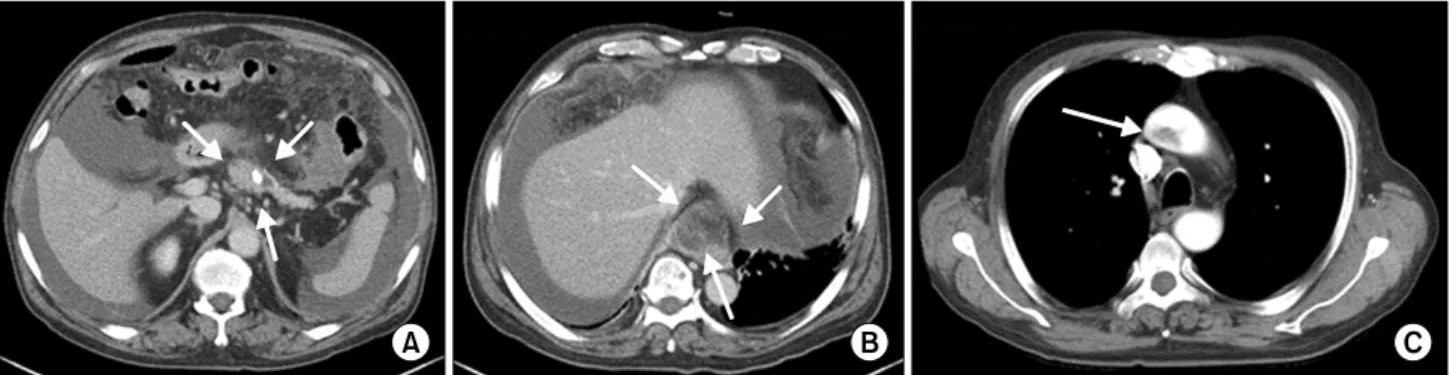
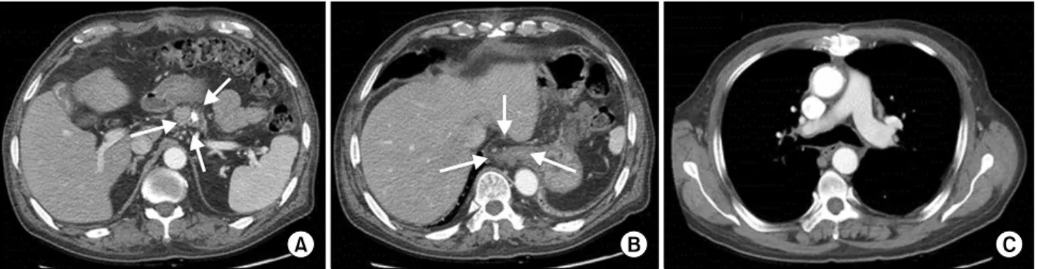
Multiple Ascending Aortic Mural Thrombi and Acute Necrotizing Mediastinitis Secondary to Acute Pancreatitis
Byung Kwon Chong, M.D. 1 , Jae Kwang Yun, M.D. 1 , Joon Bum Kim, M.D., Ph.D. 1 , Do Hyun Park, M.D., Ph.D. 2
1