INTRODUCTION
Duodenal duplication cyst (DDC) is a rare congenital ano- maly (1-13), which can appear during the neonatal period or later in life depending on the degree of the gastric outlet obstruction. About thirty percent of patients diagnosed with duplication cysts are older than twelve years of age, and the oldest patient with DDCs reported was 73 yr old (1). These lesions often present a diagnostic and therapeutic challenge to surgeons. Duplications of the duodenum may be either cystic or tubular and are composed of a muscular wall with a gastrointestinal epithelial lining. Duplication cysts may be found at any level from the mouth to the anus and are usu- ally attached intimately to some portions of the gastrointesti- nal tract. Only 4-12% of these duplications are located in the duodenum. Here, we describe the first Korean case of a DDC associated with bowel obstruction, obstructive jaun- dice and acute pancreatitis in the later period of life.
CASE REPORT
A 65-yr-old man was referred to our hospital complaining of pain in the right upper quadrant and vomiting for previous 3 days. His past medical history was unremarkable except for a spinal operation performed due to a falling-down injury 10 yr ago. A physical examination demonstrated tenderness
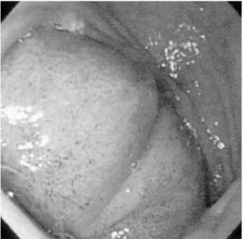
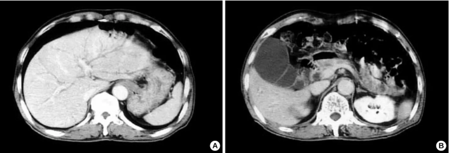
and a soft palpable mass on the right upper quadrant of the abdomen. The laboratory examinations showed a hemoglobin level of 15.3 g/dL (normal range, 14 to 18), a white blood cell count of 12,590/ L (normal range, 4,000 to 10,000), and a platelet count of 254,000/ L (normal range, 140,000 to 400,000). The liver function tests showed a total serum biliru- bin level of 3.3 mg/dL (normal range, 0.1 to 1.2), a direct bilirubin level of 2.1 mg/dL (normal range, 0.1 to 0.4), alka- line phosphatase level of 323 IU/L (normal range, 20 to 90), a gamma glutamyl transpeptidase level of 371 IU/L (normal range, 0 to 50), an aspartate aminotransferase level of 267 IU/L (normal range, 10 to 35); alanine aminotransferase, 228 IU/L (normal range, 0 to 40); amylase, 972 IU/L (normal range, 50 to 160) and a lipase level of 2,767 IU/L (normal range, 0 to 200). A duodenoscopy revealed a huge extrinsic mass with an intact mucosa causing almost complete lumi- nal obstruction in the second portion of the duodenum. On palpation with a biopsy forcep, the mass was found to be soft and easily compressible (Fig. 1). A computerized tomography scan showed a 12×6 cm sized elliptic cystic lesion with a distinct wall at the second-to-third portion of the duodenum.
The lesion occupied the intramural portion of the duodenal wall. These findings above were consistent with a DDC (Fig.
2). The intrahepatic duct and common bile duct were dilat- ed, and the gallbladder was markedly distended (Fig. 3). A percutaneous transhepatic biliary drainage for the relief of obstructive jaundice was performed one day after admission.
Young Chul Jo, Kwang Ro Joo*, Do Ha Kim, Jong Ho Park, Jae Hee Suh�, Young Min Kim�, Chang Woo Nam�
Departments of Internal Medicine, Pathology�and Surgery�, Ulsan University Hospital, University of Ulsan College of Medicine, Ulsan; Department of Internal Medicine, Kyung Hee University College of Medicine*, Seoul, Korea
Address for correspondence Kwang Ro Joo, M.D.
Department of Internal Medicine, Kyung Hee University College of Medicine, 1 Hoigi-dong, Dongdaemoon-gu, Seoul 130-702, Korea Tel : +82.2-958-8099, Fax : +82.2-968-1848 E-mail : [email protected]
604 J Korean Med Sci 2004; 19: 604-7
ISSN 1011-8934
Copyright � The Korean Academy of Medical Sciences
Duodenal Duplicated Cyst Manifested by Acute Pancreatitis and Obstructive Jaundice in an Elderly Man
A duodenal duplication cyst is an uncommon congenital anomaly that is usually encountered during infancy or in early childhood. Duodenal duplication cysts gen- erally appear on the first or second portion of the duodenum and may cause duo- denal obstruction, hemorrhage or pancreatitis. Here, we report a case of a duode- nal duplication cyst on the second and third portion of the duodenum in an old aged man with obstructive jaundice and acute pancreatitis, which was treated suc- cessfully by a surgical excision.
Key Words : Abnormalities; Duplication Cyst; Duodenum; Pancreatitis, Acute Necrotizing; Jaundice, Obstruc- tive
Received : 24 July 2003 Accepted : 1 September 2003
Duodenal Duplication Cyst in an Old Man 605
A pylorus-preserving pancreaticoduodenectomy (modified Whipple’s operation) was performed 24 days after support- ive treatment. The surgical specimens included the seg- ments of the duodenum and the duplication cyst, the gall- bladder and some portions of the pancreas. The duplication cyst was located along the antimesenteric border and was 9 cm in length and 2.8 cm in the inner diameter (Fig. 4).
There was no luminal communication between the cyst and the duodenum. The inner surface of the cyst was flat and showed multiple erosions and ulcers. Histologically, the cyst showed denudation of the mucosal layer, which was replaced by granulation tissue, due to the mucosal inflam- mation, but most parts of the cyst contained a smooth mus-
cle layer (Fig. 5). The post-operative course of the patient was uneventful, and the patient has been doing well for 6 months after surgery without any complication.
DISCUSSION
Duplication cysts are congenital anomalies that are formed during the embryonic period of the development of the human digestive system. The most common site for a gastrointesti- nal duplication cyst is the distal ileum followed by the esoph- agus and duodenum (2). Duplication cysts must adhere to a portion of the gastrointestinal tract, contain a smooth mus- cle layer in their walls and be lined with an alimentary epithe- lium. The epithelial lining of the duplication cysts may be that of the adjacent bowel, and ectopic gastric or pancreatic tissue may sometimes be present within (2, 3). Gastric dupli- cations occur along the greater curvature, and intestinal dupli- cation cysts are found on the mesenteric side of the bowel.
The cysts may be spherical or tubular, and there may be infre- quent communications with the gastrointestinal tract. DDCs are diagnosed more often in children. They are mostly lim- ited to the first or second part of the duodenum usually adja- cent to the pancreatic surface sharing a common wall and blood supply with the duodenum. DDCs may be silent for many years before they cause any symptoms including bowel obstruction, pain, distention or gastrointestinal bleeding.
Bleeding, ulceration, and perforation may occur in the cysts containing ectopic gastric mucosa (2, 3). The clinical presen- tation depends on the size and location of the cysts. In our case, the patient was old and had accompanying symptoms of bowel obstruction, obstructive jaundice, and acute pan- creatitis. The compression caused by the duplicated segment of the duodenum attributed to the above symptoms. The cyst was in the second and third portions of the duodenum and had an ulceration on the inner side.
Fig. 2.Computerized tomographic finding. A cystic lesion with a distinct wall in the second-to-third portion of the duodenum (A, B) is shown.
The duodenum (closed arrow) is compressed by the cystic lesion and is displaced to the medial side of the lesion (open arrow) (A).
A B
Fig. 1.Duodenoscopic finding. A huge extrinsic mass causing a luminal obstruction in the proximal second portion of the duode- num, with an intact mucosa.
606 Y.C. Jo, K.R. Joo, D.H. Kim, et al.
The three criteria for the diagnosis of a duplication cyst are the presence of an intimate attachment to the gastrointesti- nal tract, a smooth muscle coat, and an alimentary mucosal
lining (2). Of above three criteria, the presence of a smooth muscle coat is absolutely essential for the diagnosis of a dupli- cation cyst. In our case, although the lining epithelial cells were entirely denuded and replaced by inflamed granulation tissue due to increased intraluminal pressure and inflamma- tion, most of the cyst contained smooth muscle coats with foci of a neural plexus, which supported the diagnosis of a duplication cyst.
The differential diagnosis of DDCs and choledochoceles demands particular attention. The principal distinguishing features between these two conditions are their histological characteristics. DDCs are covered both inside and outside by duodenal mucosa containing a distinct layer of smooth
Fig. 3.Computerized tomographic finding. Dilated intrahepatic duct (A) and a common bile duct and markedly distended the gallbladder (B) is shown.
A B
Fig. 4.Gross finding of the surgical specimen. Segments of the duodenum, duplication cyst, gallbladder and some portions of the pancreas are shown. Resection along the left margin of the cyst (arrow) (A) reveals the inner side of the cystic wall (B). A Kelly is protruding into the duodenum (B).
A
B
Fig. 5.Microscopic finding of the cyst wall obtained from the sep- tum of the duplicated segment. Mucosal denudation and replace- ment by granulation tissue are seen. The cyst shares a common muscular layer with the duodenum (bottom) (H&E stain, ×100).
Denuded mucosa layer
Muscle layer
Submucosa
Mucosa
Duodenal Duplication Cyst in an Old Man 607
muscles. In contrast, choledochoceles are lined by either a bile duct or gallbladder mucosa and lack a smooth muscle layer (14).
DDCs have been traditionally treated with surgery. The surgical approach should be made in accordance with the relationship between the cyst wall and the biliary and pan- creatic drainage systems. The surgical goal is the free drainage of the cyst into the duodenum without damaging the biliary and pancreatic ducts. Occasionally, the close relationship bet- ween the DDC and the pancreaticobiliary system makes sur- gery difficult and risky. In these cases an endoscopic proce- dure appears to be a reasonable option (4, 5). A total excision of the cyst prevents further hazards including the develop- ment of a malignancy that may arise from the residual ectopic gastric mucosa (6). Thus, our case was treated with a pylorus- preserving pancreaticoduodenectomy, which cured the patient.
REFERENCES
1. Browning RW. Duodenal duplications. Rev Surg 1963; 20: 226-9.
2. Gross RE, Holcomb GW Jr, Farber S. Duplications of the alimenta- ry tract. Pediatrics 1952; 9: 449-68.
3. Rubin RB, Saltzman JR, Zawacki JK, Khan A, Swanson R. Duode- nal duplication cyst with massive gastrointestinal bleeding. J Clin Gastroenterol 1995; 21: 72-4.
4. Wada S, Higashizawa T, Tamada K, Tomiyama T, Ohashi A, Satoh Y, Sugano K, Nagai H. Endoscopic partial resection of a duodenal duplication cyst. Endoscopy 2001; 33: 808-10.
5. Sezgin O, Altiparmak E, Yilmaz U, Saritas U, Sahin B. Endoscopic management of a duodenal duplication cyst associated with biliary obstruction in an adult. J Clin Gastroenterol 2001; 32: 353-5.
6. Falk GL, Young CJ, Parer J. Adenocarcinoma arising in a duodenal duplication cyst: a case report. Aust N Z J Surg 1991; 61: 551-3.
7. Narlawar RS, Rao JR, Karmarkar SJ, Gupta A, Hira P. Sonographic findings in a duodenal duplication cyst. J Clin Ultrasound 2002; 30:
566-8.
8. Zamir G, Gross E, Shmushkevich A, Bar-Ziv J, Durst AL, Jurim O.
Duodenal duplication cyst manifested by duodeno-jejunal intussus- ception and hyperbilirubinemia. J Pediatr Surg 1999; 34: 1297-9.
9. Arbell D, Lebenthal A, Blashar A, Shmushkevich A, Gross E. Dupli- cation cyst of the duodenum as an unusual cause of massive gastroin- testinal bleeding in an infant. J Pediatr Surg 2002; 37: E8.
10. Fan ST, Lau WY, Pang SW. Infarction of a duodenal duplication cyst. Am J Gastroenterol 1985; 80: 337-9.
11. Ogura Y, Kawarada Y, Mizumoto R. Duodenal duplication cyst com- municating with an accessory pancreatic duct of Santorini. Hepato- gastroenterology 1998; 45: 1613-8.
12. Richer JP, Faure JP, Maillot N, Silvain C, Levillain P, Carretier M.
Duodenal duplication cyst communicating with the bile duct with a long common biliary-pancreatic channel. Eur J Surg 2000; 166:
504-7.
13. Keller MS, Weber TR, Sotelo-Avila C, Brink DS, Luisiri A. Duo- denal duplication cysts: A rare cause of acute pancreatitis in chil- dren. Surgery 2001; 130: 112-5.
14. Reinus FZ, Weingarten G. Choledochocele of the common bile duct.
Am J Surg 1976; 132: 646-8.