Brief Report
Vol. 30, No. 1, 2018 91
Received June 21, 2016, Revised November 15, 2016, Accepted for publication December 18, 2016
Corresponding author: Moon Kyun Cho, Department of Dermatology, Soonchunhyang University Seoul Hospital, 59 Daesagwan-ro, Yongsan-gu, Seoul 04401, Korea. Tel: 82-2-709-9368, Fax: 82-2-709-9374, E-mail:
This is an Open Access article distributed under the terms of the Creative Commons Attribution Non-Commercial License (http://creativecommons.
org/licenses/by-nc/4.0) which permits unrestricted non-commercial use, distribution, and reproduction in any medium, provided the original work is properly cited.
Copyright © The Korean Dermatological Association and The Korean Society for Investigative Dermatology
pISSN 1013-9087ㆍeISSN 2005-3894
Ann Dermatol Vol. 30, No. 1, 2018 https://doi.org/10.5021/ad.2018.30.1.91
BRIEF REPORT
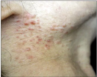
Fig. 1. Multiple erythematous, firm, non-tender papules of various sizes (3∼7 mm) on the right side of the neck.
A Case of Multiple Cutaneous Piloleiomyomas on the Neck
Yoon Jin Lee
1,2, Jin Ho Bae
1, Kyu Uang Whang
1, Moon Kyun Cho
11Department of Dermatology, Soonchunhyang University Seoul Hospital, Seoul, 2Molecular Cancer Research, Soonchunhyang University College of Medicine, Cheonan, Korea
Dear Editor:
A healthy 65-year-old woman with protruding papules on her neck which had been slowly expanding over the past 10 years visited Soonchunhyang University Seoul Hospital.
At first, there were two nodules on the lower right side of her neck. Over the course of 10 years, the lesions spread slowly, and at the time of presentation, were found on the left side of her neck. None of her family members had similar lesions. The lesions were not associated with pain or any symptoms. Also, the patient had no notable past medical history. On physical examination, multiple eryth- ematous, firm, non-tender papules of various sizes (3∼7 mm) were observed on the right side of her neck (Fig. 1).
A punch biopsy was conducted under local anesthesia.
Microscopic examination of the sections stained with hematoxylin and eosin (H&E) showed poorly demarcated tumor intermingling with bundles of dermal collagen fibers. The tumor was composed of smooth muscle fibers that had straight, blunt-ended nuclei with no evidence of nuclear atypia. Neither mitotic activity nor pleomorphism were observed (Fig. 2A, B). Immunohistochemical exami- nation revealed that the tumor cells were positive for actin (Fig. 2C) and desmin (Fig. 2D). Based on clinical and his-
tological examination, the lesion on the patient’s neck was diagnosed as multiple cutaneous piloleiomyomas. However, the patient declined surgical or medical intervention.
Multiple piloleiomyomata are known as the most com- mon type of cutaneous leiomyoma1. The condition con- sists of multiple (on rare occasion, hundreds) of lesions that are small, slowly growing papules. They are typically painful or tender, particularly when compressed or ex- posed to a cold environment2. Women with multiple pilo- leiomyomas may also develop uterine leiomyomas. Renal cell cancer also develop in a subset of affected individuals.
Hereditary leiomyomatosis and renal cell cancer is caused by germline mutation in the gene encoding fumarate hy- dratase on chromosome 1q42.3∼433. While piloleiomyo- ma is the most common type of cutaneous leiomyoma in Caucasians, angioleiomyoma is the most common type of cutaneous leiomyoma in Koreans. According to previous reports, cutaneous leiomyomas have been found only on
Brief Report
92 Ann Dermatol
Fig. 2. (A) Dermal proliferation of ill-defined smooth muscle fibers surrounded by varying amounts of collagen fibers (H&E, ×40). (B) Dermal tumor composed of smooth muscle fibers with spindle-shaped, blunt-ended nuclei (H&E, ×400).
(C) Tumor cells showing strong positively for smooth muscle actin (smooth muscle actin stain, ×200), and (D) desmin (desmin stain,
×200).
the trunk, lips, and limbs, and to date, there have been no reports on cutaneous leiomyomas on the neck in Korea.
On biopsy specimens, piloleiomyomas appear to be com- posed of poorly circumscribed smooth muscle fibers that are located in the dermis and merge imperceptibly with the surrounding connective tissue4. The tumor is com- posed of uniform spindle-shaped cells showing interlacing bundle formation or irregular collections of elongated cells with brightly eosinophilic cellularity and blunt-ended or cigar-shaped nuclei. Piloleiomyomata may show very low mitotic activity, one or less mitotic figure/10 high pow- er field (HPF). Tumor cells are usually positive for smooth muscle actin, calponin, desmin, and h-caldesmon.
Piloleiomyomas may resemble other painful subcutaneous tumors including eccrine spiradenoma, neuroma, glomus tumor, angiolipoma, neurilemmoma, dermatofibroma. A differential diagnosis between cutaneous leiomyosarcoma and leiomyoma can be made depending on the presence of mitosis5. To summarize, this study reports a rare case of cutaneous leiomyomas that occurred on the neck of a middle-aged woman with no family medical history of the condition.
ACKNOWLEDGMENT
This work was supported in part by the Soonchunhyang University research fund.
CONFLICTS OF INTEREST
The authors have nothing to disclose.
REFERENCES
1. Malhotra P, Walia H, Singh A, Ramesh V. Leiomyoma cutis:
a clinicopathological series of 37 cases. Indian J Dermatol 2010;55:337-341.
2. Thyresson HN, Su WP. Familial cutaneous leiomyomatosis. J Am Acad Dermatol 1981;4:430-434.
3. Menko FH, Maher ER, Schmidt LS, Middelton LA, Aittomäki K, Tomlinson I, et al. Hereditary leiomyomatosis and renal cell cancer (HLRCC): renal cancer risk, surveillance and treatment. Fam Cancer 2014;13:637-644.
4. Ghanadan A, Abbasi A, Kamyab Hesari K. Cutaneous leiomyoma: novel histologic findings for classification and
Brief Report
Vol. 30, No. 1, 2018 93
Received September 20, 2016, Revised November 28, 2016, Accepted for publication December 22, 2016
Corresponding author: Moon-Bum Kim, Department of Dermatology, Pusan National University Hospital, 179 Gudeok-ro, Seo-gu, Busan 49241, Korea. Tel:
82-51-240-7338, Fax: 82-51-245-9467, E-mail: [email protected]
This is an Open Access article distributed under the terms of the Creative Commons Attribution Non-Commercial License (http://creativecommons.org/
licenses/by-nc/4.0) which permits unrestricted non-commercial use, distribution, and reproduction in any medium, provided the original work is properly cited.
Copyright © The Korean Dermatological Association and The Korean Society for Investigative Dermatology diagnosis. Acta Med Iran 2013;51:19-24.
5. Pijpe J, Broers GH, Plaat BE, Hundeiker M, Otto F, Mastik MF, et al. The relation between histological, tumor-biolo-
gical and clinical parameters in deep and superficial leiomyosarcoma and leiomyoma. Sarcoma 2002;6:105-110.
https://doi.org/10.5021/ad.2018.30.1.93
Hypopigmented Mycosis Fungoides Treated with 308 nm Excimer Laser
Min-Young Yang
1, Hyunju Jin
1, Hyang-Suk You
1, Woo-Haing Shim
1, Jeong-Min Kim
1, Gun-Wook Kim
1, Hoon-Soo Kim
1, Hyun-Chang Ko
1, Byung-Soo Kim
1,2, Moon-Bum Kim
1,21Department of Dermatology, Pusan National University School of Medicine, 2Biomedical Research Institute, Pusan National University Hospital, Busan, Korea
Dear Editor:
Hypopigmented mycosis fungoides (HMF) is an atypical, rare clinical variant of MF characterized by hypopigmented to achromic patches alone or, more commonly, in combi- nation with studding erythematous papules or plaques.
The epidemiologic features that distinguish HMF from classical MF are related to its high prevalence among younger patients, such as children and adolescence, as well as patients with high skin phototypes (usually Fitzpat- rick skin scale IV∼V)1. Psoralen plus ultraviolet A (PUVA) was the main treatment used in the previously reported lit- erature2. Reports on narrowband UVB (NBUVB) photo- therapy have recently demonstrated this therapy to be a successful alternative for PUVA therapy3.
A 10-year-old Korean boy presented to Pusan National University Hospital for evaluation of asymptomatic hypo- pigmented patches of skin and studded erythematous pap- ules on the left upper back and flank (Fig. 1A). Wood light examination did not show pronounced attenuation. The histopathology results were compatible with MF. The mi- croscopic description revealed epidermotrophism of ha- loed atypical lymphocytes (Fig. 1B). Additional studies on
the rearrangement of the T cell receptor gamma gene showed monoclonality (Fig. 2). Considering limited le- sional distribution in this patient and higher effectiveness of excimer laser than local NBUVB in focal vitiligo, we chose and performed 308 nm excimer laser therapy once a week for the diagnosis of HMF (stage IA). This treatment resulted in the clearance of the lesions and repigmentation of the hypopigmented areas after about 1 year (Fig. 1C).
The mean fluence emitted was 340 mJ/cm2, and the total cumulative dose was 17.7 J/cm2.
The prognosis for HMF is usually good compared with that for classical MF. Infiltrative atypical CD8+ cells are postulated to play a role in preventing the usual patch stage disease from progressing to advanced plaque and tu- mour stages1. Also, the cytotoxic effect of CD8+ T lym- phocytes are believed to influence melanocyte stability and melanogenesis resulting in hypopigmented patches clinically. In respect to treatment, HMF recurs frequently, even if there is a long period of complete remission.
Although there were some reports of 308 nm excimer use for patch or plaque stages of MF4, there has been no re- port of 308 nm excimer for HMF. 308 nm excimer laser