MR Imaging for the Differentiation of Early Infectious Spondylitis and Modic Type I Change in the Lumbar Spine
1Jong Won Kwon, M.D., Young Cheol Yoon, M.D., Sang-Hee Choi, M.D., Jee Young Jung, M.D., Bong Keun Choe, M.D.2
1Department of Radiology and Center for Imaging Science, Samsung Medical Center, Sungkyunkwan University School of Medicine, Korea
2Department of Preventive Medicine, School of Medicine, Kyung Hee University, Korea Received December 15, 2009 ; Accepted February 16, 2010
Address reprint requests to : Young Cheol Yoon, M.D., Department of Radiology, Samsung Medical Center, Sungkyunkwan University School of Medicine, 50, Ilwon-dong, Kangnam-gu, Seoul 135-710, Korea.
Tel. 82-2-3410-6454 Fax. 82-2-3410-0084 E-mail: [email protected]
Purpose: To evaluate magnetic resonance (MR) imaging findings for the differentiation of early infectious spondylitis and Modic type I change in the lumbar spine.
Materials and Methods: Contrast-enhanced lumbar spine MR images with bone mar- row edema adjacent to the endplates of 25 patients (14 men, 11 women; mean age 53.7 years) were evaluated. Margin and enhancement in the bone marrow changes, erosion or destruction of endplates on T1-weighted sagittal MR images, T2 signal intensity and enhancement in the intervertebral disks, as well as the presence of paravertebral soft tissue were evaluated. The final diagnoses were 11 cases of infectious spondylitis and 14 cases of Modic type I change.
Results: The margin of bone marrow changes was more blurred in infectious spondylitis than in Modic type I change. The endplates for infectious spondylitis showed destructions, and Modic change erosions. On postcontrast images, all the cases except for only one with degenerative disease were enhanced in the vertebral bodies.
Intervertebral disks showed T2 hyperintensities and were enhanced in both the infec- tious spondylitis and Modic change. Paravertebral soft tissue was seen more common- ly in infectious spondylitis.
Conclusion: The useful findings of early infectious spondylitis include endplate de- struction, blurred margins of the bone marrow changes on T1-weighted images, and the presence of paravertebral soft tissue.
Index words :Spine
Magnetic Resonance Imaging Infection
Spondylitis
Bone marrow abnormalities of the lumbar spine adja- cent to the endplates are commonly seen on MR images.
These abnormalities are most frequently associated with degeneration. A diagnosis of degenerative subchondral abnormalities is usually not problematic. However, some cases of degenerative bone marrow changes are al- so present with inflammatory symptoms and imaging findings that may simulate infectious spondylitis (1, 2).
Modic et al. (3) defined a commonly used MR imaging classification system for degenerative disk diseases.
However, the type I signal alterations (hypointensity on T1-weighted and hyperintensity on T2-weighted im- ages) may mimic MR imaging findings associated with infection (1, 2).
The MR findings of infectious spondylitis and degen- erative disk disease have been well described in each (3- 16). However, there are few reports comparing the imaging findings for both diseases. Moroever, no studies have compared the MR findings of early infectious spondylitis without abscess formation and a Modic type I change. The purpose of this study was to evaluate the MR imaging findings for the differentiation of degenera- tive disk disease with Modic type I change from early infectious spondylitis in the lumbar spine.
A B
C D
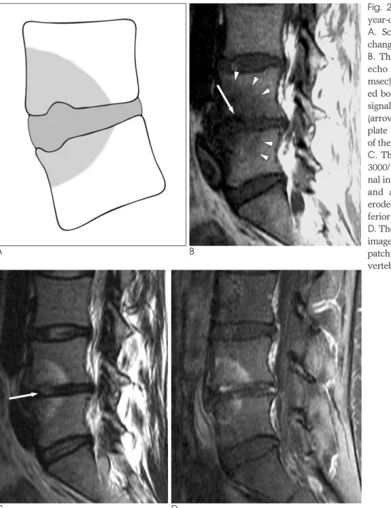
Fig. 1. Infectious spondylitis in a 33- year-old woman with lower back pain.
A. Schematic picture illustrating the change of infectious spondylitis.
B. The T1-weighted sagittal image (TR/TE = 350/17 msec) shows de- creased signal intensities of the verte- bral bodies of L3 and L4. There are fo- cally destructed vertebral endplates at the inferior aspect of L3 (arrow) and the superior aspect of the L4 vertebral body. The margins of bone marrow changes in the L3 and L4 vertebral bodies are not demarcated.
C. The T2-weighted sagittal image (TR/TE = 2200/102 msec) shows in- creased signals of the adjacent por- tions of the L3 and L4 vertebral bod- ies. Hyperintensity equivalent to fluid is seen in the L3-4 disk.
D. The contrast enhanced T1-weighted sagittal image (TR/TE = 350/17 msec) shows discrete patchy enhancement in the vertebral bodies of L3 and L4.
Materials and Methods
Patients
This retrospective study included the evaluation of 25 patients. Our institutional review board granted study approval and waived informed consent. This study was Health Insurance Portability and Accountability Act compliant.
Patients criteria for inclusion in this study consisted of the presence of bone marrow abnormalities adjacent to the endplates with low signal intensity (SI) on T1- weighted images and high SI on T2-weighted images,
the availability of MRI with pre- and post-contrast T1- and T2-weighted images before a biopsy or antibiotic treatment, a clinical follow-up of at least six months af- ter presentation, and clinical or pathological confirma- tion. Patients that underwent previous surgery, a recent fracture, and metastatic disease were excluded. For the selection of early infectious spondylitis, we excluded the advanced infectious spondylitis with abscess formation as seen in the MR imaging.
Eleven patients were diagnosed with infectious spondylitis, while 14 patients were diagnosed with de- generative disk disease. Infectious spondylitis was con- sidered to be present when blood culture results were
A B
C D
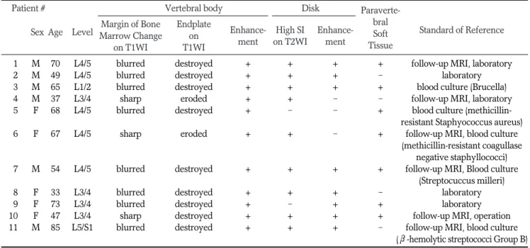
Fig. 2. Modic type I change in a 37- year-old woman with low back pain.
A. Schematic picture illustrating the changes of Modic type I change.
B. The sagittal T1-weighted fast spin- echo MR image (TR/TE = 650/17 msec) shows relatively well-demarcat- ed bone marrow abnormalities of low signal intensity at the L4 and L5 bodies (arrowheads). There is an eroded end- plate with an irregular low signal line of the inferior aspect of L4 (arrow).
C. The T2-weighted image (TR/TE = 3000/110 msec) reveals increased sig- nal intensity involving the disk (arrow) and adjacent vertebral bodies. An eroded endplate is also seen at the in- ferior aspect of L4.
D. The contrast enhanced T1-weighted image (TR/TE = 650/17 msec) shows patchy enhancement in the L4 and L5 vertebral bodies.
positive (n = 5). In five cases, follow-up MRI showed an increase in paravertebral inflammatory tissue along with an epidural abscess and bone destruction (Figs. 1, 2). In three cases, infectious spondylitis was diagnosed on the basis of clinical and laboratory findings (elevated erythrocyte sedimentation rate and C-reactive protein).
Percutaneous bone biopsies were performed in five pa- tients but no infectious agent was found in the tissue culture. Degenerative disk disease was diagnosed if a fa- vorable outcome without antibiotic treatment was seen as well as when the findings from the laboratory data and the clinical signs were negative after 6 months.
Bone biopsies were performed in two patients with de- generative disk disease and the histology results showed no inflammatory tissue.
MR Imaging Protocol
MR imaging was performed using a 1.5-T supercon- ducting system (Signa; General Electric Medical Systems, Milwaukee, WI USA) with a dedicated phased- array coil (CTL; GE Medical Systems). Sagittal T1- weighted (TR/TE, 380-720/9-23) and sagittal T2-weight- ed fast spin-echo (2200-4500/96-126) sequences were obtained with an image acquisition matrix of 512 192, 512 256, or 512 294, a field of view of 260 260 mm or 290 290 mm, and a section thickness of 4 mm. In addition, axial T1-weighted and axial T2-weight- ed fast spin echo sequences were obtained with an im- age acquisition matrix of 256 256, a field of view of
200 200 mm, and a slice thickness of 4 mm. Contrast- enhanced axial and sagittal T1-weighted MR images were obtained in all patients by using a spin echo se- quence. Gadopentetate dimeglumine (Magnevist;
Schering AG, Berlin, Germany) at a dose of 0.1 mmol per kilogram of body weight was used as an intravenous contrast agent.
Evaluation of the MR Images
MR images of 25 lumbar spines in 25 patients (11 women, 14 men; mean age: 53.7 years; age range: 28-85 years) were reviewed. All MR images were evaluated in conference by two investigators (J.W.K. and Y.C.Y.).
The observers were unaware of the clinical and other imaging findings. Images obtained with different se- quences in one MR examination were reviewed togeth- er as a group.
We evaluated the sharpness of the margin of bone marrow changes in the affected vertebral bodies on pre- contrast T1-weighted sagittal images, and determined whether the image was blurred or sharply demarcated.
The vertebral endplates of the affected spinal segments were graded on precontrast T1-weighted MR images as intact, eroded (focal areas of bone loss without disrup- tion of the hypointense cortical signal), or destroyed (dis- ruption of the cortical signal). On contrast-enhanced im- ages, the presence of any enhancement of the vertebral bodies was evaluated.
At the affected level, the presence of hyperintense
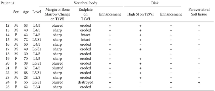
Table 1. Data on Eleven Patients with Infectious Spondylitis
Patient # Vertebral body Disk Paraverte-
Sex Age Level Margin of Bone Endplate
Enhance- Enhance- bral
Standard of Reference Marrow Change on
ment
High SI
ment Soft
on T1WI T1WI on T2WI Tissue
01 M 70 L4/5 blurred destroyed + + + + follow-up MRI, laboratory
02 M 49 L4/5 blurred destroyed + + + - laboratory
03 M 65 L1/2 blurred destroyed + + + + blood culture (Brucella)
04 M 37 L3/4 sharp eroded + + - - follow-up MRI, laboratory
05 F 68 L4/5 blurred destroyed + - - + blood culture (methicillin-
resistant Staphyococcus aureus)
06 F 67 L4/5 sharp eroded + + - + follow-up MRI, blood culture
(methicillin-resistant coagullase negative staphyllococci)
07 M 54 L4/5 blurred destroyed + + + + follow-up MRI, Blood culture
(Streptocuccus milleri)
08 F 33 L3/4 blurred destroyed + + + - laboratory
09 F 73 L3/4 blurred destroyed + - + + laboratory
10 F 47 L3/4 sharp destroyed + + + + follow-up MRI, operation
11 M 85 L5/S1 blurred destroyed + + + - follow-up MRI, blood culture
(�-hemolytic streptococci Group B) Note.─ Plus sign (+) = present, minus sign (-) = absent.
disk signal was equivalent to that of fluid and was evalu- ated from T2-weighted images. On contrast-enhanced images, the presence of enhancement in the disk was noted. We also evaluated the soft tissue changes of the paravertebral area on axial and sagittal images. MR imaging criteria for the presence of paravertebral in- flammatory tissue were discrete hypointensity on T1- weighted MR images and hyperintensities on T2- weighted MR images, as well as contrast enhancement (5, 8).
Review of the Clinical Data
One of investigators who were not involved in image analysis reviewed the medical records and laboratory reports. For each patient, it was noted whether antibiot- ic treatment was administered or a surgical intervention was performed after MR imaging. Associated medical conditions were reviewed from the patient records. The pathology and microbiology reports for the collected biopsy samples and the results of blood cultures were reviewed.
Statistical Analysis
Mean and standard deviation values were calculated for continuous data. Differences for dichotomous vari- ables were tested using Fisher’s exact test and the Pearson’s Chi-squared test. Statistical calculations were performed with the SPSS software package (SPSS, ver- sion 13; SPSS, Chicago, IL USA). A p-value of less than 0.05 was considered to indicate a statistically significant difference.
Results
Tables 1 and 2 summarize the MRI features of all en- rolled patients. The signal alteration of the bone marrow in the affected vertebral body on T1-weighted sagittal images were sharp in three (27.3%) and blurred in eight (72.7%) patients with infectious spondylitis. In contrast, the images were sharp in ten (71.4%) and blurred in four (28.6%) patients with a degenerative disk disease (p = 0.028). Endplates of infectious spondylitis showed ero- sions in two cases (18.2%) and destructions (Fig. 1) in nine cases (81.2%) as seen on T1-weighted sagittal im- ages. For the degenerative disk disease, the vertebral bodies had intact endplates in two cases (14.3%) and eroded endplates (Fig. 2) in 11 cases (78.6%). Endplate destruction with cortical disruption was seen in only one degenerative disk disease case (7.1%) (p = 0.01). On postcontrast images, all the cases except for one degen- erative disk disease case were homogeneously en- hanced in the low T1 signal areas of the vertebral bodies (p = 1.00).
Hyperintensity equivalent to fluid on T2-weighted im- ages was seen in nine (81.2%) intervertebral disks of the affected levels for the infectious spondylitis cases. In the degenerative disk disease, 10 (71.4%) intervertebral disks showed hyperintensity (p = 0.661) On post-con- trast images, intervertebral disks were focally or periph- erally enhanced in eight cases (72.7%) of infectious spondylitis. Intervertebral disks of Modic type I change were enhanced in 11 cases (78.6%) (p = 1.00).
Paravertebral soft tissue changes on axial and sagittal
Table 2. Data on Fourteen Patients with Modic Type I Change
Patient # Vertebral body Disk
Sex Age Level Margin of Bone Endplate Paravertebral
Marrow Change on Enhancement High SI on T2WI Enhancement Soft tissue
on T1WI T1WI
12 M 53 L4/5 blurred eroded + + - +
13 M 40 L4/5 sharp eroded + + + -
14 F 42 L4/5 sharp intact + + + -
15 M 72 L5/S1 sharp intact + + + -
16 M 50 L4/5 sharp eroded + - + -
17 M 49 L5/S1 sharp eroded + + + -
18 M 30 L4/5 sharp eroded + - + -
19 F 70 L4/5 sharp eroded + + + -
20 F 38 L5/S1 blurred eroded + + + -
21 F 37 L4/5 blurred eroded + + + -
22 M 68 L5/S1 sharp eroded + + - -
23 M 28 L2/3 sharp eroded - + - -
24 F 55 L5/S1 blurred destroyed + - + +
25 F 62 L3/4 sharp eroded + - + -
images were seen in seven cases (63.6%) of infectious spondylitis and two cases (14.3%) of a degenerative disk disease, respectively (p = 0.017).
Discussion
The differential diagnosis for early infectious spondyli- tis without an abscess and a Modic type I change is sometimes difficult in both an imaging and clinical set- ting (1, 2). In patients with the clinical possibility of the infectious spondylitis, MR imaging should be consid- ered as the imaging technique of choice. Plain films, ra- dionuclide scans, and CT scans may provide false-nega- tive findings, and if the scans show signs of infectious spondylitis, most patients will undergo MR imaging to assess the full soft-tissue extent and potential neural compromises (13, 15, 17).
Bone marrow abnormalities adjacent to the endplates are commonly associated with the degeneration of lum- bar disks. In the presence of Modic type I changes, pre- sumably relating to active remodeling of bone with loose vascularized fibrous tissue, these abnormalities may simulate the involvement of the bone marrow by infectious spondylitis. Although the characteristic histo- logical findings are present in infection, such as an in- creased number of polymorphonuclear leukocytes, there are some similarities in the histological appear- ance of Modic type I degenerative disk disease and in- fectious spondylitis (2, 3, 5, 6, 12, 18). In the resolved or resolving infection, the absorption or resolution of the inflammatory exudate and ischemia with healing results in a degenerated disk with fibrous scar tissue, are sur- rounded by vertebral bodies with some degree of new bone formation (5). Many of Modic type I changes pre- sent with inflammatory symptoms may mimic infec- tious spondylitis (1, 2). Toyone et al. (19) stated that 73%
of patients with a Modic type I change had significant lower back pain.
The differential diagnosis between infectious spondylitis and degenerative disk disease is usually based on clinical presentation (septic or not) and the presence of vertebral body abnormalities. Erosions that are small, located near areas of endplate sclerosis, and with sharp margins, suggest a diagnosis of degenerative change (20). Degenerative disk disease is suspected in patients with sclerosis of the adjacent endplates that may extend into the vertebral bodies with sharp mar- gins (1). Sclerosis occurs less frequently with infectious spondylitis than with degenerative disease. In the pre-
sent study, the endplates of degenerative change had ei- ther eroded or maintained intact margins. In the case of infectious spondylitis, destruction of the endplates was evident. The signal alteration in the affected vertebral bodies had blurred margins in infectious spondylitis, but relatively sharp margins in degenerative changes. We think these findings result from the difference of disease progression. Modic type I changes are assumed to be as- sociated with an ongoing active degenerative process (3).
However, infectious spondylitis progresses more rapid- ly. Thus, reactive scleroses of the endplate tend to devel- op less often and the margins of the affected bone mar- row changes are blurred. In addition, it is likely that the degenerative changes may be more reflective of biome- chanical stress and micro motion rather than true in- flammatory changes. It has been reported that abnormal SI in the vertebral bodies usually involves the whole vertebra in infectious spondylitis (14, 21). Most of our patients with infectious spondylitis, however, had par- tial involvement of the adjacent vertebral bodies as seen on T1-weighted, T2-weighted, and contrast-enhanced MR images, because we included only the early stage of infectious spondylitis that did not have abscess forma- tion.
On T2-weighted MR images, infected disks have been considered hyperintense compared to normal adjacent disks (4, 5, 8, 10, 14). Most of the patients in the present study also had hyperintensity or fluid-equivalent SI on T2-weighted MR images. Only two infected disks in this study were seen as not being hyperintense. However, hyperintensity of the disks on T2-weighted MR images is not specific to infectious spondylitis (3, 6, 9). In our study, 10 of 14 disks with degenerative changes also showed hyperintensity. Degenerated disks usually show hypointensity on T2-weighted images, and are presumed to be secondary to the dehydration (6).
However, there can be linear or focal areas of high sig- nal intensity within the decreased signal intensity on T2- weighted images that are thought to represent free fluid within the cracks or fissures of the degenerated complex (6). After contrast material administration, the infected disks almost invariably enhance. Only three patients in our study group with proven infectious spondylitis did not have disk enhancement (Fig. 2) (7). Lack of enhance- ment of infected disks has been rarely reported (7, 10).
Enhancement of the disk can increase reader confidence for the diagnosis of infection when there are equivocal findings from non-enhanced MR imaging (7). However, this is also not a specific finding for infectious spondyli-
tis, and intervertebral disks of degenerative change are enhanced after contrast administration. Most of our pa- tients showing degenerative change, except for three, showed enhancement of the intervertebral disks. Disk enhancement in patients with degenerative disk disease is a well known phenomenon (3, 6, 9). This enhance- ment probably is due to the vascularization of the disk tissue secondary to the endplate or annular degenera- tion (11). In some patients, disk degeneration is followed by ingrowth of vascularized granulation tissue causing secondary vascularization of the disk material (11). It is important to note that areas of enhancement are hyper- intense on T2WI. Therefore, these hyperintense areas correspond to areas of inflammation, and not an ab- scess.
The presence of paravertebral inflammatory tissue is often described for a spinal infection, and its presence may help considerably in establishing the diagnosis (4, 8). In patients with infectious spondylitis, seven cases showed evidence of paravertebral inflammatory tissue, which is a reliable finding. Ledermann et al., however, reported that more than 90% of infectious spondylitis patients showed evidence of paravertebral inflammato- ry tissue, which was a higher level than found in the present study (15). This difference was thought to occur, since our study included only patients with an early stage of infectious spondylitis without abscess forma- tion. In our study, two cases with a Modic change showed paravertebral soft tissue change. Modic type I changes are related with an active degenerative process, resulting in paravertebral soft tissue change (3, 22).
There are some limitations of this study. First, our study was retrospective. Therefore, we could not obtain STIR (short-tau inversion recovery) images or fat satu- rated T2-weighted images, which are useful in demon- strating bone marrow change. Second, the sample size (n
= 25) was small. However, we set strict criteria for in- clusion in the study. To include only early infectious spondylitis patients, we excluded infectious spondylitis cases with rim enhancing abscesses of even a very small size. Third, because our hospital is a tertiary referral center, it was impossible to confirm whether our pa- tients had been treated with oral antibiotics before their arrival at our hospital. Thus we could not exclude the possibility that the culture results were influenced by previous treatment elsewhere.
In conclusion, the most useful differential findings for early infectious spondylitis without abscess formation are endplate destruction and a blurred margin of mar-
row signal alteration. When the endplates are intact or eroded and the margin of the bone marrow change is relatively sharply demarcated, a follow-up MRI may be sufficient and can replace a biopsy.
References
1. Resnick D, Niwayama G. Degenerative disease of the spine. In Resnick D. Diagnosis in bone and joint disorders. 3rd ed.
Philadelphia: Saunders, 1995:1372-1462
2. Stumpe KD, Zanetti M, Weishaupt D, Hodler J, Boos N, Von Schulthess GK. FDG positron emission tomography for differenti- ation of degenerative and infectious endplate abnormalities in the lumbar spine detected on MR imaging. AJR Am J Roentgenol 2002;179:1151-1157
3. Modic MT, Steinberg PM, Ross JS, Masaryk TJ, Carter JR.
Degenerative disk disease: assessment of changes in vertebral body marrow with MR imaging. Radiology 1988;166:193-199 4. Modic MT, Pavlicek W, Weinstein MA, Boumphrey F, Ngo F,
Hardy R, et al. Magnetic resonance imaging of intervertebral disk disease. Clinical and pulse sequence considerations. Radiology 1984;152:103-111
5. Modic MT, Feiglin DH, Piraino DW, Boumphrey F, Weinstein MA, Duchesneau PM, et al. Vertebral osteomyelitis: assessment using MR. Radiology 1985;157:157-166
6. Modic MT, Masaryk TJ, Ross JS, Carter JR. Imaging of degenera- tive disk disease. Radiology 1988;168:177-186
7. Post MJ, Sze G, Quencer RM, Eismont FJ, Green BA, Gahbauer H.
Gadolinium-enhanced MR in spinal infection. J Comput Assist Tomogr 1990;14:721-729
8. Thrush A, Enzmann D. MR imaging of infectious spondylitis.
AJNR Am J Neuroradiol 1990;11:1171-1180
9. Ross JS, Modic MT. Current assessment of spinal degenerative dis- ease with magnetic resonance imaging. Clin Orthop Relat Res 1992;279:68-81
10. Dagirmanjian A, Schils J, McHenry M, Modic MT. MR imaging of vertebral osteomyelitis revisited. AJR Am J Roentgenol 1996;167:
1539-1543
11. Stabler A, Weiss M, Scheidler J, Krodel A, Seiderer M, Reiser M.
Degenerative disk vascularization on MRI: correlation with clini- cal and histopathologic findings. Skeletal Radiol 1996;25:119-126 12. Braithwaite I, White J, Saifuddin A, Renton P, Taylor BA.
Vertebral end-plate (Modic) changes on lumbar spine MRI: corre- lation with pain reproduction at lumbar discography. Eur Spine J 1998;7:363-368
13. Dagirmanjian A, Schils J, McHenry MC. MR imaging of spinal in- fections. Magn Reson Imaging Clin N Am 1999;7:525-538
14. Stabler A, Reiser MF. Imaging of spinal infection. Radiol Clin North Am 2001;39:115-135
15. Ledermann HP, Schweitzer ME, Morrison WB, Carrino JA. MR imaging findings in spinal infections: rules or myths? Radiology 2003;228:506-514
16. Mitra D, Cassar-Pullicino VN, McCall IW. Longitudinal study of vertebral type-1 end-plate changes on MR of the lumbar spine. Eur Radiol 2004;14:1574-1581
17. Forrester DM. Infectious spondylitis. Semin Ultrasound CT MR 2004;25:461-473
18. Michel SC, Pfirrmann CW, Boos N, Hodler J. CT-guided core biop- sy of subchondral bone and intervertebral space in suspected spondylodiskitis. AJR Am J Roentgenol 2006;186:977-980
19. Toyone T, Takahashi K, Kitahara H, Yamagata M, Murakami M, Moriya H. Vertebral bone-marrow changes in degenerative lum- bar disc disease. An MRI study of 74 patients with low back pain. J Bone Joint Surg Br 1994;76:757-764
20. Battikha JG, Garcia JF, Wettstein P. Aspects of atypical degenera- tive lesions of vertebrae. Skeletal Radiol 1981;6:103-107
21. Smith AS, Blaser SI. Infectious and inflammatory processes of the spine. Radiol Clin North Am 1991;29:809-827
22. Jinkins JR. Acquired degenerative changes of the intervertebral segments at and suprajacent to the lumbosacral junction. A ra- dioanatomic analysis of the nondiscal structures of the spinal col- umn and perispinal soft tissues. Eur J Radiol 2004;50:134-158
대한영상의학회지 2010;62:563-570
요추의 초기 감염성 척추염과 Modic I형 변화의 감별:
자기공명영상 소견을 중심으로1
1성균관대학교 의과대학 삼성서울병원 영상의학과
2경희대학교 의학전문대학원 예방의학교실
권종원∙윤영철∙최상희∙정지영∙최봉근2
목적: 요추에서 초기 감염성 척추염과 Modic I형 퇴행성 척추 변화는 자기공명영상에서 유사한 신호강도를 보여 감 별이 어려울 수가 있다. 본 연구에서는 초기 감염성 척추염과 Modic I형 퇴행성 척추변화를 감별하는 도움이 되는 자기공명영상 소견을 알아보고자 하였다.
대상과 방법: 조영 증강 자기공명영상에서 척추종판 주위 골수에 부종이 있는 25명의 환자(11예의 감염성 척추염과 14예의 Modic I형 퇴행성 척추)를 대상으로 하였다. 골수 변화의 경계와 조영증강 여부, T1 강조영상에서 척추 종 판의 미란 또는 파괴 여부, T2 강조영상에서 추간판의 신호강도 변화와 조영증강 여부, 그리고 척추 주위 연부조직 병변 여부 등을 분석하였다.
결과: 감염성 척추염에서 골수 병변의 경계가 더 흐리게 보였으며, 척추 종판에 파괴가 보였고, Modic 변화에서는 척추 종판의 미란이 보였다. 조영 증강 영상에서 퇴행성 척추 1예를 제외하고 모두 척추체에 조영증강이 보였다. 감 염성 척추염과 퇴행성 변화 모두 추간판은 T2 강조영상에서 신호강도가 증가하여 있고 조영증강이 되었다. 척추주 위 연부조직 병변은 감염성 척추염에서 더 많이 보였다.
결론: 초기 감염성 척추염의 유용한 소견으로는 척추 종판의 파괴, T1 강조영상에서 흐린 골수 병변의 경계, 그리고 척추 주위 연부 조직 병변 등이 있다.