submit.radiology.or.kr J Korean Soc Radiol 2011;65(3):213-216
213 INTRODUCTION
Warthin’s tumor (WT) is the second most common benign salivary gland tumor, occurring almost exclusively in the pa- rotid gland and is usually located at the superficial lobe of the tail end of the parotid gland (1). Multifocal WT is uncom- mon and is reported in 2% of cases (1). Histologically, WT is an adenomatous epithelial proliferation from the entrapment of heterotopic salivary gland ductal epithelial tissue within the intraparotid and periparotid lymph nodes. WT has lym- phoid stroma; therefore, lymph node pathology, such as lym- phoma, metastasis, and tuberculosis (TB) can develop within a WT (2-5). To the best of our knowledge, only a few cases of parotid gland TB within a WT have been reported (6).
We report on a rare case of two WTs in the upper and lower posterior aspect of the superficial lobe of the left parotid gland, which differed in appearance; one was composed only of a WT and the other was composed of TB within a WT.
CASE REPORT
A 51-year-old female was admitted to the otorhinolaryn- gology outpatient ward complaining of a palpable nontender mass at the left infra-auricular area, which had been growing slowly over a period of two months. The patient had no other symptoms or complaints. All laboratory tests were within normal range. However, a chest X-ray revealed linear and nodular opacities in both upper lobes, suggesting inactive TB.
Her sputum culture and acid-fast bacilli (AFB) smear were negative for mycobacterium TB. A neck sonographic exami- nation demonstrated a well-demarcated mixed hypoechoic and anechoic intra-parotid lesion measuring 3.0 × 2.4 cm within the inferior aspect of the left parotid tail. No definite color signals were noted in the mass on power Doppler imag- ing (Fig. 1A). However, computed tomography (CT) revealed two masses in the same parotid gland that differed in appear- ance. The mass detected by sonogram showed central low
Case Report
pISSN 1738-2637
J Korean Soc Radiol 2011;65(3):213-216
Received June 8, 2011; Accepted July 4, 2011 Corresponding author: Sang-Il Suh, MD Department of Radiology, Korea University Guro Hospital, Korea University College of Medicine, 97 Gurodong-gil, Guro-gu, Seoul 152-703, Korea.
Tel. 82-2-2626-3212 Fax. 82-2-6280-9076 E-mail: [email protected]
Copyrights © 2011 The Korean Society of Radiology
Warthin’s tumor (WT) is a common benign tumor of the parotid gland. Because WT has a lymphoid stroma, lymph node pathology can be superimposed on a WT, and its appearance can differ from that of a typical WT on imaging studies. We report on a rare case of a WT and tuberculosis within another WT in the same parotid gland, with a good correlation between histological sections and computed tomography scans. The patient underwent a superficial parotidectomy and received anti-tubercu- lous treatment. The patient recovered without complication. In summary, clinicians and radiologists should be concerned about lymph node pathology superimposed on a WT, particularly when an unusual imaging finding of a WT is suspected.
Index terms Parotid Gland Tuberculosis Warthin’s Tumor CT
Tuberculosis Infection within a Warthin’s Tumor of the Parotid Gland: A Case Report
Warthin씨 종양 내에서 발생한 폐외결핵: 증례 보고
Hyelarn Lee, MD, Sang-Il Suh, MD
Department of Radiology, Korea University Guro Hospital, Korea University College of Medicine, Seoul, Korea
Tuberculosis Infection within a Warthin’s Tumor of the Parotid Gland
submit.radiology.or.kr
J Korean Soc Radiol 2011;65(3):213-216
214
mor (1). WT is bilateral in 10-15% and multifocal in 2% of cases (1). On ultrasonographic examination, WTs are oval, hypoechoic, well-defined tumors, which often contain multi- ple anechoic areas (2). Many of these tumors were shown as low density in plain CT and homogeneous enhancement or thin rim enhancement with contrast material (2). Since the lymphoid stroma of WT shares features with normal or reac- tive lymph nodes, lymph node pathology, such as TB, lym- phoma, and metastasis can develop within a WT. However, reported cases are rare (3-7). When lymph node diseases de- velop within a WT, its appearance may differ from that of a typical WT.
TB of the parotid gland is uncommon, comprising 2.5% to 10.0% of parotid gland lesions (8). Incidence of TB of the pa- rotid gland has increased due to the increased prevalence of human immunodeficiency virus (9). TB is of particular im- portance because it is contagious by the respiratory route and preventable with chemoprophylaxis (10). Even with FNA cy- tology, it is impossible to distinguish TB from a parotid gland neoplasm. Most cases are diagnosed after histopathologic ex- amination.
TB within a WT is an even more rare disease entity. Ac- cording to Ozcan et al. (6), only 5 cases of parotid gland TB within a WT have been reported in the literature. Our case showed two multifocal WTs in the same parotid gland that differed in appearance. One appeared as a solid WT and the density, including necrosis, and peripheral irregular thick rim
enhancement in the lower posterior aspect of the parotid gland, without internal calcification (Fig. 1B, C). The other mass showed homogeneous enhancement in the upper posterior aspect of the parotid gland, and showed no internal calcifica- tion (Fig. 2A). No pathologic lymph nodes, including TB or metastasis, were noted in the neck. Sonography-guided fine- needle aspirates (FNA) revealed epithelial cells with oncocytic change; WT was considered. Under the impression of parotid WT, a superficial parotidectomy was performed. On a serial section, the cut surface showed two well-demarcated myxoid yellowish discolored lesions confined within the parotid gland, and measuring 3 × 2.5 cm and 1.5 × 1.8 cm (Fig. 2B).
The smaller mass consisted of proliferative oncocytic colum- nar epithelium and lymphocytic infiltration within the stro- ma, characteristic of WT (Fig. 3A). In the larger mass, TB was detected in the lymphoid stroma intermingled with WTs (Fig.
3B). AFB were found within TB granuloma (Fig. 3C). There- fore, our patient was diagnosed with TB within a WT. The pa- tient was stable postoperatively and was discharged 7 days af- ter surgery. The patient received antituberculous treatment and recovered without complication.
DISCUSSION
WT is the second most common benign salivary gland tu-
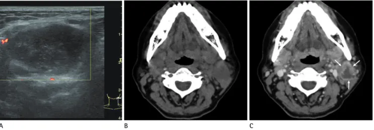
Fig. 1. A 51-year-old female with multifocal Warthin's tumors.
A. Axial neck sonogram shows a mixed hypoechoic and anechoic, well defined mass measuring 3.0 × 2.4 cm in the left inferior aspect of the pa- rotid tail.
B. Non-contrast enhanced axial CT scan reveals a low density mass without internal calcification.
C. Contrast enhanced study shows a well-defined lesion with central low density, including necrosis and thick irregular rim-enhancement (ar- rows).
A B C
Hyelarn Lee, et al
submit.radiology.or.kr J Korean Soc Radiol 2011;65(3):213-216
215
scans. Although the definitive diagnosis requires a histologic confirmation after surgery, clinicians and radiologists should be concerned about lymph node pathology superimposed on a WT, particularly when an unusual imaging finding of a WT is suspected.
REFERENCES
1. Barnes L. Pathology and genetics of head and neck tu- mours. Lyon: IARC Press, 2005
other as a cystic WT with irregular thick rim enhancement, which is a different imaging finding from that of a typical WT. The solid aspect was composed only of a WT and the other was composed of TB within a WT. No discernable TB lymphadenitis was observed in the neck on imaging, surgical, or pathological findings. Our patient showed no evidence of concomitant active TB elsewhere in the body.
In conclusion, we report on a rare case of a TB infection within a WT of the parotid gland with good correlation be- tween findings of the histological sections and those of CT
Fig. 3. A. The homogeneously enhancing nodule in Fig. 2 is composed of proliferative oncocytic columnar epithelium and lymphocytic infiltra- tion within the stroma, which was consistent with a solid WT (H&E stain, original magnification × 12).
B. Mass with central low density in Fig. 2 shows TB granuloma (asterisk) in the lymphoid stroma, intermingled with WT (W) (H&E, original magni- fication × 100).
C. AFB were found within TB granuloma (Ziehl-Neelsen stain, original magnification × 1,000).
Note.-AFB = acid-fast bacilli, TB = tuberculosis, WT = Warthin's tumor
A B C
Fig. 2. A. Contrast-enhanced coronal salivary gland CT shows two lesions that differed in appearance: the upper lesion is a small homogenous enhancing solid nodule (arrowheads) and the lower lesion, detected by sonogram, is an irregular, thick-rim enhanced complex mass (arrows) in the superficial lobe of the left parotid gland.
B. Corresponding gross specimen of parotid lesions (arrowheads; upper solid nodule, arrows; lower necrotic complex mass).
A B
Tuberculosis Infection within a Warthin’s Tumor of the Parotid Gland
submit.radiology.or.kr
J Korean Soc Radiol 2011;65(3):213-216
216
rium tuberculosis infection within parotid gland Warthin tumor. J Craniofac Surg 2008;19:1561-1565
7. Wen YH, Chen PR, Wu HP. Tuberculosis infection within a Warthin’s tumor of the parotid gland. Tzu Chi Med J 2008;
20:332-334
8. Franzen A, Franzen CK, Koegel K. [Tuberculosis of the pa- rotid gland: a rare differential diagnosis of parotid tumor].
Laryngorhinootologie 1997;76:308-311
9. Oh JH, Cho JH, Kim BG, Nam SK, Kim MS. A case of War- thin’s tumor associated with primary tuberculosis of the parotid gland in a patient with acquired immunodeficien- cy syndrome. Korean J Otorhinolaryngol-Head Neck Surg 2009;52:376-379
10. Barnes PF, Bloch AB, Davidson PT, Snider DE Jr. Tuberculo- sis in patients with human immunodeficiency virus infec- tion. N Engl J Med 1991;324:1644-1650
2. Ikarashi F, Nakano Y, Nonomura N, Kawana M. Radiologi- cal findings of adenolymphoma (Warthin’s tumor). Auris Nasus Larynx 1997;24:405-409
3. Watanabe M, Nakayama T, Koduka Y, Katoh H, Hirokawa Y, Inoue R, et al. Mycobacterium tuberculosis infection with- in Warthin’s tumor: report of two cases. Pathol Int 2001;
51:797-801
4. Seifert G, Bull HG, Donath K. Histologic subclassification of the cystadenolymphoma of the parotid gland. Analysis of 275 cases. Virchows Arch A Pathol Anat Histol 1980;
388:13-38
5. Marioni G, Marchese-Ragona R, Marino F, Poletti A, Otta- viano G, de Filippis C, et al. MALT-type lymphoma and Warthin's tumour presenting in the same parotid gland.
Acta Otolaryngol 2004;124: 318-323
6. Ozcan C, Apa DD, Aslan G, Gülhan S, Görür K. Mycobacte-
Warthin씨 종양 내에서 발생한 폐외결핵: 증례 보고
이혜란 · 서상일
Warthin씨 종양은 이하선에 생기는 흔한 양성종양이다. Warthin씨 종양은 림프 세포성 간질이 있으므로 림프절 관련 질 환들이 동반되어 생길 수 있으며 이때는 전형적인 Warthin씨 종양과 다르게 보일 수 있다. 이하선에 Warthin씨 종양만으 로 이루어진 병변과 Warthin씨 종양 내에 결핵이 동반된 경우를 조직 소견과 컴퓨터단층촬영 소견을 비교 분석하였다. 환 자는 표층적 이하선 절제술을 시행하였으며 항결핵제 복용 후 합병증 없이 퇴원하였다. 전형적인 Warthin씨 종양과 다르 게 보이는 병변이 있으면 림프절 관련 질환이 Warthin씨 종양에 동반되어 있을 가능성도 염두해 두어야 한다.
고려대학교 의과대학 구로병원 영상의학과학교실