Introduction
As the interest in health and fitness increases, gluteal muscle exercise has received much attention from clinicians, athletes, and general people as well in recent years. Gluteus medius (Gmed) and gluteus maximus (Gmax) contribute to improving athletic performance, preventing injuries of lower extremity, supplying structural stability, and maintaining lower extremity alignment (Al-Hayani, 2009; Gottschalk et al, 1989). Gluteal weakness has a close relationship
(r=-.74) with knee pathology, such as iliotibial band syndrome, patellofemoral pain syndrome, genu val- gum, and anterior cruciate ligament injury (Bolgla and Uhl, 2005; Reiman et al, 2009). Moreover, it can cause trochanteric bursitis and chronic ankle sprains (Presswood et al, 2008; Taunton et al, 2002).
Clinicians generally aim at the hip abductors while treating these lower extremity injuries (Akuthota and Nadler, 2004). A wide variety of hip abduction ex- ercises have been investigated to strengthen gluteal muscles (Macadam et al, 2015; Reiman et al, 2012).
Corresponding author: Sang-hyun Cho [email protected]
Effect of Hip Flexion and Internal Rotation on the Hip Abductor Muscle Activity During Side-Lying Hip Abduction in
Subjects With Gluteus Medius Weakness
Hye-jin Park1, BPT, PT, Sang-hyun Cho2,3, PhD, MD
1Dept. of Physical Therapy, The Graduate School, Yonsei University
2Dept. of Physical Therapy, College of Health Science, Yonsei University
3Dept. of Ergonomic Therapy, The Graduate School of Health and Environment, Yonsei University
Abstract
1)Background: Many previous studies recommended the side-lying hip abduction (SHA) exercise for targeting the gluteus medius (Gmed) and gluteus maximus (Gmax) muscle activity while the decreasing tensor fasciae latae (TFL) activation. Mischoice of hip position and angle in SHA may increase the risk of lower extremity injuries and undesirable muscle activation. However, information is limited on the effect of composite hip flexion angles and hip rotation on the gluteal muscle activity during SHA.
Objects: This study aimed to compare muscle activity (Gmed, TFL, and Gmax) and activity ratios (Gmed/TFL, Gmax/TFL, and Gmed/Gmax) using surface electromyography (EMG) during SHA exercise at three different hip flexion angles either with or without internal rotation (IR) in subjects with Gmed weakness. We hypothesized that applying hip flexion and IR during SHA would increase gluteal muscle activity and decrease TFL activity.
Methods: Muscle activity and activity ratios in 20 volunteers with Gmed weakness during 6 different SHA were investigated with surface EMG. One-way repeated-measures analysis of variance was used to determine the statistical significance.
Results: Significant differences were found among the six different exercises for Gmed (F2,41=11.817, p<.001) and Gmax (F3,52=5.513, p=.003) muscle activity, and Gmed/TFL (F3,54=8.735, p<.001) and Gmax/TFL (F2,37=4.019, p=.028) activity ratios.
Conclusion: Applying hip flexion is an effective method for increasing gluteal activity, and it elicits great Gmed/TFL and Gmax/TFL activity ratios during SHA in subjects with Gmed weakness.
Key Words: Gluteus medius weakness; Hip flexion; Hip internal rotation; Side-lying hip abduction exercise.
Among the exercises, many previous studies recom- mended side-lying hip abduction (SHA) exercise for targeting Gmed muscle activity while decreasing tensor fasciae latae (TFL) activation (Ekstrom et al, 2007;
McBeth et al, 2012; Willcox and Burden, 2013).
According to Distefano et al (2009), Gmed activity was greater than almost 16% of maximal voluntary iso- metric contraction (MVIC) than band walk, side-way hop exercise, single-limb squat and single-limb deadlift.
Muscle activity is generally quantified with electro- myography (EMG), and exercises that generate high activation are the best for strengthening (Andersen et al, 2006). Different hip positions and angles in SHA can likely affect EMG activity because they are asso- ciated with external torque, gravitational moment arm, and direction of muscle fiber (Distefano et al, 2009;
Kang et al, 2013; Soderberg, 1983). Mischoice of hip position and angle in exercise may increase the risk of lower extremity injuries and undesirable muscle activa- tion (Willcox and Burden, 2013). According to Fredericson and Wolf (2005), Gmed weakness may arise from compensation using the TFL to dominant, thus leading to tightness and hypertonicity of the TFL.
According to the concept of synergist dominance, in- dividuals with Gmed weakness should rely on TFL to accomplish the hip abduction required for gait and ac- tivities of daily living, thus contributing to “TFL domi- nance” associated with further weakness of Gmed due to disuse (Sahrmann, 2002).
Previous studies compared SHA exercises in vari- ous hip positions, such as traditional SHA and clam exercise (Boren et al, 2011; Selkowitz et al, 2013), SHA with internal (IR) or external rotation (ER) (Lee et al, 2014; McBeth et al, 2012; Philippon et al, 2011), and clam with various hip flexion (FLX) an- gles (Distefano et al, 2009; Willcox and Burden, 2013), to increase Gmed activity. The clam exercise is commonly used for hip abductor rehabilitation, but a low gluteal activity was reported (Distefano et al, 2009). Lee et al (2014) reported that the SHA with IR (SHA-IR) could be effective to increase gluteal activity, and McBeth et al (2012) reported the TFL
was more active than the gluteal muscles when per- forming SHA with ER (SHA-ER). The reason is that the hip external rotator (gluteal muscles) becomes more active because of gravity and extended moment arm, but the internal rotator (TFL) becomes under- active because of less gravity and an active in- sufficiency during SHA-IR than during SHA-ER.
Through the length–tension relationship, the shortened TFL reaches an active insufficiency that is difficult to produce force during SHA-IR (Neumann, 2010).
SHA exercise has been used a common therapeutic exercise to generate adequate neuromuscular control and hip abductor strength (McBeth et al, 2012). Of the sev- eral studies that have investigated hip abductor recruit- ment during the SHA, however, limited information is available on the effect of composite hip positions and angles on the sagittal, transverse, and frontal planes.
SHA with FLX (SHA-F) extends the gravitational mo- ment arm of the gluteal muscles similar to SHA-IR (Neumann, 2010). To overcome the great external tor- que, the gluteal muscles contract more than the TFL.
However, studies experimenting on the effects of hip FLX angles on the hip abductor activity during SHA are difficult to find. Moreover, we could not confirm any study considering SHA with FLX and IR together.
This study aimed to compare Gmed, TFL, and Gmax muscle activity and activity ratios (Gmed/TFL, Gmax/TFL and Gmed/Gmax) using surface EMG during SHA exercise at three different FLX angles either with or without IR in subjects with Gmed weakness.
Particularly, we sought to ascertain which position could best activate the gluteal muscles. We hypothesized that applying hip FLX and IR during SHA would increase gluteal muscle activity and decrease TFL activity.
Methods
Subjects
A power analysis performed using the results of a pilot study with 5 subjects demonstrated that this study would require at least 6 subjects to satisfy a
level of .05, power of .80, and effect size of .19 (G-power software 3.1.2, Franz Faul, University of Kiel, Kiel, Germany). A total of 86 subjects took the manual muscle testing (MMT) to determine Gmed weakness, and 20 subjects (6 males and 14 females) voluntarily participated in this study. Through MMT by Kendall’s muscle grading, the subjects that were graded 3/5 or less were considered to have Gmed weakness (Bewyer et al, 2009; Kendall et al, 2005).
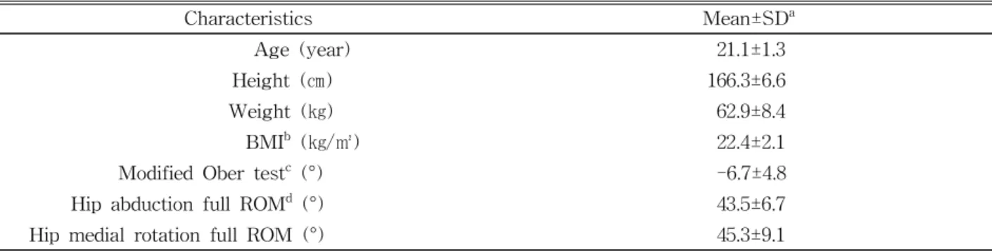
The leg used to kick the soccer ball was determined as the dominant (test) leg (Reimer and Wikstrom, 2010). Subjects who could hold isometric hip abduc- tion for 5 sec in the side-lying position and had a normal range of motion (ROM) of hip were included (Kim et al, 2011). Subjects who met one or more of the following conditions were excluded: (1) history of lower extremity injury or surgery within the past two years (2) cardiopulmonary, neurological, or mus- culoskeletal diseases or pain, (3) shortness of TFL (Ober test), and (4) body mass index above 25 (Flegal et al, 1998; McBeth et al, 2012). The general characteristics of the subjects are presented in Table 1. Prior to the test, all subjects read the experimental protocol and signed an informed consent form ap- proved by the Yonsei University Wonju Institutional Review Board (1041849-201608-BM-043-02).
Surface EMG recording and data processing
Surface EMG (TeleMyo DTS, Noraxon Inc., Scottsdale,
AZ, USA) with a wireless telemetry system was used.
Disposable bipolar electrodes (Ag/AgCl) were adhered with a 2 ㎝ inter-electrode distance after skin prepara- tion to reducing skin impedance (Cram et al, 1998).
The positions and all details of electrodes were set as instructions by Cram (Criswell, 2010). For the Gmed, the electrodes were set to the proximal third of the range between the iliac crest and the greater trochanter. The TFL electrodes were set approx- imately 2 ㎝ under the anterior superior iliac spine.
For the upper Gmax, the electrodes were set to half of the range between the sacral vertebrae and the trochanter of the femur at the level of the trochanter.
The Noraxon MyoResearch 1.06 software (Noraxon Inc., Scottsdale, AZ, USA) was used for the ampli- tude of EMG signal processing. The MVIC was used to normalize the EMG data and to confirm muscle crosstalk. To collect the MVIC data, we used stand- ard positions based on the MMT (Kendall et al, 2005). Subjects repeated the MVIC three times with a 10 sec rest between each trial to obtain the peak value (Boren et al, 2011). Subjects maintained each trial for 5 sec with a 3 min rest between each mus- cle contraction (Soderberg and Knutson, 2000). The average value of the middle 3 sec of the 5 sec peri- od was used for data collection (Ayotte et al, 2007).
All EMG data were expressed as percentages of the MVIC. The intra-class correlation coefficient (ICC) for MVICs of Gmed, TFL, and Gmax were .94 [95%
confidence interval (CI)=.89-.97], .86 (.74-.93), and .89
Characteristics Mean±SDa
Age (year) 21.1±1.3
Height (㎝) 166.3±6.6
Weight (㎏) 62.9±8.4
BMIb (㎏/㎡) 22.4±2.1
Modified Ober testc (°) -6.7±4.8
Hip abduction full ROMd (°) 43.5±6.7
Hip medial rotation full ROM (°) 45.3±9.1
amean±standard deviation,bbody mass index, ca negative number in modified Ober test indicates hip adduction and a positive number indicates abduction, drange of motion.
Table 1. Demographic characteristics of subjects (N=20)
(.79-.94), respectively. The EMG data were sampled at 1000 ㎐, amplified, band-pass filtered (20∼450 ㎐, Lancosh FIR), and notch filtered (92 ㏈ rejection ra- tio at 60 ㎐). Then, the root mean square with a 50
㎳ moving window was calculated.
Three-dimensional (3D) motion analysis system
To control the ROM and positions of hip, a 3D motion analysis system (MyoMotion Research Pro, Noraxon Inc., Scottsdale, AZ, USA) was used. The wireless sensors include inertial measurement units consisting of tri-axial accelerometers, gyroscopes, and a magnetometer (Poepel et al, 2014). Three sensors were attached to a subject’s bony area of the sacrum (frontal plane) and thigh (sagittal attachment to the greater trochanter level and slightly above the knee cap in line with the femur) according to the Noraxon standard manual (Noraxon Inc., Scottsdale, AZ, USA).
Calibration in the standing position was performed to define the 0° of ROM (Struzik et al, 2015). Sampling frequency was set to 100 ㎐, and the MyoMotion Research ver. 3.6 software (Noraxon Inc., Scottsdale, AZ, USA) was used. The Vicon 3D motion capture system is used as the gold standard reference, and the correlation coefficient between Vicon and MyoMotion was .99 (Balasubramananian, 2013).
Procedures
Before the data collection, all subjects stretched their lower extremities and jogged at a self-selected moderate speed for 5 min to decrease possible incon- venience or pain (McBeth et al, 2012). Subjects were instructed on the correct way to perform 6 different SHA and practiced until they felt comfortable (Distefano et al, 2009). To prevent substitution from the quadratus lumborum, the Stabilizer Pressure Biofeedback Unit (PBU) was used (Cairns et al, 2000;
Mills et al, 2005). The PBU was positioned under the waist between the distal ribs and the iliac crest with the air bag inflated. The subject was asked to sus- tain a stable pressure of 40±5 ㎜Hg during SHA. To
use the PBU without the pelvic compensation, enough familiarization time was allowed.
After a 10 min rest, a modified Ober test and an active ROM of hip were measured with a smartphone inclinometer application called Clinometer (Plaincode Software Solutions, Stephanskirchen, Germany) on a Galaxy S6 (Samsung Electronics, Suwon, Gyeonggido, Korea) (Shin et al, 2012). The application, which uses a three-axis linear accelerometer, is a slope finder tool and shows good intra-observer reliability (ICC>.80) (Milani et al, 2014). In the same position of the Gmed MMT, the principal investigator (PI) conducted a modified Ober test, and the other investigator placed the Clinometer on the lateral epicondyle of the subject and read scale (Kendall et al, 2005; Reese and Bandy, 2003). The mean value of two repetitions was used. If the leg did not drop 10° below the horizontal, the subject was regarded as TFL shortness and excluded.
Also full ROM of hip abduction, FLX and IR were measured using the Clinometer.
Three hip FLX angle variations (0°, 15°, and 30°) of the SHA were performed without rotation of hip (neutral hip). The three FLX angle variations were re- peated with the IR. A target bar was positioned at 35°
of the hip abduction and adjusted to each subject (McBeth et al, 2012). The subjects were instructed to abduct until they touched the bar and down over 3 sec in each position to the beat of an electronic met- ronome (Nyland et al, 2004). The subject repeated each position three times and held the SHA for 5 sec. The EMG average value of the middle 3 sec was used for data analysis (Ayotte et al, 2007). A 2 min rest be- tween each exercise was given (Boren et al, 2011).
The order of exercises was randomized by drawing lots to prevent muscle fatigue or learning effect.
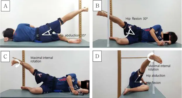
SHA with neutral hip and flexion 0°(SHA-NF 0)
Subjects lay on their non-dominant side down with the hip and knee flexed for stability. The trunk, pelvis, and test leg were straightened with full knee extension. Then subjects abducted the test
leg at 35° along with the axis of the target bar with toes pointing forward until the lateral ankle touched the bar (Boren et al, 2011; McBeth et al, 2012) (Figure 1A). The position was maintained for 5 sec while maintaining a neutral hip and knee extension. The PI monitored every hip motion and angle targeted and the pelvic compensation through the 3D motion analysis system, MyoMotion. The subjects controlled the lumbar and pelvis monitoring by the PBU.
SHA with neutral hip and flexion 15°and 30°(SHA-NF 15 and 30)
To perform these exercises, the subjects were positioned in the same way as in SHA-NF 0 and asked to flex test hip at 15° or 30° in each exercise, respectively, before starting the SHA. A plastic ref- erence bar guided the FLX angle, and MyoMotion controlled the angle. The subjects flexed their hip until the front ankle drew close to the bar, and they performed SHA in the same way as in SHA-NF 0 (Figure 1B).
SHA with hip internal rotation and flexion 0°(SHA-IRF 0)
The subjects performed SHA-IRF 0 in the same way as in SHA-NF 0, excluding the hip rotation.
The subjects were asked to internally rotate the hip as far as possible and direct toes down toward the table (McBeth et al, 2012). They were instructed to remain in the neutral pelvic position and not to ro- tate the pelvis forward. The subjects abducted their hip until their posterolateral ankle touched the target bar (Figure 1C).
SHA with hip internal rotation and flexion 15°and 30°(SHA-IRF 15 and 30)
The subjects performed SHA-IRF 15 or 30 in the same way as SHA-NF 15 or 30, excluding the IR.
The subjects were asked to internally rotate the hip as far as possible, flex it at 15° or 30° until it is close to the reference bar, and perform SHA (Figure 1D). The PI controlled the subjects’ IR, FLX, abduc- tion, and any compensatory movement through the MyoMotion.
A B
C D
Figure 1. Side-lying hip abduction exercise. A: side-lying hip abduction with neutral hip and hip flexion 0° (SHA-NF 0), B: side-lying hip abduction with neutral hip and hip flexion 30° (SHA-NF 30), C: side-lying hip abduction with internal rotation and hip flexion 0° (SHA-IRF 0), D: side-lying hip abduction with internal rotation and hip flexion 30° (SHA-IRF 30).
Statistical analysis
The test-retest reliability of the EMG data was as- sessed by calculating ICC, CI based on the following criteria: <.69=poor, .70∼.79=moderate, .80∼.89=good, and .90∼.99=excellent (T’Jonck et al, 1996).
One-way repeated-measures analysis of variance was used to test the differences in the EMG activity (Gmed, TFL, and Gmax) and activity ratios (Gmed/TFL, Gmax/TFL, and Gmed/Gmax) among the six differ- ent exercises (SHA-NF 0, 15, and 30 and SHA-IRF 0, 15, and 30). The statistical significance level was set to α=.05. All statistical analyses were performed us- ing SPSS ver. 21.0 (SPSS Inc., Chicago, IL, USA). The Holm-Bonferroni method was performed to reduce type I error when a significant difference was found (Berry et al, 2015; Holm, 1979). This test can significantly decrease type II error compared with the conservative Bonferroni correction (Eichstaedt et al, 2013; Perneger, 1998).
Results
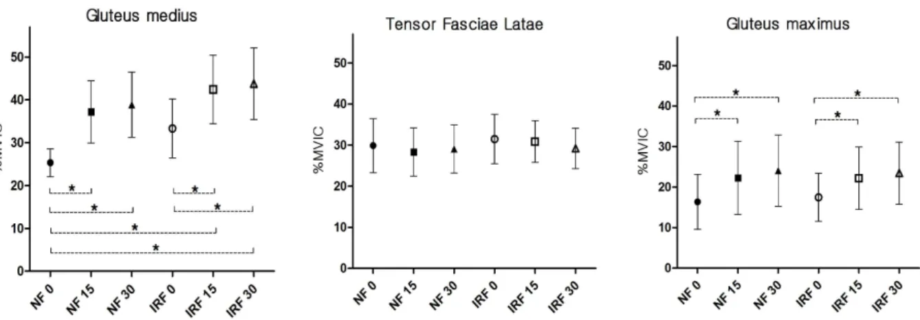
Muscle activity of Gmed, TFL and Gmax Significant differences were found among the six
different exercises for Gmed (F2,41=11.817, p<.001) and Gmax (F3,52=5.513, p=.003) muscle activity as shown on Figure 2. SHA-NF 15 (p<.001) and SHA-NF 30 (p<.001) produced a significantly greater Gmed activ- ity than SHA-NF 0. SHA-IRF 15 (p<.001) and SHA-IRF 30 (p<.001) showed a significantly greater Gmed activity than SHA-NF 0 and SHA-IRF 0. The Gmax activity during SHA-NF 15 (p=.010) and SHA-NF 30 (p=.005) was significantly greater than that during SHA-NF 0. The Gmax activity during SHA-IRF 15 (p=.002) and SHA-IRF 30 (p=.001) was significantly greater than that during SHA-IRF 0. No significant difference was found among the six ex- ercises for TFL activity (F2,33=.237, p=.637).
Muscle activity ratios of Gmed/TFL, Gmax/TFL and Gmed/Gmax
Significant differences were found among the six different exercises for Gmed/TFL (F3,54=8.735, p<.001) and Gmax/TFL (F2,37=4.019, p=.028) muscle activity ratios as shown on Figure 3. SHA-NF 15 (p<.001) and SHA-NF 30 (p<.001) showed significantly great- er Gmed/TFL ratios than SHA-NF 0. SHA-IRF 15 (p<.001) and SHA-IRF 30 (p=.001) showed sig-
Figure 2. The muscle activity of gluteus medius, tensor fasciae latae, and gluteus maximus for six different side-lying hip abduction exercises. (CI: confidence interval, MVIC: maximal voluntary isometric contraction, NF 0, 15, and 30: side-lying hip abduction with neutral hip and flexion 0°, 15°, and 30°, IRF 0, 15, and 30: side-lying hip abduction with hip internal rotation and flexion 0°, 15°, and 30°), *denoted a significant difference on one-way repeated-measures analysis of variance with Holm-Bonferroni method.
nificantly greater Gmed/TFL ratios than SHA-IRF 0.
The Gmax/TFL activity ratios during SHA-NF 15 (p=.010) and SHA-NF 30 (p=.006) were significantly greater than those during SHA-NF 0. Moreover, the ratio during SHA-IRF 30 (p=.002) was significantly greater than that during SHA-IRF 0. No significant difference was found (F1,26=.955, p=.367) among the six exercises for Gmed/Gmax ratios.
Discussion
This study investigated the effect of varying hip FLX angles and IR on hip abductor activity during SHA in subjects with Gmed weakness. The Gmed and Gmax activity and the Gmed/TFL and Gmax/TFL ra- tios with hip FLX at 15° or 30° were significantly greater than at 0° regardless of IR. The findings re- vealed that the application of SHA-F could increase the gluteal activity and activity ratios to the TFL.
These results partially supported our research hypotheses. To our knowledge, this study is the first to examine the effect of FLX angles and IR on hip abductor activity during SHA.
Distefano et al (2009) and Willcox and Burden
(2013) studied the effect of different hip FLX angles during clam exercise on the gluteal activity. Distefano et al (2009) reported no significance difference in glu- teal activity as the hip angle changed. Nevertheless, Willcox and Burden (2013) found that the Gmed ac- tivity with the FLX at 60˚ was greater than at 0°
because the moment arm also changed as the FLX angle shifted from 0° to 60°. In current study, both Gmed and Gmax activity during SHA-F at 15° or 30° were significantly greater than those at 0° re- gardless of IR. The gravitational moment arms of both muscles extended during SHA-F. As the mo- ment arm is lengthened, muscle activation becomes greater to surmount external torque (Delp et al, 1999;
Neumann, 2010). Gluteal activity during SHA-F was higher than that during clam exercise as the moment arms were longer because of the maintenance of the knee extension unlike in the clam exercise with knee flexion (Hoy et al, 1990). Furthermore, Gmax activa- tion during SHA-F was significantly greater than without FLX unlike in the clam exercise. During clam exercise, the gluteal muscles act as external rotators and are less affected by gravity. However, during SHA-F, the center of gravity of the leg is positioned anteriorly as the hip is flexed (Willcox Figure 3. The muscle activity ratios of gluteus medius, tensor fasciae latae, and gluteus maximus
for six different side-lying hip abduction exercises. (CI: confidence interval, Gmed: gluteus medius, TFL: tensor fasciae latae, Gmax: gluteus maximus, NF 0, 15, and 30: side-lying hip abduction with neutral hip and flexion 0°, 15°, and 30°, IRF 0, 15, and 30: side-lying hip abduction with hip internal rotation and flexion 0°, 15°, and 30°), *denoted a significant difference on one-way repeated-measures analysis of variance with Holm-Bonferroni method.
and Burden, 2013), and the muscles are challenged to go against gravity, thus leading to more activation.
Therefore, SHA-F is an effective method to induce gluteal muscles activation.
The gluteal activity and activity ratios of Gmed/TFL and Gmax/TFL during SHA-IRF 15 or 30 significantly increased compared with those during SHA-IRF 0 and were similar to those during SHA-NF except the Gmax/TFL ratio during SHA-IRF 15. The average value of Gmax/TFL ratio during SHA-IRF 15 (.744) also increased compared with that during SHA-IRF 0 (.596), but no statistical difference was found. These findings indicate that SHA-IRF could increase the glu- teal activity and ratios in the same way as SHA-NF.
Lee et al (2014) reported that SHA-IR significantly in- creased the Gmed activity and Gmed/TFL ratio com- pared with SHA-ER or SHA with neutral hip. However, we could not confirm the statistical differences between SHA-NF and SHA-IRF 0, 15, and 30 at each FLX angle, respectively. There were significant increases only in the Gmed muscle activity during SHA-IRF 15 and 30 than SHA-NF 0. But the results could not make sure that these were caused by hip IR or FLX.
Although no statistical difference was found, most of the average values during SHA-IRF (except Gmax ac- tivity and Gmax/TFL ratio during SHA-IRF 15 and 30) tended to increase compared with those during SHA-NF in terms of each FLX angle. According to previous studies, an increment in hip FLX increases IR and causes a potential switching of the moment arm of the gluteal muscles (Delp et al, 1999; Neumann, 2010).
Therefore, SHA-F inducing IR can extend the leverage of gluteal muscles and facilitate the muscles.
The activity ratios of Gmed/TFL and Gmax/TFL during SHA-F significantly increased unlike those without FLX regardless of IR. The increased activity of Gmed and Gmax was assumed to affect the ratios even though the TFL activity did not decrease significantly. Practically, the average value of the TFL activity during SHA-F 15 and 30 decreased compared with that during SHA-F 0 regardless of IR, but the decrease had no statistical significance
(Figure 2). As the hip is flexed, total muscle torque necessary for SHA-F become increased, thus the hip abductors are more challenged to go against external torque, leading to more activation (Neumann, 2010).
The TFL is primary hip abductor (Gottschalk et al, 1989). There were no statistical significances of the TFL activity among the SHA-F 0, 15, and 30, how- ever the activity ratios of Gmed/TFL and Gmax/TFL during SHA-F significantly increased. Although we did not compare contribution levels of each muscle among 6 SHA exercises, our results clearly show that the TFL was activated to a lesser extent than the Gmed or Gmax. This finding is consistent with Willcox and Burden (2013) who found that in- cremental FLX angle increases gluteal activation while limiting TFL activity during the clam exercise.
The Gmed/Gmax ratios of the six exercises were not significantly different. We assume that the result was due to the similarity in tendency of variations in both Gmed and Gmax activity. Because both the muscle activity increased synchronistically as the FLX angle increased.
The abductors were nearly fully shortened (contracted) at 35° of abduction when we collected data. In the length-tension relationship, fully short- ened abductors reach an active insufficiency and be- come difficult to produce force (Neumann et al, 1988).
Performing the SHA-IRF allows the shortened gluteal muscles to be placed at an appropriate length to pro- duce force. Consequently, it can result in more acti- vation of the gluteal muscles because of gravity and extended moment arm (Arnold et al, 2000). Similarly, a fully shortened TFL reaches an active insufficiency and becomes underactive because of less gravity.
This study has several limitations. First, this work is a cross-sectional study. Longitudinal studies are warranted to determine the long-term effect of hip FLX and IR on hip abductor activity during SHA.
Second, we used one-way repeated-measures analy- sis of variance to assess the EMG activity in 6 dif- ferent exercises. However, we could not determine the interaction between the hip FLX and IR. Further
research needs to perform more appropriate analysis to determine a significant interaction for hip angle by position. Third, only gluteal and TFL activity was measured in this study. The role of other recruited abductor activity (including gluteus minimus, sartorius, rectus femoris, etc.) was not measured. Finally, This study compared gluteal muscle activity during SHA;
however, we could not ensure that applying hip FLX is an effective method to induce gluteal activation in a closed chain position. Future research should com- pare the effect of hip FLX and IR on the hip abduc- tor activity between open chain and closed chain exercises.
Conclusion
This study measured the Gmed, TFL, and Gmax muscle activity and the Gmed/TFL, Gmax/TFL, and Gmed/Gmax muscle activity ratios to demonstrate the effect of varying hip FLX angles and IR during SHA on subjects with Gmed weakness. The Gmed and Gmax activity and the Gmed/TFL and Gmax/TFL activity ratios with hip FLX at 15° or 30° during SHA were significantly greater than those at 0° re- gardless of IR. Therefore, applying hip FLX is an ef- fective method for increasing gluteal activity and eliciting greater Gmed/TFL and Gmax/TFL activity ratios during SHA in subjects with Gmed weakness.
References
Akuthota V, Nadler SF. Core strengthening. Arch Phys Med Rehabil. 2004;85(3 Suppl 1):S86-S92.
Al-Hayani A. The functional anatomy of hip abductors.
Folia Morphol (Warsz). 2009;68(2):98-103.
Andersen LL, Magnusson SP, Nielsen M, et al.
Neuromuscular activation in conventional ther- apeutic exercises and heavy resistance exercises:
Implications for rehabilitation. Phys Ther. 2006;
86(5):683-697.
Arnold AS, Salinas S, Asakawa DJ, et al. Accuracy of muscle moment arms estimated from MRI-based musculoskeletal models of the lower extremity. Comput Aided Surg. 2000;5(2):108-119.
Ayotte NW, Stetts DM, Keenan G, et al.
Electromyographical analysis of selected lower ex- tremity muscles during 5 unilateral weight-bear- ing exercises. J Orthop Sports Phys Ther.
2007;37(2):48-55.
Balasubramanian S. Comparison of angle measure- ments between Vicon and MyoMotion systems.
Arizona State University. 2013.
Berry JW, Lee TS, Foley HD, et al. Resisted side step- ping: The effect of posture on hip abductor muscle activation. J Orthop Sports Phys Ther. 2015;45(9):
675-682. http://dx.doi.org/10.2519/jospt.2015.5888 Bewyer KJ, Bewyer DC, Messenger D, et al. Pilot
data: Association between gluteus medius weak- ness and low back pain during pregnancy. Iowa Orthop J. 2009;29:97-99.
Bolgla LA, Uhl TL. Electromyographic analysis of hip rehabilitation exercises in a group of healthy subjects. J Orthop Sports Phys Ther. 2005;35(8):
487-494.
Boren K, Conrey C, Le Coguic J, et al.
Electromyographic analysis of gluteus medius and gluteus maximus during rehabilitation exercises.
Int J Sports Phys Ther. 2011;6(3):206-223.
Cairns MC, Harrison K, Wright C. Pressure biofeed- back: A useful tool in the quantification of ab- dominal muscular dysfunction? Physiotherapy.
2000;86(3):127-138.
Cram JR, Kasman GS, Holtz J. Electrode Placement.
Introduction to surface electromyography. 1st ed.
Gaithersburg MD, Aspen Publishers, 1998:360-371.
Criswell E. Cram’s Introduction to Surface Electromyography. 2nd ed. Sudbury, MA, Jones
& Bartlett Publishers, 2010:353-360.
Delp SL, Hess WE, Hungerford DS, et al. Variation of rotation moment arms with hip flexion. J Biomech. 1999;32(5):493-501.
Distefano LJ, Blackburn JT, Marshall SW, et al. Gluteal
muscle activation during common therapeutic exercises. J Orthop Sports Phys Ther. 2009;
39(7):532-540. http://dx.doi.org/10.2519/jospt.2009.2796 Eichstaedt KE, Kovatch K, Maroof DA. A less con-
servative method to adjust for familywise error rate in neuropsychological research: The Holm’s sequen- tial Bonferroni procedure. NeuroRehabilitation. 2013;
32(3):693-696. http://dx.doi.org/10.3233/NRE-130893 Ekstrom RA, Donatelli RA, Carp KC. Electromyographic analysis of core trunk, hip, and thigh muscles during 9 rehabilitation exercises. J Orthop Sports Phys Ther. 2007;37(12):754-762. http://dx.doi.org/
10.2519/jospt.2007.2471
Flegal KM, Carroll MD, Kuczmarski RJ, et al.
Overweight and obesity in the united states:
Prevalence and trends, 1960–1994. Int J Obes Relat Metab Disord. 1998;22(1):39-47.
Fredericson M, Wolf C. Iliotibial band syndrome in runners: Innovations in treatment. Sports Med.
2005;35(5):451-459.
Gottschalk F, Kourosh S, Leveau B. The functional anatomy of tensor fasciae latae and gluteus medius and minimus. J Anat. 1989;166:179-189.
Holm S. A simple sequentially rejective multiple test procedure. Scand J Statist. 1979;6(2):65-70.
Hoy MG, Zajac FE, Gordon ME. A musculoskeletal model of the human lower extremity: The effect of muscle, tendon, and moment arm on the mo- ment-angle relationship of musculotendon actua- tors at the hip, knee, and ankle. J Biomech.
1990;23(2):157-169.
Kang SY, Jeon HS, Kwon O, et al. Activation of the gluteus maximus and hamstring muscles during prone hip extension with knee flexion in three hip abduction positions. Man Ther. 2013;18(4):
303-307. http://dx.doi.org/10.1016/j.math.2012.11.006 Kendall F, McCreary E, Provance P, et al. Muscles;
Testing and function with posture and pain. 5th ed. Baltimore, Williams & Wilkins, 2005:19-24, 391-394, 425, 433-436.
Kim SJ, Kwon OY, Yi CH, et al. Comparison of ab- dominal muscle activity during a single-legged
hold in the hook-lying position on the floor and on a round foam roll. J Athl Train. 2011;46(4):
403-408.
Lee JH, Cynn HS, Kwon OY, et al. Different hip rota- tions influence hip abductor muscles activity dur- ing isometric side-lying hip abduction in subjects with gluteus medius weakness. J Electromyogr Kinesiol. 2014;24(2):318-324. http://dx.doi.org/10.1016/
j.jelekin.2014.01.008
Macadam P, Cronin J, Contreras B. An examination of the gluteal muscle activity associated with dynamic hip abduction and hip external rotation exercise: A systematic review. Int J Sports Phys Ther. 2015;10(5):573-591.
McBeth JM, Earl-Boehm JE, Cobb SC, et al. Hip muscle activity during 3 side-lying hip-strength- ening exercises in distance runners. J Athl Train. 2012;47(1):15-23.
Milani P, Coccetta CA, Rabini A, et al. Mobile smart- phone applications for body position measurement in rehabilitation: A review of goniometric tools.
PM R. 2014;6(11):1038-1043. http://dx.doi.org/10.1016/
j.pmrj.2014.05.003
Mills JD, Taunton JE, Mills WA. The effect of a 10-week training regimen on lumbo-pelvic sta- bility and athletic performance in female ath- letes: A randomized-controlled trial. Phys Ther Sport. 2005;6(2):60-66.
Neumann DA. Kinesiology of the hip: A focus on mus- cular actions. J Orthop Sports Phys Ther. 2010;
40(2):82-94. http://dx.doi.org/10.2519/jospt.2010.3025 Neumann DA, Soderberg GL, Cook TM. Comparison of maximal isometric hip abductor muscle tor- ques between hip sides. Phys Ther. 1988;68(4):
496-502.
Nyland J, Kuzemchek S, Parks M, et al. Femoral anteversion influences vastus medialis and glu- teus medius emg amplitude: Composite hip ab- ductor emg amplitude ratios during isometric combined hip abduction-external rotation. J Electromyogr Kinesiol. 2004;14(2):255-261.
Perneger TV. What’s wrong with Bonferroni
adjustments. BMJ. 1998;316(7139):1236-1238.
Philippon MJ, Decker MJ, Giphart JE, et al.
Rehabilitation exercise progression for the glu- teus medius muscle with consideration for iliop- soas tendinitis an in vivo electromyography study. Am J Sports Med. 2011;39(8):1777-1785.
http://dx.doi.org/10.1177/0363546511406848
Poepel C, Feitsch J, Strobel M, et al. Design and evaluation of a gesture controlled singing voice installation. NIME. 2014:359-362.
Presswood L, Cronin J, Keogh JW, et al. Gluteus medius: Applied anatomy, dysfunction, assess- ment, and progressive strengthening. Strength Cond J. 2008;30(5):41-53.
Reese NB, Bandy WD. Use of an inclinometer to measure flexibility of the iliotibial band using the Ober test and the modified Ober test:
Differences in magnitude and reliability of measurements. J Orthop Sports Phys Ther.
2003;33(6):326-330.
Reiman MP, Bolgla LA, Lorenz D. Hip function’s in- fluence on knee dysfunction: A proximal link to a distal problem. J Sport Rehabil. 2009;18(1):
33-46.
Reiman MP, Bolgla LA, Loudon JK. A literature re- view of studies evaluating gluteus maximus and gluteus medius activation during rehabilitation exercises. Physiother Theory Pract. 2012:28(4);
257-268. http://dx.doi.org/10.3109/09593985.2011.604981 Reimer RC, Wikstrom EA. Functional fatigue of the
hip and ankle musculature cause similar alter- ations in single leg stance postural control. J Sci Med Sport. 2010;13(1):161-166. http://dx.doi.org/
10.1016/j.jsams.2009.01.001
Sahrmann S. Diagnosis and treatment of movement impairment syndromes. 1st ed. St Louis, Mosby, 2002:35-39.
Selkowitz DM, Beneck GJ, Powers CM. Which ex- ercises target the gluteal muscles while mini- mizing activation of the tensor fascia lata?
Electromyographic assessment using fine-wire electrodes. J Orthop Sports Phys Ther. 2013;
43(2):54-64. http://dx.doi.org/10.2519/jospt.2013.4116 Shin SH, Lee OS, Oh JH, et al. Within-day reliability of shoulder range of motion measurement with a smartphone. Man Ther. 2012;17(4):298-304.
http://dx.doi.org/10.1016/j.math.2012.02.010 Soderberg GL. Muscle mechanics and pathomechanics.
Their clinical relevance. Phys Ther. 1983;63(2):
216-220.
Soderberg GL, Knutson LM. A guide for use and in- terpretation of kinesiologic electromyographic data.
Phys Ther. 2000;80(5):485-498.
Struzik A, Konieczny G, Grzesik K, et al. Relationship between lower limbs kinematic variables and ef- fectiveness of sprint during maximum velocity phase. Acta Bioeng Biomech. 2015;17(4):131-138.
Taunton JE, Ryan MB, Clement D, et al. A retro- spective case-control analysis of 2002 running injuries. Br J Sports Med. 2002;36(2):95-101.
T’Jonck L, Lysens R, Grasse G. Measurements of scapular position and rotation: A reliability study.
Physiother Res Int. 1996;1(3):148-58.
Willcox EL, Burden AM. The influence of varying hip angle and pelvis position on muscle recruitment patterns of the hip abductor muscles during the clam exercise. J Orthop Sports Phys Ther. 2013;
43(5):325-331. http://dx.doi.org/10.2519/jospt.2013.4004
This article was received July 22, 2016, was re- viewed July 22, 2016, and was accepted August 24, 2016.