INTRODUCTION
Hypertrophic obstructive cardiomyopathy (HOCM) involves the ventricluar septum and less frequently segments of the right ventricle (1). Outflow obstruction of the left ventricle is the usual hemodynamic finding in HOCM and hypertrophic cardiomyopathy has right ventricular obstruction in 15% of cases (2). However, it is uncommon to find infundibular steno- sis of the right ventricle in the presence of left ventricular out- flow tract obstruction (2, 3). Management options for patients with HOCM accompanying infundibular stenosis of the right ventricle may include surgical removal of hypertrophied mus- cle bundles (4-6). Alcohol ablation therapy, known as a non- surgical therapy of HOCM, has been reported in the treatment of hypertrophic cardiomyopathy or infundibular pulmonary stenosis (7-9). However, alcohol ablation therapy in HOCM with infundibular stenosis of the right ventricle has not been reported. We report a case of HOCM with infundibular steno- sis of the right ventricle treated by alcohol ablation therapy, in a 28-yr-old male patient presenting with dyspnea on exer- tion.
CASE REPORT
A 28-yr-old male patient was referred to Inha Medical Center in July, 2000 for an echocardiogram. The patient had a 6-yr history of exertional dyspnea, for which he was treated medically. He had no prior history of medical and surgical diseases. His family members were healthy and had no his- tory of congenital heart disease. On physical examination, the peripheral pulses and blood pressure measurements were nor- mal. A 5/6 harsh, systolic ejection murmur accompanied by a thrill was heard between the apex and the lower left sternal border.
Basic laboratory tests, including CBC, lipid profiles and liver function were not remarkable. There was no significant change in the levels of cardiac enzymes. Urinalysis revealed no abnormalities. The chest roentgenogram showed a normal heart size. Electrocardiogram revealed increased amplitude of the P wave in lead II exceeding 2.5 mV and increased QRS voltages with inverted T waves in the precordial chest leads.
The echocardiogram showed asymmetric hypertrophy of the interventricular septum and systolic anterior motion of the mitral valve (Fig. 1), accompanying the left ventricular outflow tract obstruction and mitral incompetence. The
Sung Hak Park, Keum Soo Park, Hoon Gi Park, Hyo Jung Lee, Jeong Kee Seo, Ki Hoon Lee, Dae Hyeok Kim, Woo Hyung Lee, Cheol Whan Lee*, Myung Ki Hong*, Seong-Wook Park*, Seung Jung Park*
Department of Internal Medicine, Inha Medical Center, Inha University, Inchon; *Department of Medicine, Asan Medical Center, University of Ulsan, Seoul, Korea
Address for correspondence Hoon-Gi Park, M.D.
Department of Medicine, Inha Medical Center, Inha University, 7336 Taepyung-dong, Sujung-gu, Sungnam 461-192, Korea Tel : +82.31-755-2812, Fax : +82.31-720-5305 E-mail: [email protected]
585 J Korean Med Sci 2003; 18: 585-8
ISSN 1011-8934
Copyright � The Korean Academy of Medical Sciences
Hypertrophic Obstructive Cardiomyopathy with Infundibular Stenosis Treated by Alcohol Ablation Therapy
This report describes an uncommon case of hypertrophic obstructive cardiomyo- pathy (HOCM) accompanying infundibular stenosis of the right ventricle treated by alcohol ablation therapy, in a 28-yr-old male patient presenting with dyspnea on exertion. HOCM with infundibular stenosis was detected by echocardiogram and cardiac catheterization and patient has dynamic obstructions of both ventricu- lar outflow tracts. We performed alcohol ablation therapy to improve clinical symp- toms and to relieve dynamic obstructions of both ventriclular outflow tracts. This is the first case in which HOCM with infundibular stenosis of the right ventricle was treated by alcohol ablation therapy.
Key Words : Cardiomyopathy, Hypertrophic; Pulmonary Subvalvular Stenosis; Ablation, Alcoholic
Received : 16 July 2002 Accepted : 16 August 2002
586 S.H. Park, K.S. Park, H.G. Park, et al.
pressure gradient across the left ventricular outflow tract was 100 mmHg. The right ventricular outflow tract obstruction was also observed in the parasternal short axis view (Fig. 2).
The ejection fraction was increased to 83%.
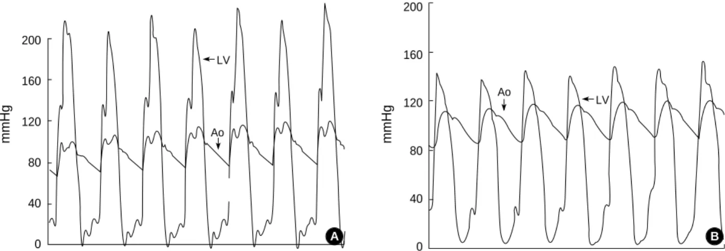
Left and right heart catheterization was carried out under local anesthesia. Simultaneous hemodynamic tracings of left ventricular and aortic pressures during pullback of the catheter from the left ventricle showed a large aortic-left ventricular
pressure gradient. Peak-to-peak aortic-left ventricular pres- sure difference was 100 mmHg at baseline and 200 mmHg after an extrasystolic beat (Fig. 3A). The selective right ven- tricular angiography revealed infundibular narrowing of the right ventricle due to hypertrophied muscle bundles. The systolic pressure in the right ventricle was elevated up to 80 mmHg and right ventricular diastolic pressure was increased to 10 mmHg (Fig. 4A). An intracardiac shunt was not observed during oxymetry procedure.
Surgical treatment was offered to the patient, but he was reluctant to undergo operation. After he had been offered all other surgical and non-surgical treatment option, he agreed to have an alcohol ablation therapy. The right ventricular angiography displayed that the hypertrophied muscle mass, extending from the infundibulum to the portion of the adja- cent interventricular septum, bulged toward the outflow tract during the systolic phase and obstructed the blood flow across the right ventricular outflow tract. According to these find- ings, we thought that the obstruction across the right ven- tricular outflow tract was mainly attributed to the infundibu- lar stenosis and the hypertrophied septal mass was partially involved in the obstructions of right ventricular outflow tract.
The patient was transferred to the cardiac catheterization laboratory for alcohol ablation therapy. A 5-Fr multipurpose catheter was placed at the apex of the left ventricle through the left femoral artery in order to measure pressure gradients of the left ventricular outflow tract. A catheter of the tempo- rary pacemaker was positioned at the apex of the right ven- tricle. Through a femoral approach, a 7- Fr Judkins type guiding catheter was introduced into the left coronary ostium.
After passing a 0.014′′extra-support long guidewire to the
Fig. 1.Asymmetric septal hypertrophy (A) and systolic anterior motion of the anterior mitral leaflet (B) is shown in the parasternal long axis view. RV: right ventricle; VS: ventricular septum; LV: left ventricle; MV: mitral valve; LA: left atrium; PM: papillary muscle; SAM: systolic anterior motion.
A B
RV RV
VS VS
LV LV SAM
MV
LA PM
Fig. 2.Right ventricular outflow tract obstruction (RVDT) in the pa- rasternal short axis view. PV: pulmonary valve; AO: aorta; RA:
right atrium.
RVOT
PV
RA
LA AO
Hypertrophic Cardiomyopathy with Infundibular Pulmonary Stenosis 587
first septal artery, a 1.5-mm angioplasty balloon was advanced over the guidewire to the first septal artery. Prior to the alco- hol ablation, pressure tracing was done by occlusion of septal artery with balloon and the pressure decrease in both LVOT and RVOT were observed. To perform a contrast echocardio- gram, we injected 10 mL of agitated saline through the cen- tral lumen of the balloon catheter. Under contrast echocardio- gram, enhancement was observed up to RVOT of septal area.
After fulfilling the contrast echocardiogram, we performed a prolonged balloon inflation to obstruct the ostium of the first septal artery with 4 atmospheres and 5 mL absolute alcohol was administered directly into the first septal artery through the central lumen of the balloon catheter. After 5 min of con- tinuous inflation, the balloon catheter was deflated and a sub- sequent angiography showed the first septal artery to be occlud- ed at its origin, without evidence of alcohol extravasation. Im- mediately after the procedure, the pressure gradient between the left ventricle and the aorta was decreased to 20 mmHg at baseline and to 60 mmHg after extrasystole (Fig. 3B). The systolic pressure in the right ventricle was also decreased to 32 mmHg (Fig. 4B). The patient remained hemodynami-
cally stable and complained of a mild chest pain without any change in the ECG findings. The patient was transferred to the coronary care units and the subsequent hospital course was uneventful and the patient was discharged after 7 days of in-hospital observation.
DISCUSSION
Hypertrophic obstructive cardiomyopathy with infundibu- lar stenosis of the right ventricle is an uncommon congenital anomaly in which outflow tract obstructions of both ventri- cles may occur due to the hypertrophied muscle bundles. In such cases, outflow tracts of both ventricles may have dynam- ic obstructions via hypertrophied muscle bundles. Hyper- trophied muscle bundles alter the dynamic outflow by caus- ing narrowing of the outflow tracts and the dynamic gradi- ents are produced through the outflow tracts. In the present case, HOCM with infundibular stenosis was detected by echocardiography and cardiac catheterization and the patient had dynamic gradients of both ventriclular outflow tracts.
mmHg
200
160
120
80
40
0
Fig. 3. Simultaneous aortic and left ventricular outflow tract pressure tracings before (A) and after (B) alcohol ablation.
LV
Ao
A
mmHg
200
160
120
80
40
0
Ao LV
B
mmHg
200
160
120
80
40
0
Fig. 4. Right ventricle pressure tracings before (A) and after (B) alcohol ablation.
RV RV
A
mmHg
100
80
60
40
20
0 B
The patient represents a unique example of both ventricular outflow obstructions due to hypertrophied muscle bundles.
In patient with obstruction of right ventricular outflow tract, the ventricular septal defect is sometimes accompanied (10, 11). In the present case, there was no evidence of intracardiac shunts by cardiac catheterization.
Reduction of hypertrophied muscle bundles is necessary not only for lowering dynamic gradients of the outflow tracts, but also for improving the clinical symptoms. Treatment for HOCM accompanying infundibular stenosis of the right ventricle may include surgical removal of anomalous muscle bundles. In our patient, due to his refusal to any surgical ther- apy, we decided to adapt non-surgical therapy under his con- sent. The hypertrophied septal mass was partially involved in the obstructions of right ventricular outflow tract accord- ing to right ventricular angiography and the pressure trac- ing prior to alcohol ablation revealed that occlusion of sep- tal artery with balloon decreased the pressure in both LVOT and RVOT. So we thought that reduction of the septal mass had led to relief of dynamic obstructions across the left ven- tricular outflow tract and partially across the right ventricu- lar outflow tract. For HOCM patients, a couple of mechanisms are involved in the relief of the obstruction after alcohol abla- tion. However, concerning no significant septal thinning after the ablation and also pressure decrease in both LVOT and RVOT, we assume that the decreased septal motion might be the main mechanism in the reliet of obstruction. To the best of our knowledge, this is the first case of which HOCM with infundibular stenosis of the right ventricle treated by alco- hol ablation therapy. At a 1-yr follow-up, the patient was still asymptomatic without any complications from the treatment.
In conclusion, alcohol ablation therapy is an emerging treat- ment modality, and it may be used on behalf of surgical ther- apy in patients with hypertrophic obstructive cardiomyopathy accompanying infundibular stenosis. However, more experi- ences are needed to establish it as an effective therapeutic approach in this disease.
REFERENCES
1. Lockhart A, Charpentier A, Bourdarias JP, Ben Ismail M, Ourbak P, Scebat L. Right ventricular involvement in obstructive cardiomyopa- thies: hemodynamic studies in 13 cases. Br Heart J 1966; 28: 122-33.
2. Frank S, Braunwald E. Idiopathic hypertrophic subaortic stenosis.
Clinical analysis of 126 patients with emphasis on the natural history.
Circulation 1968; 37: 759-88.
3. Falcone DM, Moore D, Lambert EC. Idiopathic hypertrophic car- diomyopathy involving the right ventricle. Am J Cardiol 1967; 19:
735-40.
4. Maron BJ. Hypertrophic cardiomyopathy. Lancet 1997; 350: 127-33.
5. Ford DK, Bollaboy CA, Derkac WM, Hopkins RA, Jennings RB, Johnson DH. Transatrial repair of double-chambered right ventricle.
Ann Thorac Surg 1988; 46: 412-5.
6. Shyu KG, Tseng CD, Chiu IS, Hung CR, Chu SH, Lue HC, Tseng YZ, Lieu WP. Infundibular pulmonic stenosis with intact ventricu- lar septum: a report of 15 surgically corrected patients. Int J Cardi- ol 1993; 41: 115-21.
7. Sigwart U. Non-surgical myocardial reduction for hypertrophic obs- tructive cardiomyopathy. Lancet 1995; 346: 211-4.
8. Knight CJ, Kurbaan AS, Seggewiss H, Hennein M, Gunning M, Harrington D, Fassbender D, Gleichmann U, Sigwart U. Nonsurgical septal reduction for hypertrophic obstructive cardiomyopathy: out- come in the first series of patients. Circulation 1997; 95: 2075-81.
9. Park SJ, Lee CW, Hong MK, Song JK, Park SW, Kim JJ. Transcoro- nary alcohol ablation of infundibular hypertrophy in patients with idiopathic infundibular pulmonic stenosis. Am J Cardiol 1997; 80:
1514-6.
10. Maron BJ, Ferrans VJ, White RI Jr. Unusual evolution of acquired infundibular stenosis in patients with ventricular septal defect. Clin- ical and morphologic observations. Circulation 1973; 48: 1092-103.
11. Jain V, Subramanian S, Lambert EC. Concomitant development of infundibular pulmonary stenosis and spontaneous closure of ven- tricular septal defect. An unusual variant in the natural history of ventricular septal defect. Am J Cardiol 1969; 24: 247-54.
588 S.H. Park, K.S. Park, H.G. Park, et al.