INTRODUCTION
Wind-blown dust originating from the arid deserts of Mongolia and China, so called Asian dust, is a well-known springtime meteorological phenomenon in Korea (1). Pub- lic concerns about the possible adverse effects of this dust have increased because the occurrence of these dust events has become more frequent and more erratic during the last decade (2).
Asian dust events are a form of air pollution episode. Expo- sure to ambient air pollutants has been associated with the impairment of respiratory health (3). Results of epidemio- logic studies have shown that asthmatic subjects are more susceptible to air pollutants than are other groups (4, 5). A recent study has reported altered respiratory symptoms and peak expiratory flow (PEF) outcomes during the Asian dust events in adult asthmatic patients (6). However, little is known about their effects on asthmatic children.
The severity of asthma has been assessed based on symp- tom diary recordings and measurements of pulmonary func- tion. Home PEF monitoring has been widely used for the monitoring of asthma in both epidemiological studies and clinical trials (7).
The aims of this study were to investigate the possible effects of Asian dust events on respiratory symptoms and pulmonary function by measuring PEF and to estimate the
changes of airway hyperresponsiveness (AHR) after exposure to Asian dust events in asthmatic children.
MATERIALS AND METHODS Subjects
A group of 52 children with mild asthma were recruited from the allergy clinic at Seoul National University Chil- dren’s Hospital. All subjects had a history of wheezing and dyspnea, and a methacholine PC20 (provocative concentra- tion causing a 20% fall in forced expiratory volume in one second [FEV1]) of less than 16 mg/mL. The clinical severity of asthma was assessed according to the National Asthma Education and Prevention Program (8). The subjects whose clinical severity could not be accurately assessed were exclud- ed. All patients lived in the Seoul Metropolitan area during the study period. Atopy was defined as the presence of at least one positive skin prick test result to a panel of 13 com- mon aeroallergens, in the presence of positive and negative controls. The allergens tested were: 1) house dust mites (Der- matophagoides pteronyssinus and Dermatophagoides farinae), 2) animal danders (cat epithelium and dog epithelium), 3) pol- lens (mugwort, ryegrass, ragweed, hazel, alder, and oak), 4) molds (Aspergillus fumigatus and Alternaria alternata), and 5)
Young Yoo, Ji Tae Choung, Jinho Yu*, Do Kyun Kim�, Young Yull Koh�
Department of Pediatrics, Korea University Anam Hospital, Seoul; Department of Pediatrics*, Dongguk University International Hospital, Goyang; Department of Pediatrics�, Seoul National University Hospital, Seoul, Korea
Address for correspondence Young Yull Koh, M.D.
Department of Pediatrics, Seoul National University Hospital, 28 Yeongeon-dong, Jongno-gu, Seoul 110-744, Korea
Tel : +82.2-2072-3631, Fax : +82.2-747-5130 E-mail : [email protected]
66 DOI: 10.3346/jkms.2008.23.1.66
Acute Effects of Asian Dust Events on Respiratory Symptoms and Peak Expiratory Flow in Children with Mild Asthma
The aim of this study was to investigate the possible adverse effects of Asian dust events on respiratory health in asthmatic children. Fifty-two children with mild asth- ma were studied for eight consecutive weeks in the spring of 2004 (March 8 to May 2). During the study period, five Asian dust days were identified; we included a lag period of two days following each of the events. Subjects recorded their respiratory symptom diaries and peak expiratory flow (PEF) twice daily during the study peri- od; and they underwent methacholine bronchial challenge tests. The subjects report- ed a significantly higher frequency of respiratory symptoms during the Asian dust days than during the control days. They showed significantly more reduced morn- ing and evening PEF values, and more increased PEF variability (10.1%±±3.5%
vs. 5.5%±±2.2%) during the Asian dust days than during the control days. Metha- choline PC20was not significantly different between before and after the study peri- od (geometric mean: 2.82 mg/mL vs. 3.16 mg/mL). These results suggest that the short-term Asian dust events might be associated with increased acute respiratory symptoms and changes in PEF outcomes. However, there might be little long-term influence on airway hyperresponsiveness in children with mild asthma.
Key Words : Asian Dust Events; Asthma; Peak Expiratory Flow Rate
Received : 2 January 2007 Accepted : 2 June 2007
cockroach (Blatella germanica). Parents provided written in- formed consent for their children to participate in the study.
Respiratory symptom diary
At the beginning of the study, parents and children were instructed to complete diaries twice daily with regard to respiratory symptoms. The current day symptom scores reflected respiratory symptoms experienced that day (pres- ence or absence) and the nighttime records associated with the preceding night’s symptoms (filled out each morning).
The number of as-needed short-acting bronchodilator use was also noted.
PEF measurements
Participants were asked to record PEF each morning and each evening using a Mini-Wright adult type peak-flow meter (Clement Clarke Ltd., London, U.K.). After achieving documented reproducibility within 20 L/min, PEF record- ings were taken twice daily for eight consecutive weeks (bet- ween March 8 and May 2). Morning values were obtained at 8:00 a.m. and evening values at 8:00 p.m. On each occa- sion, the best of three attempts was recorded. Subjects inhaled two doses of 200 g salbutamol sulfate (Ventolin, Glaxo, U.K.) immediately afterwards, and all these measurements were repeated 15 min later. During the study, asthmatic patients were asked to refrain from taking any medication other than salbutamol sulfate, 200-400 g, which was allow- ed as a rescue medication, but preferably not within 4 hr before the PEF measurements. At the time of the study, all patients were free of acute respiratory tract infections or asth- ma exacerbations for 4 weeks. All patients were asked to dis- continue oral theophylline for 48 hr, long-acting 2-agonists for 48 hr, and inhaled corticosteroids for 7 days before the start of the study. Diary cards with values missing for more than 10% of the eight weeks were excluded from the final analysis.
Methacholine inhalation tests
Methacholine bronchial challenges were carried out using a modification of the method described by Chai et al. (9).
Concentrations (0.075, 0.15, 0.3, 0.625, 1.25, 2.5, 5, 10, and 25 mg/mL) of methacholine (Sigma-Aldrich, St Louis, MO, U.S.A.) were prepared by diluting with buffered saline (pH 7.4). A Rosenthal-French dosimeter (Laboratory for Applied Immunology, Baltimore, MD, U.S.A.) triggered by a solenoid valve set to remain open for 0.6 sec was used to deliver aerosol generated from a DeVilbiss 646 nebulizer with pressurized air at 20 psi. Each subject inhaled five inspi- ratory capacity breaths of buffered saline and increasing con- centrations of methacholine until the FEV1fell by >20% of the post-saline value. FEV1was measured 1.5 min after each concentration was inhaled. The methacholine PC20was ob-
tained from the log concentration-percent fall in FEV1curve by linear interpolation of the last two points. These tests were repeated within 1 month after the study period.
Asian dust event data
Daily information on Asian dust events during the study period was provided by the Korea Meteorological Adminis- tration from a station located in central Seoul. During the study period, five Asian dust days (March 10-11, March 30- 31, and April 23) were identified in the Seoul Metropolitan area.
Statistical analysis
There were five Asian dust days during the length of the study, and the effects of dust events, if any, might have been delayed or manifested over a period of several days. To inves- tigate potential effects between exposure and outcomes (time lapse), we examined a lag period of two days following each of the events (10, 11). The control days included all the days during the study period except the Asian dust days and the lag periods. Average levels of respiratory symptoms and pul- monary function parameters on the Asian dust days and dur- ing the lag period were compared with those of the control days. Daily symptom prevalence was calculated as the frac- tion (%) of children reporting symptoms or medication use, using data only from children with complete dairy informa- tion for each separate day. The proportion of decrements larg- er than 10% of morning PEF was calculated as the number of children experiencing a decrement divided by the total number of children reporting valid PEF measurements on each day of the study. The percentage of evening decrements was calculated in the same way. PEF diurnal variability, which was equal to the difference between morning and afternoon PEF values divided by the mean value, was calculated. Statis- tical comparisons of average daily reported respiratory symp- toms and the PEF indices between each period were made with the Kruskall-Wallis non-parametric test. Correlation between daily average PM10concentration and PEF variability was examined using Pearson correlation tests. Methacholine PC20values obtained before and after the study period were log-transformed for statistical analysis, and the difference between the two was calculated by the paired t-test. All data analyses were performed using SPSS version 12.0 (SPSS Inc, Chicago, IL, U.S.A.). In each case, statistical significance was accepted when the two-sided p value was <0.05.
RESULTS Clinical characteristics
Of the 57 patients who were initially enrolled in this study,
five were excluded because of unacceptable PEF records or having symptoms suggestive of respiratory infections dur- ing the study period. The characteristics of the 52 study sub- jects are shown in Table 1. The mean age of 32 boys and 20 girls was 10.5±3.2 yr. Atopy was identified in 45 (86.5%) subjects.
Respiratory symptoms and signs
The daily prevalence of acute respiratory symptoms and signs was significantly higher during the Asian dust days than during the control days (p<0.05). More as-needed bron- chodilator use was noted during the Asian dust days than during the lag period (p=0.016). Regarding the lag period, the daily prevalence of cough, sore throat, or eye irritation was significantly higher during the lag period than during the control days (p<0.05) (Table 2).
Peak expiratory flow indices
Table 3 summarizes the PEF parameters during each peri- od. The mean morning PEF value (87.9±5.2%) and evening
PEF value (90.4±5.4%) for % best during the Asian dust days were significantly (p<0.05) lower than those during the control days (morning, 94.2±3.2%; evening, 92.8±4.3%).
The mean proportion of decrements larger than 10% of morn- ing PEF was higher during the Asian dust days (57.3±16.0
%) than during the control days (21.4±5.5%, p=0.015). The evening data showed similar results (40.7±18.3% vs. 21.0
±4.3%, p=0.023). PEF diurnal variability during the Asian dust days was significantly higher than during the control days (10.1±3.5% vs. 5.5±2.2%, p=0.017), and it was aug- mented when post-bronchodilator values were included (15.8
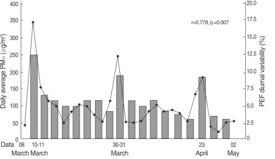
±6.8% vs. 10.9±7.2%, p=0.011). All PEF indices except the proportion of decrements of larger than 10% of evening PEF during the Asian dust days were significantly different from those during the lag period. There was a significant asso- ciation between the degree of average PM10concentration and PEF diurnal variability during the study period (r=0.778, p=0.007) (Fig. 1).
Data are presented as mean±SD or geometric mean (range of 1 SD).
*Defined as at least one positive skin prick test; �Forced expiratory vol- ume in one second. % predicted value, median (range); �Methacholine provocative concentration causing a 20% fall in FEV1.
Characteristics Asthmatic children (n=52)
Age (yr) 10.5±3.2
Sex (M/F) 32/20
No. of study days 56
(Asian dust days/lag period/control days) (5/6/45)
Atopy* (%) 45 (86.5)
FEV1�
91.1 (80.1-144.9) Methacholine PC20�
(mg/mL) 2.82 (1.17-6.76)
Table 1. Clinical characteristics of the asthmatic children
Dust days Lag period Control days Cough 42.9±20.8* 33.0±14.3* 20.2±3.4 Runny/stuffed nose 53.8±19.2* 45.2±18.4 39.1±9.3 Sore throat 24.2±13.5* 20.2±9.1* 6.1±2.6 Eye irritation 24.5±18.1* 16.1±4.9* 7.8±2.1 Limited physical activity 16.2±12.5 16.4±11.3 11.7±5.7 Nocturnal awakening 15.7±14.1 9.5±2.9 10.7±2.9 Shortness of breath 20.1±13.8* 11.3±3.0 5.4±3.4
Wheeze 16.7±7.1* 12.2±2.8 7.1±2.3
Bronchodilator use 8.7±6.0� 3.6±2.4 1.5±2.1 Table 2. Mean daily prevalence (%) of respiratory symptoms, signs, and medication use during Asian dust days, lag period, and control days in asthmatic children
Data are presented as mean±SD.
*p<0.05, compared to control days; �p<0.05; compared to lag period and control days.
Daily average PM10(g/m3) 400
350
300
250
200
150
100
50
0
PEF diurnal variability (%)
20.0
17.5
15.0
12.5
10.0
7.5
5.0
2.5 0
08 10-11 30-31 23 02
March March March April May
Data
Fig. 1. Daily average of PM10
concentration ( , g/m3) and peak expiratory flow (PEF) vari- ability ( ) in Seoul, Korea in the spring of 2004.
r=0.778, p=0.007
Changes in methacholine PC20
Among the 52 subjects with a measurable PC20both before and after the study period, five subjects exhibited an increase in PC20and five subjects showed a decrease in PC20by a two- fold magnitude (Fig. 2). The geometric mean of methacholine PC20after the study period (3.16 mg/mL) was not significant- ly different from that before the study period (2.82 mg/mL, p=0.290).
DISCUSSION
This study demonstrated that children with mild asthma reported more respiratory symptoms, as needed bronchodila- tor use, and changes in PEF outcomes during the Asian dust days than during the control days. Our findings are consis- tent with previous data that have revealed associations bet- ween respiratory outcomes and Asian dust (6, 12, 13). Al- though several measures of respiratory symptoms were signif- icantly increased up to the lag period, these findings appear- ed to be mostly transient.
The changes in airway caliber can be reliably monitored by home recordings of PEF over a period of time (7). We found decrements of both morning and evening PEF values and increases in the diurnal variability during the Asian dust days. The reduction of evening PEF might have resulted from the preceding daytime Asian dust event. However, morning decrements in PEF on the first Asian dust day may appear to be an unusual finding, because changes in morning values are not a result from the current day of Asian dust exposure.
This might be explained as a possible effect of the presence of the Asian dust before notification from the Meteorologi- cal Administration was publicly made. The reduction in PEF values could partly explain the increase in diurnal variabili-
ty during the Asian dust days, assuming parallel changes in morning and evening values (14).
We have shown that the effects of the Asian dust lasted at least two days after the exposure occurred. Several measures of respiratory symptoms and PEF parameters during the lag period were significantly different from those during the con- trol days. The findings in this study are consistent with other epidemiologic studies that have suggested an association with air pollution and delayed manifestation of respiratory effects (10, 11), indicating that pollutant-induced inflam- mation may play a role in these relations (10).
An estimation of changes in bronchial reactivity in our study subjects was made before and after the study period.
The initial values of methacholine PC20were not significant- ly different from those after the eight-week study period.
For patients with asthma, a change of at least one doubling concentration in bronchial sensitivity to a bronchoconstric- tor stimulus is considered to be clinically significant in the evaluation of disease progression (15). For the patients in this study, most were unchanged on the basis of this criteri- on, and when changes occurred, their frequency was evenly distributed in either direction. The overall unchanged AHR, before and after the study period, indicates that the Asian dust events may have little or no long-term effects on airway reac- tivity in children with mild asthma. However, these find- ings must be interpreted with caution because there was a lower concentration of the Asian dust during the study peri- od than that observed in preceding years. Also, these obser- vations could partly be explained by patients selection. In this study, subjects with mild asthma were chosen in order to avoid severe exacerbation during the study period. The difference between before and after study period might have been greater if the measurements had been done including subjects with moderate to severe asthma.
Dust days Lag period Control days Mean morning PEF, % best 87.9±5.2� 91.7±5.2* 94.2±3.2 Mean evening PEF, % best 90.4±5.4� 92.0±5.6 92.8±4.3
>10% decrements in morning 57.3±16.0�28.4±18.2 21.4±5.5 PEF
>10% decrements in evening 40.7±18.3* 29.9±13.0* 21.0±4.3 PEF
PEF diurnal variability (pre-BD) 10.1±3.5� 7.7±3.1* 5.5±2.2 PEF diurnal variability (post-BD) 15.8±6.8� 13.8±7.5* 10.9±7.2 Bronchodilator response 12.1±8.7� 7.9±8.5 7.2±7.9
(morning)
Bronchodilator response 10.1±6.9� 7.4±6.8 6.1±5.7 (evening)
Table 3. Peak expiratory flow (PEF) indices during Asian dust days, lag period, and control days in asthmatic children
Data are presented as mean±SD.
*p<0.05, compared to control days; �p<0.05, compared to lag period and control days.
BD, bronchodilator.
Methacholine PC20(mg/mL)
100
10
1
0.1
Before After
p=0.290
Fig. 2. Methacholine PC20values of the asthmatic children before and after the study period. Horizontal bars represent the geomet- ric mean and its range of 1SD.
Although exposure to an Asian dust event was associated with an altered respiratory outcome (6), the exact mechanism by which the dust causes adverse respiratory health effects is unclear. One possible explanation is an increase in the con- centration of fine or ultrafine particles during the Asian dust events (16). Fine particles are of the greatest health concern because they can be inhaled deeply into the lungs (16). A further possibility is that the aerosol properties during Asian dust events were different from those of the general atmo- spheric conditions. It has been shown that chemical compo- nents in the atmosphere during Asian dust event included NO2, SO2, and O3, (17) and that they may produce adverse effects on the pulmonary function and respiratory symptoms both in asthmatics and normal subjects (11, 18, 19).
Several limitations should be considered when interpret- ing our results. We observed consistent effects of the Asian dust on respiratory health in this panel of mainly asthmatic children. As pollutant exposure was common to all mem- bers of the cohort, a traditional ‘‘control’’ group was not need- ed; each subject acted as his or her own control, and only covariates that varied across time within an individual need- ed to be considered for analysis (20). The Asian dust might not be adequately characterized by fixed-site ambient air concentrations, since children spend a varied portion of their time outdoors (21). Misclassification of the records of respi- ratory symptoms and PEF measurements in children by their parents is also possible. This may be overestimated because parents of asthmatic children are very aware of their children’s respiratory health. Therefore, knowledge of Asian dust events by parents might affect symptom records or PEF measure- ments (22). Given that respiratory infections are related to asthma, they might have confounded the observed associa- tions. However, we took respiratory infections into account for analysis, and this inclusion did not substantially alter the association between the Asian dust and respiratory outcomes.
It is also possible that the sensitization to pollen may produce respiratory symptoms and change PEF indices. However, this is thought to be unlikely because this effect might exist not only in the control days but also in the Asian dust days.
In conclusion, our mild asthmatic children showed increas- ed upper and lower respiratory symptoms, as needed bron- chodilator use, and changes in PEF outcomes during the Asian dust days, which might also have delayed effects. Although several asthmatic children had considerable individual changes in AHR, as a group they remained similar over time. These findings indicate that Asian dust events increase the risk of acute respiratory symptoms and pulmonary function deteri- oration but do not appear to have long-term influence on AHR in children with mild asthma. Because we did not attempt to estimate these effects in subjects with moderate to severe asthma, the actual relations between the Asian dusts and respiratory outcomes may be greater than those observed in our study.
REFERENCES
1. Kwon HJ, Cho SH, Chun Y, Lagarde F, Pershagen G. Effects of the Asian dust events on daily mortality in Seoul, Korea. Environ Res 2002; 90: 1-5.
2. Hwang SS, Cho SH, Kwon HJ. Effects of the severe Asian dust events on daily mortality during the spring of 2002, in Seoul, Korea. J Prev Med Pub Health 2005; 38: 197-202.
3. Schwartz J, Dockery DW, Neas LM, Wypij D, Ware JH, Spengler JD, Koutrakis P, Speizer FE, Ferris BG Jr. Acute effects of summer air pollution on respiratory symptom reporting in children. Am J Respir Crit Care Med 1994; 150: 1234-42.
4. Vedal S, Petkau J, White R, Blair J. Acute effects of ambient inhal- able particles in asthmatic and nonasthmatic children. Am J Respir Crit Care Med 1998; 157: 1034-43.
5. Preutthipan A, Udomsubpayakul U, Chaisupamongkollarp T, Pen- tamwa P. Effect of PM10pollution in Bangkok on children with and without asthma. Pediatr Pulmonol 2004; 37: 187-92.
6. Park JW, Lim YH, Kyung SY, An CH, Lee SP, Jeong SH, Ju YS.
Effects of ambient particulate matter on peak expiratory flow rates and respiratory symptoms of asthmatics during Asian dust periods in Korea. Respirology 2005; 10: 470-6.
7. Reddel HK, Salome CM, Peat JK, Woolcock AJ. Which index of peak expiratory flow is most useful in the management of stable asth- ma? Am J Respir Crit Care Med 1995; 151: 1320-5.
8. National Asthma Education and Prevention Program. Guidelines for the diagnosis and management of asthma expert panel. Report 2.
Bethesda, Md: US Dept of Health and Human Services, 1997. NIH publication 97-4051.
9. Chai H, Farr RS, Froehlich LA, Mathison DA, McLean JA, Rosen- thal RR, Sheffer AL, Spector SL, Townley RG. Standardization of bronchial inhalation challenge procedures. J Allergy Clin Immunol 1975; 56: 323-7.
10. Romieu I, Meneses F, Ruiz S, Huerta J, Sienra JJ, White M, Etzel R, Hernandez M. Effects of intermittent ozone exposure on peak expi- ratory flow and respiratory symptoms among asthmatic children in Mexico City. Arch Environ Health 1997; 52: 368-76.
11. Ostro B, Lipsett M, Mann J, Braxton-Owens H, White M. Air pollu- tion and exacerbation of asthma in African-American children in Los Angeles. Epidemiology 2001; 12: 200-8.
12. Park JW, Lim YH, Kyung SY, An CH, Lee SP, Jeong SH, Ju YS.
Effects of ambient particulate matter (PM10) on peak expiratory flow and respiratory symptoms in subjects with bronchial asthma during Yellow Sand period. Tubercul Respir Dis 2003; 55: 570-8.
13. Min PK, Kim CW, Yun YJ, Chang JH, Chu JK, Lee KE, Han JY, Park JW, Hong CS. Effect of Yellow Sand on respiratory symptoms and diurnal variation of peak expiratory flow in patients with bron- chial asthma. J Asthma Allergy Clin Immunol 2001; 21: 1179-86.
14. Hakala K, Stenius-Aarniala B, Sovijarvi A. Effects of weight loss on peak flow variability, airways obstruction, and lung volumes in obese patients with asthma. Chest 2000; 118: 1315-21.
15. Barnes PJ. A new approach to the treatment of asthma. N Engl J Med 1989; 321: 1517-27.
16. Lei YC, Chan CC, Wang PY, Lee CT, Cheng TJ. Effects of Asian
dust event particles on inflammation markers in peripheral blood and bronchoalveolar lavage in pulmonary hypertensive rats. Envi- ron Res 2004; 95: 71-6.
17. Choi JC, Lee MH, Chun YS, Kim J, Oh S. Chemical composition and source signature of spring aerosol in Seoul Korea. J Geophys Res 2001; 106: 18067-74.
18. Blomberg A, Krishna MT, Bocchino V, Biscione GL, Shute JK, Kelly FJ, Frew AJ, Holgate ST, Sandstrom T. The inflammatory effects of 2 ppm NO2on the airways of healthy subjects. Am J Respir Crit Care Med 1997; 156: 418-24.
19. Burnett RT, Smith-Doiron M, Stieb D, Raizenne ME, Brook JR, Dales RE, Leech JA, Cakmak S, Krewski D. Association between
ozone and hospitalization for acute respiratory diseases in children less than 2 years of age. Am J Epidemiol 2001; 153: 444-52.
20. Ward DJ, Ayres JG. Particulate air pollution and panel studies in children: a systemic review. Occup Environ med 2004; 61: e13.
21. Krzyzanowski M, Quackenboss JJ, Lebowitz MD. Relation of peak expiratory flow rates and symptoms to ambient ozone. Arch Environ Health 1992; 47: 107-15.
22. Romieu I, Meneses F, Ruiz S, Sienra JJ, Huerta J, White MC, Etzel RA. Effects of air pollution on the respiratory health of asthmatic children living in Mexico City. Am J Respir Crit Care med 1996;
154: 300-7.