• 검색 결과가 없습니다.

Effects of Sasa quelpaertensis Extract on mRNA and microRNA Profiles of SNU-16 Human Gastric Cancer Cells

N/A
N/A
Protected

Academic year: 2021

Share "Effects of Sasa quelpaertensis Extract on mRNA and microRNA Profiles of SNU-16 Human Gastric Cancer Cells"

Copied!
12
0
0

로드 중.... (전체 텍스트 보기)

전체 글

(1)

Effects of Sasa quelpaertensis Extract on mRNA and microRNA Profiles of SNU-16 Human Gastric Cancer Cells

Mi Gyeong Jang

1,2

, Hee Chul Ko

2

and Se-Jae Kim

1,2

*

1

Department of Biology, Jeju National University, Jeju 63243, Korea

2

Biotech Regional Innovation Center, Jeju National University, Jeju 63243, Korea Received March 29, 2020 /Revised April 6, 2020 /Accepted April 20, 2020

Sasa quelpaertensis Nakai leaf has been used as a folk medicine for the treatment of gastric ulcer, dip- sosis, and hematemesis based on its anti-inflammatory, antipyretic, and diuretic characteristics. We have previously reported the procedure for deriving a phytochemical-rich extract (PRE) from S. quel- paertensis and how PRE and its ethyl acetate fraction (EPRE) exhibits an anticancer effect by inducing apoptosis in various gastric cancer cells. To explore the molecular targets involved in this apoptosis, we investigated the mRNA and microRNA profiles of EPRE-treated SNU-16 human gastric cancer cells. In total, 2,875 differentially expressed genes were identified by RNA sequencing, and gene ontol- ogy (GO) and Kyoto Encyclopedia of Genes and Genomes (KEGG) analyses indicated that the EPRE- modulated genes are associated with apoptosis, mitogen-activated protein kinase, inflammatory re- sponse, tumor necrosis factor signaling, and cancer pathways. Subsequently, protein-protein inter- action network analysis confirmed interactions among genes associated with cell death and apoptosis, and 27 differentially expressed microRNAs were identified by further sequencing. Here, GO and KEGG pathway analysis revealed that EPRE modified the expression of microRNAs associated with the cell cycle and cell death, as well as signaling of tropomyosin-receptor-kinase receptor, trans- forming growth factor-b, nuclear factor kB, and cancer pathways. Taken together, these results provide insight into the mechanisms underlying the anticancer effect of EPRE.

Key words : Anticancer, miRNA, RNA sequencing, Sasa quelpaertensis Nakai, SNU-16 cell

*Corresponding author

*Tel : +82-64-754-3529, Fax : +82-64-751-4406

*E-mail : [email protected]

This is an Open-Access article distributed under the terms of the Creative Commons Attribution Non-Commercial License (http://creativecommons.org/licenses/by-nc/3.0) which permits unrestricted non-commercial use, distribution, and reproduction in any medium, provided the original work is properly cited.

Introduction

Gastric cancer is the fourth most common malignancy and the second leading cause of cancer-related death worldwide [29]. Currently, the most effective treatment for gastric can- cer is surgery combined with chemotherapy and radio- therapy; however, these therapies have side effects and can lead to antitherapeutic resistance [33, 39, 40]. Certain plant extracts can ameliorate these side effects, prolong survival, and improve the quality of life by enhancing anticancer ac- tivity [5, 17, 34, 36]. Therefore, there is demand for new natu- ral products proven to be safe for the prevention and treat- ment of gastric cancer.

The genus Sasa (Poaceae) is composed of perennial plants commonly known as dwarf bamboo; various Sasa species

are distributed in Asian countries, including China, Japan, Korea, and Russia. Their leaves have been used in traditional medicine for the treatment of gastric ulcer, dipsosis, and hematemesis because of their anti-inflammatory, antipyretic, and diuretic activities [2]. Extracts of the leaves of various Sasa species have anticancer effects. For example, S. albo- marginata leaf extract has been used to treat hypertension, cardiovascular disease, and cancer [26, 28]. Two poly- saccharide preparations from S. kurilensis were found to sup- press the growth of sarcoma-180 implanted in mice [22]. An alkaline extract from S. senanensis leaves (containing poly- saccharides, chlorophyllin, lignin, and flavonoids) reportedly prevented spontaneous mammary tumorigenesis and Her2/

NeuN mammary tumorigenesis [23].

S. quelpaertensis Nakai is a dwarf bamboo grass that grows

on Mt. Halla on Jeju Island, Republic of Korea. Its leaf ex-

tracts have antiobesity, antidepressant, antifatigue, and anti-

cancer activities [12-14, 24]. In addition, we previously re-

ported the preparation method for phytochemical-rich ex-

tract (PRE) for efficient utilization of its leaf [15]. PRE and

its ethyl acetate fraction (EPRE) have been shown to inhibit

the proliferation of human gastric cancer cells (MKN-74,

(2)

MKN-45, SNU-1, and SNU-16) by inducing apoptosis [11].

The present study was performed to investigate the mRNA and microRNA (miRNA) profiles in EPRE-treated SNU-16 cells to explore the underlying mechanism of this anti- proliferative activity.

Materials and Methods

Plant material and extraction

S. quelpaertensis leaves were collected from Mt. Halla on Jeju Island, Republic of Korea. The leaves were washed and dried in a hot air dryer at 60℃ for 24 hr. The resulting pow- der was extracted for 4 hr with hot water (90℃). The hot water extract was removed, and the residue was used to prepare PRE and EPRE, in accordance with the method de- scribed by Lee et al. [15]. PRE is a mixture of such phytonu- trients as polysaccharides, amino acids, and polyphenols, in- cluding the tricin (5.35 mg/g) and p-coumaric acid (44.10 mg/g) as indicator components [16].

Cell culture and cell viability assay

SNU-16 human gastric cancer cells were obtained from the Korean Cell Line Bank (Seoul, Korea). The cells were cultured in Roswell Park Memorial Institute-1640 medium supplemented with 10% fetal bovine serum and 1% pen- icillin-streptomycin (Gibco, Grand Island, NY, USA) in a 37

℃ incubator with a humidified atmosphere containing 5%

CO

2

. To evaluate the cell viability, the cells plated in 96-well plates(2.0×10

5

–3.0×10

5

cells/ml) were cultured overnight, treated with EPRE and then incubated for an additional 48 hr. Each well was supplemented with 50 μl of MTT and in- cubated for 4 hr at 37℃. The formazan crystals formed were subsequently dissolved in 150 μl DMSO, and the optical den- sity of the resultant reaction solution was read at 540 nm using a microplate reader (Bio-Tek, VT, USA).

Western blot analysis

The harvested cells were lysed in ice-cold RIPA lysis buf- fer (Merck, Darmstadt, Germany) according to the manu- facturer’s protocol. Protein concentration was quantified us- ing the Bio-Rad Protein Assay reagent (Bio-Rad Laboratories, CA, USA). Proteins were separated on 8% to 12% poly- acrylamide gel and then transferred for PVDF membranes.

The membranes were blocked for 1hr with 5% skim milk in 0.1% Tween-20 and Tris-buffer saline (TTBS). They were incubated for overnight at 4℃ with the following primary

antibodies; B-cell lymphoma 2 (BCL2), bcl-2-associated X protein (BAX), procaspase-3, poly (ADP-ribose) polymerase (PARP) (Santa Cruz, CA, USA) and cleaved caspase-3 (Cell Signaling Technology, Beverly, MA). The membranes were washed with TTBS, and incubated with horseradish perox- idase-conjugated secondary antibodies (Jackson Immuno Research, West Grove, PA, USA) for 1 hr at room temper- ature. Immunodetection was carried out using enhanced chemiluminescence (ECL) western blotting detection reagent (Cyanagen, Bologna, Italy).

RNA extraction

The cells were treated with EPRE (100 μg/ml) for 48 hr;

total RNA was then extracted using TRIzol reagent (Invitro- gen, Carlsbad, CA, USA). RNA quality was assessed using the Agilent 2100 Bioanalyzer and the RNA 6000 Nano Chip (Agilent Technologies, Amstelveen, Netherlands); RNA was quantified using the ND-2000 spectrophotometer (Thermo Fisher Scientific, Waltham, MA, USA).

mRNA sequencing and data analysis

For mRNA sequencing, a library was constructed using SMARTer Stranded RNA-Seq Kit (Clontech Laboratories, Inc., Mountain View, CA, USA). Total RNA (2 μg) was in- cubated with magnetic beads conjugated to oligo-dT; other RNAs were removed by washing. Library production was initiated by random hybridization of starter/stopper hetero- dimers to the poly(A) RNA bound to the magnetic beads.

These starter/stopper heterodimers contained Illumina-com-

patible linker sequences. A single-tube reverse transcription

and ligation reaction extended the starter to the next hybri-

dized heterodimer, where the newly synthesized cDNA in-

sert was ligated to the stopper. Second strand synthesis was

performed to release the library from the beads, and the li-

brary was then amplified. Barcodes were introduced when

the library had been amplified. High-throughput sequencing

was performed as paired-end 100-bp sequencing using a

HiSeq 2500 (Illumina, Inc., San Diego, CA, USA). mRNA-seq

reads were mapped using TopHat software [32] to obtain

alignment files. Differentially expressed genes (DEGs) were

determined based on counts from unique and multiple

alignments using coverage in Bedtools [21]. Read count data

were processed based on the quantile normalization method

using EdgeR within R (R Development Core Team, 2016)

and Bioconductor [7]. Alignment files were used for assem-

bly of transcripts, estimation of abundances, and detection

(3)

Table 1. The primer sequences of the genes used in Real-time PCR analysis

Gene Primer sequence

MAP3K5 Forward

Reverse

5’- GAA ATC TCC CTG GTG ACA GA -3’

5’- AGC AAC TTG TCC TTC GCT TT -3’

BCL2L14 Forward

Reverse

5’- ATG AAG CTG TGG ACC CCA AA -3’

5’- TGG TGGTGG CCA GGA GTA AA -3’

FLCN Forward

Reverse

5’- CTG CTC CCG AGG TTA ACA TT -3’

5’- CAA ACA TAA GGC CCA GAA AC -3’

MYC Forward

Reverse

5’- AGA TCA GCA ACA ACC GAA AA -3’, 5’- TTG TGT GTT CGC CTC TTG AC -3’

FADD Forward

Reverse

5’- AGG TGG AGA ACT GGG ATT TG -3’

5’- AGA ACG CCA CAG TGG TTG AG -3’

GAPDH Forward

Reverse

5’- CGA GAT CCC TCC AAA ATC AA -3’

5’- CCT TCT CCA TGG TGG TGA A -3’

of differential expression of genes or isoforms using Cuf- flinks. The fragments per kilobase of exon per million frag- ments method was used to determine gene expression levels.

Gene classification was based on searches performed in DAVID (http://david.abcc.ncifcrf.gov/). The Search Tool for the Retrieval of Interacting Genes/Proteins (STRING) data- base was used to provide a comprehensive protein inter- actome that included known and predicted protein-protein interactions, which were given confidence scores; accessory information (e.g., protein domains and 3D structures) was also searched within this stable and consistent identifier space. Networks were visualized and analyzed using Cyto- scape 3.3.0 visualization software (http://www.cytoscap- e.org/).

miRNA sequencing and data analysis

For miRNA sequencing, a library was constructed using a NEBNext Multiplex Small RNA Library Prep Kit (New England BioLabs, Inc., Ipswich, MA, USA). Total RNA (1 µg) was ligated to the adaptors and cDNA was synthesized using reverse transcriptase with adaptor-specific primers.

PCR was performed for library amplification and libraries were cleaned-up using the QIAquick PCR Purification Kit (Qiagen, Inc., Hilden, Germany) and AMPure XP beads (Beckman Coulter, Inc., Brea, CA, USA). The yields and size distributions of small RNA libraries were assessed by high-sensitivity DNA assays using the Agilent 2100 Bioana- lyzer (Agilent Technologies, Inc.). High-throughput se- quences were produced by the NextSeq 500 system for sin- gle-end 75 sequencing (Illumina). Sequence reads were map- ped by Bowtie2 software to obtain a bam alignment file. A mature miRNA sequence was used as a reference for map-

ping. Read counts mapped on the mature miRNA sequence were extracted from the alignment file using Bedtools (v.

2.25.0) and Bioconductor, which utilizes the R (v. 3.2.2) stat- istical programming language (R Development Core Team, 2011). Read counts were assessed to determine the levels of miRNAs. The quantile normalization method was used for comparisons between samples. For miRNA target analyses, miRWalk 2.0 software was used. Functional gene classi- fication was performed using DAVID (http://david.abcc.

ncifcrf.gov/). For pathway analysis, genes were selected and input into the KEGG mapper (http://www.genome.jp/

kegg/tool/map_pathway2.html). Significant pathways that included selected genes were recorded. CluePedia software, based on the mirTarBase.validated.miRNAs_15.06.2016in Cytoscape (v. 3.7.1), was used to investigate interactions be- tween miRNAs and their target genes. The criteria for net- work interaction formation were analyzed using the default value for each database.

Quantitative real-time PCR

For quantification of mRNA, reverse transcription was

performed using SuperScript II RTase (Invitrogen). Real-time

PCR was performed on the StepOnePlus™ Real-Time PCR

System with the SYBR Green PCR Kit (Applied Biosystems

Inc., Foster City, CA, USA), using the primers listed in Table

1. Thermal cycling conditions were 95℃ for 10 min, followed

by 40 cycles of 95℃ for 15 s and 59℃ for 30 s. Data were

analyzed using StepOne software v. 2.2.2 (Applied Biosys-

tems). The expression level of each gene was normalized to

the level of the endogenous control (GAPDH) and calculated

using the 2

−ΔΔCt

method. For quantification of miRNA,

cDNA synthesis and real-time PCR were performed using

(4)

A B

C D

Fig. 1. The cell viability and expression of apoptosis-related proteins in EPRE-treated SNU-16 cells. (A) The cells were incubated with different solvent fractions (100 μg/ml). (B) Cells were incubated with different concentration of EPRE. (C) Western blot analysis. (D) HS-68 cells were incubated with different concentration of EPRE. The data expressed means ± standard deviation (SD) of three independent experiments. *p<0.05, **p<0.01, and ***p<0.001 compared to the untreated group. Western blotting results are representative of three independent experiments.

the miScript PCR system (Qiagen). cDNA was synthesized from total RNA using the miScript II RT Kit with HiSpec buffer. cDNA was amplified from miRNA using the follow- ing primer pairs: hsa-miR-136-5p, hsa-miR-320d, hsa-miR- 132-5p, hsa-miR-1260b, and has-miR-92a-1-5p; hsa-U6 was used as the internal control. Real-time PCR was performed on the StepOnePlus Real-Time PCR System (Applied Biosys- tems) with QuantiTect SYBR Green PCR Master Mix and miScript Primer Assay (Qiagen). Thermal cycling conditions were 95℃ for 15 min, followed by 40 cycles of 94℃ for 15 s, 55℃ for 30 s, and 70℃ for 30 s. Data were analyzed using StepOne software v. 2.2.2 (Applied Biosystems). The ex- pression level of each microRNA was normalized to the level of an endogenous small RNA U6 and calculated using the 2

-ΔΔCt

method.

Statistical analysis

Statistical analysis was performed using SPSS Statistics v.

12.0 for Windows (SPSS Inc., Chicago, IL, USA). Data are expressed as means ± standard deviations. Differences be-

tween groups were examined by one-way analysis of var- iance. Differences with p<0.05 were considered statistically significant.

Results and Discussion

The leaves of Sasa species have been used in Eastern Asia as a potential source of natural drug since hundreds of years ago. S. quelpaertensis leaves are considered as one of the use- ful sources for food, nutraceuticals, and cosmetics because of their therapeutic potential [12-16, 24]. To expend the ap- plicability of S. quelpaertensis leaves as cancer preventive agent, we investigated the effects of EPRE on cell viability, and evaluated the mRNA and miRNA profiles of EPRE- treated SNU-16 cells.

Cell viability

The cytotoxicity of PRE solvent fractions was evaluated

in the human gastric cancer cell (SNU-16) and human skin

fibroblasts (Hs-68) using the MTT assay. As shown in Fig. 1,

(5)

A

B

Fig. 2. The GO and KEGG pathway analysis of differentially expressed genes (DEGs) in EPRE-treated SNU-16 cells. (A) The 280 annotated genes regulated by EPRE were assigned into biological process, cellular component, and molecular function catego- ries based on gene ontology annotations. (B) KEGG pathway analysis.

EPRE inhibited the most effectively the proliferation of SNU-16 cells (IC

50

= 21.1 μg/ml), but it did not show cytotox- icity to normal HS-68 cells. EPRE decreased the expressions of Bcl-2 and procaspase-3, but increased the expressions of Bax, cleaved caspase-3, and cleaved PARP in a dose-depend- ent manner. These results suggested that EPRE inhibited cell growth by inducing apoptosis of SNU-16 cells through mi- tochondrial mediated pathway. Apoptosis can be activated

through two main pathways: the mitochondrial-dependent pathway and the death receptor dependent pathway [35].

The Bcl-2 family, which acts on mitochondrial-dependent

pathways, includes pro-apoptotic (Bax, Bad, Bak) and an-

ti-apoptotic members (Bcl-2) [6]. Caspase-3 is activated from

pro-caspase-3 by the nuclear enzyme PARP, and is known

as a major player in apoptosis [30].

(6)

Table 2. List of differentially expressed miRNAs in EPRE-treat- ed SNU-16 cells (fold change >2.7, p-value <0.05)

NO miRNAs Fold

change Accession ID 1

2 3 4 5 6 7 8 9 10 11 12 13 14 15 16 17 18 19 20 21 22 23 24 25 26 27

hsa-miR-25-5p hsa-miR-27a-5p hsa-miR-92a-1-5p hsa-miR-29b-1-5p hsa-miR-128-1-5p hsa-miR-132-5p hsa-miR-136-5p hsa-miR-365a-5p hsa-miR-1290 hsa-miR-1246 hsa-miR-1261 hsa-miR-320d hsa-miR-1260b hsa-miR-320e hsa-miR-4301 hsa-miR-4454 hsa-miR-548aj-5p hsa-miR-4483 hsa-miR-6073 hsa-miR-6131 hsa-miR-7704 hsa-miR-7974 hsa-miR-32-3p hsa-miR-181b-3p hsa-miR-210-3p hsa-miR-191-3p hsa-miR-219a-2-3p

0.284 0.297 0.163 0.250 0.312 0.375 3.682 0.298 4.661 5.379 0.368 3.179 0.359 3.180 6.025 0.054 0.318 0.135 5.177 4.846 3.825 0.081 0.252 0.359 0.214 0.266 2.512

MIMAT0004498 MIMAT0004501 MIMAT0004507 MIMAT0004514 MIMAT0026477 MIMAT0004594 MIMAT0000448 MIMAT0009199 MI0006352 MI0006381 MI0006396 MIMAT0006764 MI0014197 MI0014234 MI0015828 MI0016800 MIMAT0022739 MI0016844 MI0020350 MI0021276 MI0025240 MI0025750 MIMAT0004505 MIMAT0022692 MIMAT0000267 MIMAT0001618 MIMAT0004675 mRNA profile

RNA-seq was performed to identify DEGs in EPRE-treat- ed cells, compared to untreated SNU-16 cells. Among 25,737 genes, 2,875 DEGs (2.7-fold cut-off) were identified; of these, 1,200 were upregulated and 1,675 were downregulated. GO analysis revealed that EPRE significantly enriched 280 GO terms in the biological process category, including apoptosis, cell proliferation, and inflammatory response; 37 GO terms in the cellular component category, including extracellular exosome, nucleoplasm, intracellular and perinuclear cyto- plasm; and 78 GO terms in the molecular function category, including protein binding, DNA binding, and ATP binding (Fig. 2A). KEGG pathway analysis showed that the cancer, MAPK signaling, and TNF signaling pathways were highly enriched by EPRE (Fig. 2B). Liu et al. [18] reported that acti- vation of the reactive oxygen species(ROS)/ASK1/MAPK pathway induces apoptosis in human lung cancer cells.

Certain cancer preventive substances induced apoptosis by activation of p38 MAPK in hepatocellular carcinoma cells [4]. Considering these previous studies, EPRE seems to influ- ence the expression of genes that are closely related to apop- tosis in SNU-16 cells.

A protein-protein interaction(PPI) analysis using Cyto- scape v. 3.0 (STRING database, cut-off >0.4) showed that the protein-protein interaction network consisted of 154 nodes and 403 edges; moreover, v-myc avian myelocytomatosis vi- ral oncogene homologous (MYC), Fas-associated via death domain (FADD), BCL2-like 14 (BCL2L14), mitogen-activated protein kinase kinase kinase 5 (MAP3K5), and BCL-2-asso- ciated X protein (BAX) were located at the major key nodes (Fig. 3). SNU-16 human gastric cancer cells have an abnor- mal p53 gene and express MYC [20]. MYC controls cell pro- liferation, differentiation, and apoptosis; inhibition of MYC expression induces brain tumor cell death [41]. BCL2L14 is a member of the Bcl-2 family; it is associated with apoptosis in laryngeal squamous cell carcinoma, prostate cancer, and acute leukemia cancer cells [38]. FADD transmits cell death signals by activation of procaspase-8 [1, 19]. MAP3K5 is also known as apoptosis pathway control kinase 1 (ASK1); it is activated by ROS and Fas, as well as other factors [8, 31].

Downregulation of ASK1 expression reportedly promotes cancer cell apoptosis [9, 10]. Therefore, EPRE may exert the anti-cancer effects by modulating the expression of genes that regulate SNU-16 cell apoptosis.

miRNA profile

MiRNA sequencing was performed to examine the effect of EPRE on the miRNA profile of SNU-16 gastric cancer cells.

Compared with untreated control cells, 27 differentially ex- pressed miRNAs (DEMs) were identified in EPRE-treated cells (fold-change >2.7). Among these 27 DEMs, 10 were up- regulated and 17 were downregulated (Table 2). GO analysis showed that 222 biological process GO terms were sig- nificantly enriched (miRTbase); the top 25 GO terms were related to the cell cycle, cell death, and the tropomyosin-re- ceptor-kinase receptor signaling pathway (Fig. 4A). KEGG pathway analysis revealed 50 significant pathways (p<0.05) (Table S1). Among the top 20 pathways, DEMs were asso- ciated with the transforming growth factor-β pathway, nu- clear factor-kappaB (NF-κB) pathway, wingless-related in- tegration site (Wnt) signaling pathway, and cancer signaling pathway (Fig. 4B).

To evaluate the interactions between 27 DEMs and their

target genes, network analysis was performed using Cyto-

(7)

Fig. 3. The protein–protein interaction networks (PPI) among differentially expressed genes (DEGs) in EPRE-treated SNU-16 gastric cancer cells. PPI network was constructed by the String online tool with a confidence score of >0.4. The nodes represent proteins, edges represent interactions between proteins, and the colors of the nodes represent the log2 fold change in the expression level. Red node stands for up-regulated gene and blue node stands for down-regulated gene. The disconnected nodes in the network were hidden.

scape v. 3.7.1 (Fig. 5). The network comprised 21 nodes that consisted of 21 DEMs: miR-136-5p, miR-320d, miR-6131, miR-132-5p, miR-92a-1-5p, miR-320e, miR-6073, miR-32-3p, miR-1260b, miR-4483, miR-4301, miR-548aj-5p, miR-1261, miR-181b-3p, miR-1290, miR-25-5p, miR-27a-5p, miR-29b-1- 5p, miR-4454, miR-365a-5p, and miR-1246. Potential target genes of the 21 DEMs involved in the network are listed in Table S2.

It is well known that various cellular activities like cell growth, proliferation, and differentiation are regulated by miRNAs through their regulatory effects on particular RNA species. In many tumors, up- or down-regulation of different miRNAs has been reported. MiR-136-5p was highly upregu- lated in EPRE-exposed SNU-16 cells. Expression of miR-136- 5p was reportedly reduced in ovarian cancer and glioma cell lines; moreover, it promoted chemotherapy-induced death of glial cells [37, 42]. MiR-136-5p suppresses tumor growth

and migration, while induces apoptosis [3]. Also, it has been reported that the overexpression of miR-320d has been shown to inhibit proliferation of breast cancer cells and in- duce apoptosis of these cells [27]. In addition, it was reported that the apoptosis of human acute promyelocytic leukemia cell line (HL-60) can be induced by inhibiting miR-92a [25].

Therefore, it is suggested that the anticancer effect of EPRE may be mediated by modulating key miRNA expressions in SNU-16 cells.

Validation of RNA-seq data

To validate the mRNA-seq data, qRT-PCR analysis was

performed for randomly selected genes. The levels of MYC,

MAP3K5, and BCL2L14 mRNAs in EPRE-treated cells were

significantly lower, while the level of FADD mRNA tended

to be lower comparing to untreated control cells. These qRT-

PCR results are consistent with the mRNA-seq data (Fig.

(8)

A

B

Fig. 4. GO and KEGG pathway analysis of differentially expressed miRNAs (DEMs) in EPRE-treated SNU-16 cells. The top 20 GO terms (A) and the top 10 KEGG pathway (B) significantly enriched by DEMs.

6A). Also, to validate the miRNA-seq data, we performed qRT-PCR analysis for five randomly selected miRNAs. We found that miR-136-5p and miR-320d were significantly up- regulated in EPRE-treated cells, compared to untreated con- trol cells. In contrast, miR-132-5p, miR-1260b, and miR-92a-1- 5p were significantly downregulated in EPRE-treated cells.

These qRT-PCR results are consistent with the miRNA-seq

data (Fig. 6B).

In summary, EPRE inhibited the proliferation of SNU-16

cells by activating apoptosis. Total 2,875 DEGs and 27 DEMs

were identified in EPRE-treated SNU-16 cells, compared to

untreated control cells. Most DEGs and DEMs were asso-

ciated with biological processes and signaling pathways,

such as the cell death, apoptosis, NF-κB, cancer, MAPK, and

(9)

Fig. 5. Network between differentially expressed miRNAs and potential target genes. The yellow diamond-shapes node indicate target genes, gray circles indicate target miRNAs.

A B

Fig. 6. Validation of differentially expressed mRNAs and miRNAs using qRT-PCR. (A) Cells were incubated with 100 μg/ml of EPRE for 48 hr. Expression levels of the genes (MAP3K5, MYC, FADD, BCL2L14) were analyzed by quantitative RT-PCR and normalized by GAPDH. (B) Expression levels of miRNAs (miR-136-5p, miR-320d, miR-132-5p, miR-1260b, miR-92a-1-5p) were analyzed by quantitative RT-PCR and normalized by has-U6. The data are expressed as means ± standard deviation (SD) of three determinations. *p<0.05, **p<0.01, and ***p<0.001 compared to untreated SNU-16 cancer cell.

TNF signaling pathways. The collective findings suggest that EPRE exerts an anticancer effect through regulation of sev- eral key genes and miRNAs involved in these pathways.

Further research is needed to identify the molecular mecha- nisms underlying the anticancer effects of EPRE.

Acknowledgment

This research was supported by Basic Science Research

Program through the National Research Foundation of

Korea (NRF) funded by the Ministry of Education

(2017R1D1A3B03029845 and 2019R1A6A10072987).

(10)

The Conflict of Interest Statement

The authors declare that they have no conflicts of interest with the contents of this article.

References

1. Aggarwal, B. B. 2003. Signaling pathways of the TNF super- family: a double-deged sword. Nat. Rev. Immunol. 3, 745-756.

2. Bae, K. 2003. The medicinal plants of Korea, pp 565-567, Kyo- Hak Publishing Company, Seoul, Korea.

3. Chen, P., Zhao, L., Pan, X., Jin, L., Lin, C., Xu, W., Xu, J., Guan, X., Wu, X., Wang, Y., Yang, S., Wang, T. and Lai, Y. 2018. Tumor suppressor microRNA-136-5p regulates the cellular function of renal cell carcinoma. Oncol. Lett. 15, 5995- 6002.

4. Chiba, T., Suzuki, E., Yuki, K., Zen, Y., Oshima, M., Miyagi, S., Saraya, A., Koide, S., Motoyama, T., Ogasawara, S., Ooka, Y., Tawada, A., Nakatsura, T., Hayashi, T., Yamashita, T., Kaneko, S., Miyazaki, M., Iwama, A. and Yokosuka, O. 2014.

Disulfiram eradicates tumor-initiating hepatocellular carci- noma cells in ROS-p38 MAPK pathway-dependent and -independent manners. PLoS ONE 9, e84807.

5. Chung, M. Y., Lim, T. G. and Lee, K. W. 2013. Molecular mechanisms of chemopreventive phytochemicals against gastroenterological cancer development. World J. Gastroen- terol. 19, 984-993.

6. Danial, N. N. and Korsmeyer, S. J. 2004. Cell death: critical control points. Cell 116, 205-219.

7. Gentleman, R. C., Carey, V. J., Bates, D. M., Bolstad, B., Dettling, M., Dudoit, S., Ellis, B., Gautier, L., Ge, Y., Gentry, J., Hornik, K., Hothorn, T., Huber, W., Iacus, S., Irizarry, R., Leisch, F., Li, C., Maechler, M., Rossini, A. J., Sawitzki, G., Smith, C., Smyth, G., Tierney, L., Yang, J. Y. and Zhang, J. 2004. Bioconductor: open software development for com- putational biology and bioinformatics. Genome Biol. 5, R80.

8. Gotoh, Y. and Cooper, J. A. 1998. Reactive oxygen species- and dimerization-induced activation of apoptosis signal- regulating kinase 1 in tumor necrosis factor-α signal trans- duction. J. Biol. Chem. 273, 17477-17482.

9. Hayakawa, Y., Hirata, Y., Sakitani, K., Nakagawa, H., Nakata, W., Kinoshita, H., Takahashi, R., Takeda, K., Ichijo, H., Maeda, S. and Koike, K. 2012. Apoptosis signal-regulat- ing kinase-1 inhibitor as a potent therapeutic drug for the treatment of gastric cancer. Cancer Sci. 103, 2181-2185.

10. Ichijo, H., Nishida, E., Irie, K., ten Dijke, P., Saitoh, M., Moriguchi, T., Takagi, M., Matsumoto, K., Miyazono, K. and Gotoh, Y. 1997. Induction of apoptosis by ASK1, a mamma- lian MAPKKK that activates SAPK/JNK and p38 signaling pathways. Science 275, 90-94.

11. Jang, M. G., Ko, H. C. and Kim, S. J. 2018. Effect of Sasa quelpaertensis Nakai extracts and its constituent p-coumaric acid on the apoptosis of human cancer cell lines. Nat. Prod.

Sci. 24, 1-5.

12. Kang, H. and Lee, C. 2015. Sasa   quelpaertensis Nakai extract

suppresses porcine reproductive and respiratory syndrome virus replication and modulates virus-induced cytokine production. Arch. Virol. 160, 1977-1988.

13. Kang, S. I., Shin, H. S., Kim, H. M., Hong, Y. S., Yoon, S.

A., Kang, S. W., Kim, J. H., Ko, H. C. and Kim, S. J. 2012.

Anti-obesity properties of a Sasa quelpaertensis extract in high-fat diet-induced obese mice. Biosci. Biotechnol. Biochem.

76, 755-761.

14. Kim, M., Kim, Y. S., Kim, K. M., Ko, H. C., Kim, S. J., Kim, J. H. and Kim, Y. 2014. Combination of Sasa   quelpaerten- sis Nakai leaf extract and cisplatin suppresses the cancer stemness and invasion of human lung cancer cells. Integr.

Cancer Ther. 13, 529-540.

15. Lee, J. Y., Ko, H. C., Jang, M. K. and Kim, S. J. 2016.

Preparation and characterization of phytochemical-rich ex- tract from Sasa quelpaertensis leaf. J. Life Sci. 26, 1130-1335.

16. Lee, J. Y., Song, H., Ko, H. C., Jang, M. G. and Kim, S. J.

2018. Anti-oxidant and anti-inflammatory potentials of Sasa quelpaertensis leaf residue extracts. J. Life Sci. 28, 738-744.

17. Liu, J., Wu, N., Ma, L. N., Zhong, J. T., Liu, G., Zheng, L.

H. and Lin, X. K. 2014. p38 MAPK signaling mediates mi- tochondrial apoptosis in cancer cells induced by oleanolic acid. Asian Pac. J. Cancer Prev. 15, 4519-4525.

18. Liu, K., Li, X., Cao, Y., Ge, Y., Wang, J. and Shi, B. 2015.

MiR-132 inhibits cell proliferation, invasion and migration of hepatocellular carcinoma by targeting PIK3R3. Int. J.

Oncol. 47, 1585-1593.

19. Muppidi, J. R., Tschopp, J. and Siegel, R. M. 2004. Life and death decisions: secondary complexes and lipid rafts in TNF receptor family signal transduction. Immunity 21, 461-465.

20. Park, J. G., Frucht, H., LaRocca, R. V., Bliss, D. P. Jr., Kurita, Y., Chen, T. R., Henslee, J. G., Trepel, J. B., Jensen, R. T., Johnson, B. E., Bang, Y. J., Kin, J. P. and Gazdar, A. F. 1990.

Characteristics of cell lines established from human gastric carcinoma. Cancer Res. 50, 2773-2780.

21. Quinlan, A. R. and Hall, I. M. 2010. BEDTools: a flexible suite of utilities for comparing genomic features. Bioinformatics 26, 841-842.

22. Raidaru, G., Ilomets, T., Mottus, A. and Maser, M. 1997.

Isolation of polysaccharides with antitumor activity from Sasa kurilensis (Fr. et Sar.). Exp. Oncol. 20, 34-39.

23. Ren, M., Reilly, R. T. and Sacchi, N. 2004. Sasa health exerts a protective effects on Her2/NeuN mammary tumorigen- esis. Anticancer Res. 24, 2879-2884.

24. Shaif, N. A., Cho, D., Jang, D., Kim, H. M., Chung, J, O., Kim, S., Seo, D. B., Kim, K. R., Shin, J. and Shim, I. 2019.

Sasa   Quelpaertensis   Nakai induced antidepressant-like effect in ovariectomized rats. Biomed. Res. Int. 2019, 1-11.

25. Sharifi, M., Salehi, R., Gheisari, Y. and Kazemi, M. 2014.

Inhibition of microRNA miR-92a induces apoptosis and ne- crosis in human acute promyelocytic leukemia. Adv. Biomed.

Res. 3, 61.

26. Shibata, M., Fujii, M. and Yamaguchi, R. 1979. Pharmacolog-

ical studies on bamboo grass. IV. toxicological and pharma-

cological effects of the extract (FIII) obtained from Sasa albo-

marginata Makino et Shibata (author's transl). Nippon Yakur-

(11)

igaku Zasshi 99, 663-668.

27. Shi, S., Hu, X., Xu, J., liu, H. and Zou, L. 2018. MiR-320d suppresses the progression of breast cancer via lncRNA JNF1A-AS1 regulation and SOX4 inhibition. RSC Adv. 8, 19196-19207.

28. Shibata, M., Yamatake, M., Sakamoto, M., Kanamori, M. and Takagi, K. 1975. Phamacological studies on bamboo grass (1). Acute toxicity and anti-inflammatory and antiulcero- genic activities of water-soluble fraction (Folin) extracted from Sasa albomarginata Makino et Shibata. Nippon Yakuriga- ku Zasshi 71, 481-490.

29. Sitarz, R., Skierucha, M., Mielko, J., Offerhaus, G. J. A., Maciejewski, R. and Polkowski, W. P. 2019. Gastric cancer:

epidemiology, prevention, classification, and treatment.

Cancer Manag. Res. 10, 239-248.

30. Soldani, C. and Scovassi, A. I. 2002. Poly (ADP-ribose) poly- merase-1 cleavage during apoptosis: an update. Apoptosis 7, 321-328.

31. Tobiume, K., Matsuzawa, A., Takahashi, T., Nishitoh, H., Morita, K., Tadeda, K., Minowa, O., Miyazono, K., Noda, T. and Ichiho, H. 2001. ASK1 is required for sustained acti- vations of JNK/p38 map kinases and apoptosis. EMBO Rep.

2, 222-228.

32. Trapnell, C., Pachter, L. and Salzberg, S. L. 2009. TopHat:

discovering splice junctions with RNA-Seq. Bioinformatics 25, 1105-1111.

33. Wagner, A. D., Grothe, W., Haerting, J., Kleber, G., Grothey, A. and Fleig, W. E. 2006. Chemotherapy in advanced gastric cancer: a systematic review and meta-analysis based on ag- gregate data. J. Clin. Oncol. 24, 2903-2909.

34. Wei, P., Zhiyu, C., Xu, T. and Xiangwei, Z. 2015. Antitumor effect and apoptosis induction of Alocasia cucullata (Lour.)

G. Don in human gastric cancer cells in vitro and in vivo.

BMC Complement. Altern. Med. 15, 33.

35. Wong, R. S. 2011. Apoptosis in cancer: from pathogenesis to treatment. J. Exp. Clin. Cancer Res. 30, 87.

36. Xia, H. H., Wong, B. C. and Lam, S. K. 2003. Chemopreven- tion of gastric cancer: current status. Chin. Med. J. (Engl).

116, 5-10.

37. Yang, Y., Wu, J., Guan, H., Cai, J., Fang, L., Li, J. and Li, M. 2012. MiR-136 promotes apoptosis of glioma cells by tar- geting AEG-1 and Bcl-2. FEBS Lett. 586, 3608-3612.

38. Yao, X., Yao, R., Yi, J. and Huang, F. 2019. Upregulation of miR-496 decreases cerebral ischemia/reperfusion injury by negatively regulating BCL2L14. Neurosci. Lett. 696, 197- 205.

39. Yoshikawa, T., Tsuburaya, A., Kobayashi, O., Sairenji, M., Motohashi, H. and Noguchi, Y. 2003. A combination im- munochemotherapy of 5-fluorouracil, cisplatin, leucovorin, and OK-432 for advanced and recurrent gastric carcinoma.

Hepatogastroenterology 50, 2259-2263.

40. Yu, W., Whang, I., Suh, I., Averbach, A., Chang, D. and Sugarbaker, P. H. 1998. Prospective randomized trial of ear- ly postoperative intraperitoneal chemotherapy as an ad- juvant to resectable gastric cancer. Ann. Surg. 228, 347-354.

41. Zhang, P., Li, H., Wu, M. L., Chen, X. Y., Kong, Q. Y., Wang, X. W., Sun, Y., Wen, S. and Liu, J. 2006. c-Myc down- regulation: a critical molecular event in resveratrol-induced cell cycle arrest and apoptosis of human medulloblastoma cells. J. Neurooncol. 80, 123-131.

42. Zhao, H., Liu, S., Wang, G., Wu, X., Ding, Y., Guo, G., Jiang,

J. and Cui, S. 2015. Expression of miR-136 is associated with

the primary cisplatin resistance of human epithelial ovarian

cancer. Oncol. Rep. 33, 591-598.

(12)

초록:SNU-16 위암 세포의 mRNA 및 miRNA 프로파일에 미치는 제주조릿대 추출물의 영향

장미경

1,2

․고희철

2

․김세재

1,2

*

(

1

제주대학교 생물학과,

2

제주대학교 생명과학기술혁신센터 )

제주조릿대 잎은 항염, 해열 및 이뇨작용을 가지고 있어 위궤양, 목마름 및 토혈 치료를 위한 민간의약으로 사

용되어 왔다. 본 저자들은 제주조리대 잎에서 분리한 피토케미칼 풍부 추출물(PRE)과 그 에틸아세테이트 분획물

(EPRE)은 여러 위암 세포주에서 세포사멸을 유도하는 항암 효과가 있다고 보고한 바 있다. 본 연구는 EPRE의

세포사멸 유도 기전에 관여하는 분자표적들을 탐색하기 위하여 EPRE을 처리한 SNU-16 세포에서 mRNA와

microRNA (miRNA)의 프로파일 변화를 분석하였다. RNA sequencing 분석을 통해 총 2,875개의 차등적으로 발

현되는 유전자들(DEGs)을 동정하였다. 유전자 온톨로지(GO)와 KEGG 경로 분석 결과, EPRE는 세포사멸, 유사

분열-활성화 단백질 키나제(MAPK) 및 염증 반응, 종양 괴사 인자(TNF) 신호 전달 및 암 경로에 관여하는 유전자

들의 발현을 조절하는 것으로 나타났다. 단백질-단백질 상호 작용(PPI) 네트워크 분석으로 세포사멸 및 세포죽음

과 관련된 유전자들 간의 상호작용들을 확인할 수 있었다. 그리고, miRNA sequencing 분석을 통해 총 27개의

차별적으로 발현되는 miRNAs (DEMs)를 동정하였다. GO와 KEGG 경로 분석 결과, EPRE는 세포주기, 세포사멸

및 tropomyosin-receptor-kinase (TRK) 수용체 신호 전달, 성장인자-β(TGF-β), 핵인자 κB (NF-κB) 및 암 경로에

관여하는 miRNAs의 발현을 조정하였다. 본 연구결과는 EPRE의 항암 효과의 근본적인 메커니즘에 대한 통찰력

을 제공한다.

수치

Table  1.  The  primer  sequences  of  the  genes  used  in  Real-time  PCR  analysis
Fig.  1.  The  cell  viability  and  expression  of  apoptosis-related  proteins  in  EPRE-treated  SNU-16  cells
Fig.  2.  The  GO  and  KEGG  pathway  analysis  of  differentially  expressed  genes  (DEGs)  in  EPRE-treated  SNU-16  cells
Table  2.  List  of  differentially  expressed  miRNAs  in  EPRE-treat- EPRE-treat-ed  SNU-16  cells  (fold  change  &gt;2.7,  p-value  &lt;0.05)
+4

참조

관련 문서

Here, we found that TAMR-MCF-7 cells had undergone EMT, evidenced by mesenchymal-like cell shape, down-regulation of the basal E-cadherin expression

Inhibitory effects of classified methanol extracts of Smilax china L on the COX-2 and iNOS expression of human colorectal cancer cell lines... Inhibitory

 Often found connected to other molecules on the outsides of cells --- cellular recognition, cell signaling, cell

4 &gt; Effects of Taro on COX-2 expression and iNOS expression(hot water) in human thyroid cancer cells. The cells were pretreated for 48hours with either

These results suggest that the bilobalide inhibits the cell proliferation and induces the apoptotic cell death in FaDu human pharyngeal squamous cell carcinoma via both

In the present study we investigated the proliferation effect of human oral cancer KB cell treated with pulsatilla koreana extract.. We analyzed the effects of this

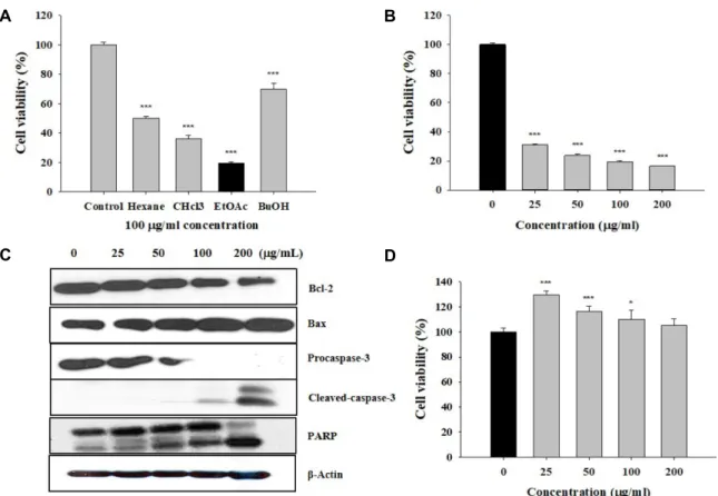
Taken together, these results suggested that latex containing the ficin inhibited the cell growth and induced apoptosis by caspase and Bcl-2 family signaling pathway in

Comparison of cell attachment on the bone grafting materials synthesized as the defined mixing ratio of Hydroxyapatite (HA) and β-tricalcium phosphate (β-TCP) by DAPI staining