Diazoxide Suppresses Mitochondria-dependent Apoptotic Signaling in Endothelial Cells Exposed to High Glucose Media
Hyun Ju Jung, Tae Hyun Kim and Jae Suk Woo*
Department of Physiology, Pusan National University School of Medicine, Yangsan 50612, Korea Received November 5, 2019 /Revised November 14, 2019 /Accepted November 14, 2019
In the present study, we examined the effect of mitochondrial K
+channel opener diazoxide on the mitochondria-dependent apoptotic signaling in endothelial cells exposed to high glucose (HG) media.
Endothelial cells derived from human umbilical veins were exposed to HG media containing 30 mM glucose, and the degree of apoptotic cell death associated with activation of the mitochondria-depend- ent apoptotic signaling pathway was determined. Exposure to HG media was seen to enhance apop- totic cell death in a time-dependent manner. In these cells, activation of caspases 3, 8, and 9 was ob- served, and while caspase-3 and -9 inhibitors suppressed the HG-induced apoptotic cell death, a cas- pase-8 inhibitor did not. The HG-treated cells exhibited disruption of mitochondrial membrane poten- tial, formation of permeability transition pores, and cytosolic release of cytochrome c. Subsequently, diazoxide was seen to attenuate the HG-induced apoptotic cell death; caspase-9 activation was sup- pressed but caspase 8 was not. Diazoxide also suppressed the depolarization of mitochondrial mem- brane potential, the formation of mitochondrial permeability transition pores, and the release of cyto- chrome c. These effects were significantly inhibited by 5-hydroxydecanoate, a selective blocker of ATP-sensitive K
+channels (K
ATP). The present results demonstrate that diazoxide exhibits a beneficial effect to ameliorate HG-induced endothelial cell apoptosis. Opening the K
ATPcould help preserve the functional integrity of mitochondria and provide an underlying mechanism to suppress HG-triggered apoptotic signaling.
Key words : Apoptosis, diazoxide, endothelial cell, high glucose, mitochondrial K+
channel
*Corresponding author
*Tel : +82-51-510-8072, Fax : +82-51-510-8076
*E-mail : [email protected]
This is an Open-Access article distributed under the terms of the Creative Commons Attribution Non-Commercial License (http://creativecommons.org/licenses/by-nc/3.0) which permits unrestricted non-commercial use, distribution, and reproduction in any medium, provided the original work is properly cited.
Journal of Life Science 2019 Vol. 29. No. 12. 1393~1400 DOI : https://doi.org/10.5352/JLS.2019.29.12.1393
Introduction
Diabetes mellitus can cause various vascular complica- tions and close correlations have been observed between blood glucose level and vascular dysfunction [2, 26].
Vascular endothelium has been suggested to be primarily involved in the vascular lesions of experimental and human diabetes. Endothelial cells exposed to high glucose (HG) en- vironment have been shown to exhibit delayed cell pro- liferation [5, 19], disturbed cell cycle [19, 21], enhanced DNA damage [20], and increased cell death [19]. In diabetes in
vivo, high glucose concentration has been found to produceendothelial pathologies [6, 15, 18] such as loss of capillary endothelium and weakening of endothelial cell junctions. It was reported that HG also triggered apoptotic cell death in
cultured human endothelial cells [3] and microvascular cells from retina [22]. Therefore, prevention of endothelial cell apoptosis in HG environment may have important im- plications for investigating pharmacological tools to alleviate diabetes-associated vascular complications.
A variety of potassium channel openers (KCOs) act on ATP-sensitive K
+channels (K
ATP). Their activities are regu- lated in connection with cellular metabolism and are sup- pressed by a rise in the intracellular ATP [7, 8]. Noma [24]
has suggested that these channels are endogenous mediators of myocardial protection in the ischemic heart. Mitochon- drial membranes also have been shown to present ATP-sen- sitive K
+conductance which has been recognized as the mi- tochontrial K
ATP[13]. Diazoxide a relatively selective opener of the mitochondrial K
ATPhas been found to reduce infarct size in the whole heart, and survival of cardiomyocytes [14, 27]. With respect to diabetes, diazoxide has been shown to delay the development of diabetes [10, 11] and prevent pan- creatic β-cell death induced by high glucose or tolbutamide [9].
An in vivo study with an established animal model of human
type 2 diabetes demonstrated that it could prevent diabetes
through suppression of pancreatic β-cell apoptosis [12].
In this study, we postulated that diazoxide could provide a beneficial effect to ameliorate HG-induced vascular complications. To test this hypothesis, human umbilical vein endothelial cells (HUVECs) were exposed to HG media in the absence and presence of diazoxide and the degree of apoptotic cell death and changes in the mitochondria- de- pendent apoptotic signaling were examined.
Materials and Methods
Cell culture
Human umbilical vein endothelial cells (HUVECs) were grown in Dulbeccos modified Eagles medium: Nitrient Mixture F-12 (Ham) (1:1) with 5% heat inactivated fetal bo- vine serum at 37℃ in humidified 95% air, 5% CO
2incubator.
When cells grew to reach confluence, they were detached using a 0.02% EDTA-0.05% trypsin solution and subculture was prepared.
Exposure to HG media
Cells were seeded and grown on 24-well culture plates or collagen-coated cover slips according to the purpose of the experiments. Experimental procedures were carried out 3 days after subculture. Cells were exposed to HG environ- ment by replacing the control media containing 5.5 mM D-glucose with media containing 30 mM D-glucose. For os- motic controls, L-glucose or mannitol (each 25 mM) was added to the control media.
Analysis of apoptotic cell death
HG-induced apoptosis was analyzed by nuclear staining with Hoechst 33,258. After exposure to experimental proce- dures, cells were fixed with 4% paraformaldehyde and stained with 10 μM Hoechst 33,258. Cells presenting frag- mented or condensed nuclei were counted as apoptotic cells.
Measurement of mitochondrial membrane potential Mitochondrial membrane potential was analyzed with DiOC6(3). This fluorochrome accumulates into the mico- thondria depending upon the mitochondrial membrane po- tential, and thus, reduction in DiOC6(3) fluorescence in- dicates loss of the mitochondrial membrane potential. Cells were loaded for 30 min in the dark with DiOC6(3) at a final concentration of 50 nM at 37℃. Cells were harvested, wash- ed and resuspended in PBS. The fluorescence intensity was analyzed with a FACsort Becton Dickinson Flow Cytometer.
Analysis of mitochondrial permeability transition pore formation
To examine the formation of mitochondrial permeability transition (MPT) pores, cells were double-stained with fluo- rescent dyes, calcein acetoxymethyl ester (calcein-AM) and tetramethylhodamine methyl ester (TMRM). Cells with mi- tochondrial permeability transition pores were analyzed un- der a confocal microscope as described by Lemasters et al [17].
Detection of cytochrom c release
Cells were washed three times with ice-cold phos- phate-buffered saline and suspened in 1 ml of extraction buf- fer composed of (in mM) PIPES 50, KCl 50, MgCl
25, EGTA 5, phenylmethylsulfonyl fluoride 1, leupeptin 10 mg/ml and pepstatin A 10 mg/ml, pH 7.0. Cells were lysed by five cy- cles of freezing in liquid nitrogen and thawing at 37℃. The cell lysates were then centrifuged for 1 hr at 100,000 g. The supernatant was then separated from the pellet containing the cellular debris. Western blot study was carried out using polyclonal anti-cytochrom c antibody.
Assay of caspase activity
A quantitative enzymatic assay of caspase activity was carried out using colorimetric assay kit (The R&D, Minneap- olis, Mn) according to the manufacturer-provided instruc- tions.
Chemicals
Reagents and medium for cell culture were purchased from GIBCO-BRL (Grand Island, NY). Ac-DEVD-CHO, Z-IETD-FMC, Z-LEHD-FMK, and Z-VAD‐FMK, were pur- chased from Calbiochem (California, USA). TMRM, Calcein/
AM and DiOC6(3) was obtained from Molecular Probes (Eugene, OR, USA). Antibody of cytochrome-c was obtained from Cell Signaling Technology Inc. (Beverly, MA, USA).
Other chemicals and reagents were obtained from Sigma- Aldrich Chemical Co. (St. Louis, MO, USA).
Statistical analysis
Data were presented as means ± S.E. The significance of
the difference between two groups was evaluated by stu-
dent's t-test. When necessary, multiple group comparison
was performed using one way analysis of variance followed
by the post hoc Tukey test. A probability level less than 0.05
was used to establish significant difference.
A
B
Fig. 1. Apoptosis of HUVECs in high glucose media. A. Repre- sentative micrographs of Hoechst 33,258-stained cell preparations after 72 hr exposure to low (LG, 5.5 mM) and high glucose (HG, 30 mM) media. Cells showing fragmented or condensed nuclei were counted as apop- totic (indicated by arrows) B. Time-dependent increase of apoptotic cell counts in HG media C. Concentration- dependent effect of D-glucose (D-Gl). The effects of L-glucose (L-Gl, 25 mM) and mannitol (Mann, 25 mM) as osmotic controls were presented together. Data are means ± S.E. of 4 experiments. *p<0.05, **p<0.01 vs. 5.5 mM D-Gl.
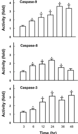
Fig. 2. Effect of high glucose on caspase activities. Cells were exposed to media containing 30 mM glucose for in- dicated time periods and assayed for caspase 3, 8, and 9 activities. Each bar represents means ± S.E. of 4 experi- ments. *p<0.01 vs. the respective control determined in cells in low (5.5 mM) glucose media.
Results
HG-induced apoptosis
When cells were assessed by Hoechst staining after 72 hr exposure to HG, 38.7±7.4% of the cells were counted as apoptotic, which were characterized by fragmented or con- densed nuclei as indicated by arrows (Fig. 1A). The time-de- pendent effect of HG to induce apoptosis was summarized in Fig. 1B. When cells were exposed to HG media the num- ber of apoptotic cells increased in a time-dependent manner.
Although, the concentration-effect relationship varied great- ly according to experimental groups, the number of apop- totic cells tended to increase when the concentration of glu- cose in media was raised (Fig. 1C). Addition of L-glucose or mannitol instead of D-glucose did not affect the rate of apoptosis suggesting that the effect of HG was not ascribable to increased osmolality (Fig. 1C).
Role of caspases in HG-induced apoptosis To examine the role of different caspases in the execution of HG-induced apoptosis, we determined caspase 3, 8 and
9 activities in the HG-treated cells. In cells exposed to HG media there was a time-dependent and sustained activation of caspase 9. The activity reached its peak (3.6 fold) in 36 hr and retained the peak level up to 48 hr. In contrast, HG-induced caspase 8 activation exhibited a transient pat- tern showing its peak activity (2.3 fold) in 24 hr then fol- lowed by a return to the basal level in 48 hr. Activation pat- tern of caspase 3 was comparable to that of caspase 9 (Fig.
2).
To delineate further the role of these caspases in the
HG-induced apoptosis, effects of different caspase inhibitors
were examined. Z-LEHD-FMK [25] and Ac-DEVD-CHO [1],
the specific inhibitors of the caspases 9 and 3 respectively,
as well as Z-VAD-FMK a broad range caspase inhibitor [29],
significantly prevented the HG-induced apoptosis. In con-
trast, the caspase 8 inhibitor Z-IETD-FMK [28] did not show
a significant protection (Fig. 3). These results suggested that
activation of caspase 9 through a mitochondria-dependent
Fig. 3. Effects of caspase inhibitors on high glucose-induced apoptosis. Cells were exposed for 48 hr to media con- taining 30 mM glucose in the presence of different cas- pase inhibitors (each 20 μM), and apoptotic cells were detected by Hoechst 33,258-staining. The number of apoptotic cells was presented as percentile of total cell counts. Each bar represents means ± S.E. of 4 experi- ments. *p<0.01 vs. control.
A
B
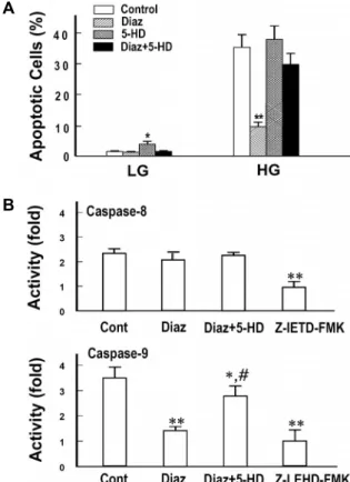
Fig. 4. Effect of diazoxide on high glucose-induced apoptosis and caspase activities. A. Cells were exposed for 72 hr to low (LG, 5.5 mM) and high (HG, 30 mM) glucose media in the presence of diazoxide (Diaz, 10 μM) with or without 5-hydroxydecanoate (5-HD, 100 μM) and apoptotic cells were detected by Hoechst 33,258-staining.
B. Cells were exposed for 24 hr to high glucose media in the presence of Diaz with or without 5-HD and ana- lyzed for caspase-8 and 9 activities. For a comparison, effects of each specific caspase inhibitors were presented together. Data are means ± S.E. of 4 experiments. *p<
0.05, **p<0.01 vs. the respective control and #p<0.01 vs.
Diaz alone.
pathway played a major role in the HG-induced apoptosis of HUVECs.
Effect of diazoxide on HG-induced apoptosis and caspase activation
Apoptotic cells were counted in different cell preparations exposed to HG in the presence of diazoxide and 5-hydrox- ydecanoate, a well-known prototype of mitochondrial K
ATPopener and blocker [27]. Diazoxide remarkably decreased the apoptotic cell counts. However, in cells pretreated with 5-hydroxydecanoate, the protective effect of diazoxide was significanly blocked. Diazoxide also showed a significant in- hibition of caspase 9 which was comparable to the effect of Z-LEHD-FMK, a specific inhibitor of caspase-9. In con- trast, caspase-8 activity was not affected by diazoxide (Fig.
4). These results strongly suggested that diazoxide exerted its anti-apoptotic effect via opening of K
ATPand suppression of the mitochondria-dependent apoptotic signaling.
HG-indiuced mitochondrial depolarization and per- meability transition
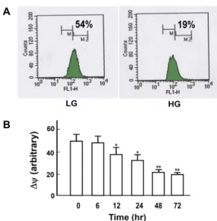
To elucidate further the role of mitochondria in the HG-induced apoptosis, changes in mitochondrial integrity was determined. As shown in the representative graph (Fig.
5A) and summarized data (Fig. 5B) of flow cytometry analy- sis of DiOC6(3)-stained cells, HG significantly depolarized the mitochondrial membrane potential in a time-dependent manner, suggesting disruption of the mitochondrial mem-
brane potential preceded the HG-induced apoptosis.
Formation of MPT pores and disruption of the perme-
ability barrier in the inner mitochondrial membrane has
been suggested to be a crucial event to initiate the mitochon-
dria-dependent apoptotic pathway. The MPT pores have
been known to mediate the release of cytochrome c [16]. In
confocal microscopic analysis, cells with intact mitochondria
were able to be discriminated by their mitochondrial con-
tours visualized as bright red spots. On the other hand, in
cells with MPT pores mitochondria tended to lose TMRM
and become permeable to calcein. As a result, it became hard
to distinguish mitochondrial contours from cytosol as in-
dicated by arrows in a representative micrograph (Fig. 6A).
A
B
Fig. 5. Effect of high glucose on mitochondrial membrane po- tential. A. Cells were exposed to low (LG, 5.5 mM) and high (HG, 30 mM) glucose media for 72 hr. Cells were then loaded with DiOC6(3), and analyzed for mitochon- drial membrane potential by flow cytometry analysis. B.
Changes in mitochondrial membrane potential were de- termined as a function of exposure time periods to HG.
Mitochondrial membrane potential was presented as ar- bitrary unit estimated by the degree of fluorescence quenching. Data are means ± S.E. of 4 experiments. *p<
0.05, **p<0.01 vs. control.
A
B
Fig. 6. Formation of mitochondrial permeability transition pores induced by exposure to high glucose. A. Cells were ex- posed to high glucose media for 72 hr and double- stained with fluorescence dyes TMRM and calcein-AM.
Intact mitochondria accumulate TMRM (bright red), whereas injured mitochondria with mitochondrial per- meability transition (MPT) pores lose TMRM and be- come permeable to and stained with calcein (green, in- dicated by arrows). B. The number of cells with MPT pores were presented as percentile of total cell counts.
Data are means ± S.E. of 6 experiments.
These results showed clear evidence that formation of MPT pores took place in the HG-treated cells. The number of cells with MPT pores increased in a time-dependent manner in cells exposed to HG media (Fig. 6B).
Diazoxide-induced suppression of mitochondrial apoptotic signaling
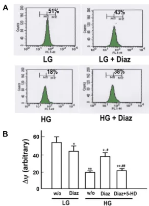
As was shown in the above results, HG-induced apoptosis was likely to proceed mostly via a mitochondria-dependent pathway. The results also suggested that diazoxide exhibited its anti-apoptotic effect via suppression of the mitochon- dria-dependent apoptotic signaling. To elucidate further the effects of diazoxide on mitochondrial events associated with the HG-induced apoptosis, effects of diazoxide on mitochon- drial membrane potential, formation of MPT pores and cyto- solic cytochrome release were examined. As presented in the representative flow cytometric analysis of DiOC6(3)-stained cells in Fig. 7A and summarized data in Fig. 7B, diazoxide significantly reversed HG-induced depolarization of the mi- tochondrial membrane potential. In accordance with this re- sult, diazoxide significantly reduced the number of cells
with MPT pores (Fig. 8A) and cytosolic release of cyto- chrome c (Fig. 8B) in cells treated with HG.
Discussion
Diabetes impacts diverse organs and systems through dif- ferent molecular mechanisms. HG-induced apoptosis has been suggested to be importantly implicated in diabetic pathologies and multiple mechanisms have been suggested to trigger and regulate the complicated signaling cascades involved in the HG-induced apoptosis [2].
Caspase enzymes play crucial roles in the process of
apoptotic pathway. Cytochrome c released from mitochon-
dria through the permeability transition pores is a main trig-
ger to activate the caspase 9. Thus activation of the caspase
9 is a crucial event to initiate the mitochondria-dependent
apoptotic signaling. On the other hand, activation of the cas-
pase 8 is achieved largely through the mitochondria-in-
dependent mechanisms [17]. In the present study, exposure
to HG resulted in activation of the caspase 8 as well as the
caspase 9 and 3. However, an inhibitor of the caspase 8 did
not show a significant effect to suppress the HG-induced
A
B
Fig. 7. Effect of diazoxide on high glucose-induced depolariza- tion of mitochondrial membrane potential. A. Cells were exposed to low (LG, 5.5 mM) and high (HG, 30 mM) glucose media for 72 hr. Cells were then loaded with DiOC6(3) and analyzed for mitochondrial membrane po- tential by flow cytometry analysis. B. Mitochondrial membrane potential was presented as an arbitrary unit estimated by the degree of fluorescence quenching. Diaz, diazoxide (10 μM); 5-HD, 5-hydroxydecanoate, 100 µM.
Data are means ± S.E. of 5 experiments. *p<0.05, **p<0.01 vs. LG without Diaz; #p<0.01 vs. HG without Diaz and 5-HD; ##p<0.01 vs. HG with Diaz.
A
B
Fig. 8. Effect of diazoxide on high glucose-induced formation of mitochondrial permeability transition pores and cyto- chrome c release. A. Cells were exposed to high glucose (HG, 30 mM) media for indicated time periods in the presence and absence of diazoxide (Diaz, 10 μM). Cells with mitochondrial permeability transition (MPT) pores were analyzed by double fluorescence imaging with TMRM and calcein-AM. B. Western blot analysis of cyto- chrom C release was performed in the cytosolic fraction of cells exposed to HG for indicated time periods. Data are means ± S.E. of 4 experiments. *p<0.01 vs. the re- spective controls.
apoptosis, suggesting that mitochondria-dependent activa- tion of the caspase 9 be a major trigger which leads to the execution of the HG-induced apoptosis.
In this study, HG caused depolarization of mitochondrial membrane potential and cytochrome c release from the mi- tochondria to the cytosol. Confocal imaging studies with TMRM and calcein-AM demonstrated that the cytosolic re- lease of cytochrome c is a result of formation of the MPT pores. These findings strongly suggested that deterioration of mitochondrial functional integrity is tightly associated with the initiation of the HG-induced apoptotic signaling.
Molecular events which link the HG-induced changes in the mitochondrial functional integrity and apoptosis are still unclear. Recent studies have suggested that uncoupling pro- teins might be key mediators of HG-induced apoptotic sig- naling [4]. Uncoupling proteins are proton carriers which
reside on inner mitochondrial membrane. They have been suggested to prevent ROS formation and maintain negative mitochondrial membrane potential. HG causes disruption of the mitochondrial membrane potential and loss of the un- coupling proteins. These events result in increased oxidative stress, cytosolic release of cytochrome c and activation of caspases [4, 30].
Opening of K
ATPby KCOs has been shown to provide cardioprotection which mimics the endogenous cardiop- rotective mechanism known as ‘ischemic preconditioning’ a paradoxical phenomenon whereby brief conditioning peri- ods of ischemia render the heart resistant to subsequent le- thal ischemia [23]. The ability to recruit such protection phar- macologically opened new prospects for limiting damage to the heart as a consequence of ischemic or oxidative insults.
It is now evident that such a protection mechanism provided by KCOs is functioning not only in the heart but also in the various organs or tissues including brain, vessels and skeletal muscle [14].
The inner mitochondrial membrane as well as the plasma
membrane harbors the K
ATP[13]. Diazoxide is a relatively
selective opener of this mitochondrial K
ATP[27]. In the pres- ent study, diazoxide protected HUVECs against HG-induced apoptosis and the protection was associated with inhibition of HG-induced activation of caspase 9 and 3. The effect of diazoxide to suppress caspase activation and apoptosis was significantly blocked when cells were pre-treated with the selective blocker of the mitochondrial K
ATP, 5-hydrox- ydecanoate [27], suggesting that the diazoxide-induced pro- tection was associated with opening of the mitochondrial K
ATP.
The results in this study consistently suggested that the beneficial effect of diazoxide to preserve mitochondrial func- tional integrity in HG-exposed cells might be a key event responsible for its protective effect against apoptosis. It help- ed mitochondria to preserve the mitochondrial membrane potential, suppressed the formation of MPT pores, and its related event, cytochrome c release. In conclusion, it was suggested that diazoxide prevented deterioration of mi- tochondrial functional integrity through opening of the mi- tochondrial K
ATPand provided a protection mechanism against HG-induced apoptosis in HUVECs.
Acknowledgement
This work was supported by a 2-Year Research Grant of Pusan National University.
References
1. Akita, K., Ohtsuki, T., Nukada, Y., Tanimoto, T., Namba, M., Okura, T., Takakura-Yamamoto, R., Torigoe, K., Gu, Y., Su, M. S., Fujii, M., Satoh-Itoh, M., Yamamoto, K., Kohno, K., Ikeda, M. and Kurimoto, M. 1997. Involvement of cas- pase-1 and caspase-3 in the production and processing of mature human interleukin 18 in monocytic THP.1 cells. J.
Biol. Chem. 272, 26595-26603.
2. Alam, U., Asghar, O., Azmi, S. and Malik, R. A. 2014.
General aspects of diabetes mellitus. Handb. Clin. Neurol.
126, 211-222.
3. Baumgartner-Parzer, S. M., Wagner, L., Pettermann, M., Grillari, J., Gessl, A. and Waldhausl, W. 1995. High-glucose- triggered apoptosis in cultured endothelial cells. Diabetes 44, 1323-1327.
4. Cardoso, S., Correia, S. C., Santos, R. X., Carvalho, C., Candeias, E., Duarte, A. I., Plácido, A. I., Santos, M. S. and Moreira, P. I. 2013. Hyperglycemia, hypoglycemia and de- mentia: role of mitochondria and uncoupling proteins. Curr.
Mol. Med. 13, 586-601.
5. Curcio, F. and Ceriello, A. 1992. Decreased cultured endo- thelial cell proliferation in high glucose medium is reversed
by antioxidants: new insights on the pathophysiological mechanisms of diabetic vascular complications. In Vitro Cell Dev. Biol. 28, 787-790.
6. Dolgov, V. V., Zaikina, O. E., Bondarenko, M. F. and Repin, V. S. 1982. Aortic endothelium of alloxan diabetic rabbits:
a quantitative study using scanning electron microscopy.
Diabetologia 22, 338-343.
7. Duty, S. and Weston, A. H. 1990. Potassium channel openers.
Pharmacological effects and future uses. Drugs 40, 785-791.
8. Edwards, G. and Weston, A. H. 1990. Structure-activity rela- tionships of K+ channel openers. Trends Pharmacol. Sci. 11, 417-422.
9. Efanova, I. B., Zaitsev, S. V., Zhivotovsky, B., Köhler, M., Efendić, S., Orrenius, S. and Berggren, P. O. 1998. Glucose and tolbutamide induce apoptosis in pancreatic beta-cells.
A process dependent on intracellular Ca2+ concentration. J.
Biol. Chem. 273, 33501-33507.
10. Guldstrand, M., Grill, V., Bjorklund, A., Lins, P. E. and Adamson, U. 2002. Improved cell function after short-term treatment with diazoxide in obese subjects with type 2 diabetes. Diabetes Metab. 28, 448-456.
11. Hansen, J. B., Arkhammar, P. O., Bodvarsdottir, T. B. and Wahl, P. 2004. Inhibition of insulin secretion as a new drug target in the treatment of metabolic disorders. Curr. Med.
Chem. 11, 1595-615.
12. Huang, Q., Bu, S., Yu, Y., Guo, Z., Ghatnekar, G., Bu, M., Yang, L., Lu, B., Feng, Z., Liu, S. and Wang, F. 2007.
Diazoxide prevents diabetes through inhibiting pancreatic beta-cells from apoptosis via Bcl-2/Bax rate and p38-beta mitogen-activated protein kinase. Endocrinology 148, 81-91.
13. Inoue, I., Nagase, H., Kishi, K. and Higuti, T. 1991. ATP-sen- sitive K+ channel in the mitochondrial inner membrane.
Nature 352, 244-247.
14. Kersten, J. R., Gross, G. J., Pagel, P. S. and Warltier, D. C.
1998. Activation of adenosine triphosphate-regulated potas- sium channels: mediation of cellular and organ protection.
Anesthesiology 88, 495-513.
15. Kohner, E. M. and Henking, P. 1970. Correlation of fluo- rescent angiogram and retinal digest in diabetic retinopathy.
Am. J. Ophthalmol. 69, 403-414.
16. Kroemer, G., Dallaporta, B. and Resche-Rigon, M. 1998. The mitochondrial death/life regulator in apoptosis and necrosis. Annu. Rev. Physiol. 60, 619-642.
17. Lemasters, J. J., Nieminen, A. L., Qian, T., Trost, L. C., Elmore, S. P., Nishimura, Y., Crowe, R. A., Cascio, W. E., Bradham, C. A., Brenner, D. A. and Herman, B. 1998. The mitochondrial permeability transition in cell death: a com- mon mechanism in necrosis, apoptosis and autophagy.
Biochim. Biophys. Acta 1366, 177-196.
18. Lorenzi, M. 1992. Glucose toxicity in the vascular complica- tions of diabetes: the cellular perspective. Diabetes Metab.
Rev. 8, 85-103.
19. Lorenzi, M., Cagliero, E. and Toledo, S. 1985. Glucose tox- icity for human endothelial cells in culture. Delayed repli- cation, disturbed cell cycle, and accelerated death. Diabetes 34, 621-627.
초록:고농도 당에 노출된 혈관 내피세포에서 미토콘드리아 의존성 세포사멸 기작 활성화에 미치는 diazoxide의 억제 효과
정현주․김태현․우재석*
(부산대학교 의과대학 생리학교실)
본 연구에서는 사람의 제대정맥 내피세포에서 고농도 당에 의해 유도되는 세포사멸과 연관된 미토콘드리아의 기능적 지표 변화에 미치는 diazoxide의 효과를 관찰하였다. 고농도 당에 노출된 내피세포에서 세포사멸이 시간 에 따라 증가하였고, caspase 3와 8, 9의 활성 증가가 동반되었다. Caspase 3와 9의 억제제들이 세포사멸을 감소시 킨 반면 caspase 8의 억제제는 효과가 없었다. 고농도 당에 노출된 세포에서 미토콘드리아 막전위의 탈분극과 막 투과도의 증가, 치토크롬 C (cytochrome C)의 유리가 동반됨을 관찰할 수 있었다. Diazoxide는 고농도 당에 의한 미토콘드리아 의존성 세포사멸 신호의 활성화를 억제하였다. Diazoxide의 이러한 효과들은 미토콘드리아막의 ATP-억제성 칼륨통로 차단제인 5-hydroxydecanoate에 의해 차단되었다. 이상의 결과들을 종합하면 diazoxide가 미토콘드리아막의 ATP-억제성 칼륨통로 개방을 통해 미토콘드리아 의존성 세포사멸 신호기작의 활성화를 차단 하여 고농도 당에 의해 유도되는 세포사멸을 억제하는 것으로 사료된다.
20. Lorenzi, M., Montisano, D. F., Toledo, S. and Barrieux, A.
1987. High glucose induces DNA damage in cultured hu- man endothelial cells. J. Clin. Invest. 77, 322-325.
21. Lorenzi, M., Nordberg, J. A. and Toledo, S. 1987. High glu- cose prolongs cell-cycle traversal of cultured human endo- thelial cells. Diabetes 36, 1261-1267.
22. Mizutani, M., Kern, T. S. and Lorenzi, M. 1996. Accelerated death of retinal microvascular cells in human and ex- perimental diabetic retinopathy. J. Clin. Invest. 97, 2883-2890.
23. Murry, C. E., Jennings, R. B. and Reimer, K. A. 1986.
Preconditioning with ischemia: a delay of lethal cell injury in ischemic myocardium. Circulation 74, 1124-1136.
24. Noma, A. 1983. ATP-regulated K+ channels in cardiac muscle. Nature 305, 147-148.
25. Ozoren, N., Kim, K., Burns, T. F., Dicker, D. T., Moscioni, A. D. and El-Deiry, W. S. 2000. The caspase 9 inhibitor Z-LEHD-FMK protects human liver cells while permitting death of cancer cells exposed to tumor necrosis factor-re-
lated apoptosis-inducing ligand. Cancer Res. 60, 6259-6265.
26. Rodriguez-Araujo, G. and Nakagami, H. 2018. Pathophysi- ology of cardiovascular disease in diabetes mellitus. Cardio- vasc. Endocrinol. Metab. 14, 4-9.
27. Szewczyk, A. and Marban, E. 1999. Mitochondria: a new target for K+ channel openers Trends. Pharmacol. Sci. 20, 157-161.
28. Talanian, R. V., Quinlan, C., Trautz, S., Hackett, M. C., Mankovich, J. A., Banach, D., Ghayur, T., Brady, K. D. and Wong, W. W. 1997. Substrate specificities of caspase family proteases. J. Biol. Chem. 272, 9677-9682.
29. Van Noorden, C. J. 2001. The history of Z-VAD-FMK, a tool for understanding the significance of caspase inhibition.
Acta Histochem. 103, 241-251.
30. Vincent, A. M, Olzmann, J. A, Brownlee, M., Sivitz, W. I.
and Russell, J. W. 2004. Uncoupling proteins prevent glu- cose-induced neuronal oxidative stress and programmed cell death. Diabetes 53, 726-734.