appear to be particularly poor with central nervous system and/or valvular involvement.
A relatively recent literature review revealed that of 39 patients with documented CNS disease, only nine were known to have survived3; no prior reports exist of survivors of endocarditis.1 Intrinsic resistance to amphotericin B, a mainstay in the treatment of most invasive fungal diseases including aspergillosis and mucormycosis, has been reported repeatedly, a trait undoubtedly associated with poor survival in patients in whom the diagnosis is delayed.1–3We describe the first immunocompetent survivor from P.
boydii native valve endocarditis complicated by multiple ischaemic and haemorrhagic strokes and peripheral embolisation.
Successful treatment of invasive disease due to P. boydii hinges upon surgical resection with institution of appropriate antifungal therapy. Miconazole, an imidazole derivative used topically for many dermatophyte infec- tions, was previously the treatment of choice in the light of this organism’s resistance to many commonly used systemic antifungals, including amphotericin B and fluconazole.
However, its poor CNS penetration,3toxicity profile, and unavailability in the USA as an intravenous formulation1–3render it less than desirable. Of the azole antifungals, voricona- zole, a newly licensed triazole derivative, appears to be the most viable due to its spectrum of activity and excellent CNS penetration. It is steadily becoming the treatment of choice.3 Although both itraco- nazole and ketoconazole have been used successfully in treatment of pulmonary pseu- dallescheriasis,3 their poor CNS penetration significantly impairs their therapeutic utility in the treatment of brain abscess.
L G Apostolova Department of Neurology, University of California, Los Angeles, CA, USA E K Johnson Saint Luke’s Hospital, Chesterfield, MO, USA H P Adams Jr Department of Neurology, University of Iowa, Iowa City, IA, USA Correspondence to: Dr L G Apostolova, Tichi Wilkerson-Kassel Dementia Fellow, Department of Neurology, University of California at Los Angeles, Reed Neurological Research Center 2–238, 710 Westwood Plaza, Los Angeles, CA 90095, USA;
References
1 O’Bryan TA, Browne FA, Schonder JF.
Scedosporium apiospermum (Pseudallescheria boydii) endocarditis. J Infect 2002;44:189–92.
2 Munoz P, Marin M, Tornero P, et al. Successful outcome of Scedosporium apiospermum disseminated infection treated with voriconazole in a patient receiving corticosteroid therapy. Clin Infect Dis 2000;31:1499–501.
3 Nesky MA, McDougal EC, Peacock JE Jr.
Pseudallescheria boydii brain abscess successfully treated with voriconazole and surgical drainage:
case report and literature review of central nervous system pseudallescheriasis. Clin Infect Dis 2000;31:673–7.
4 Travis LB, Roberts GD, Wilson WR. Clinical significance of Pseudallescheria boydii: review of
10 years’ experience. Mayo Clin Proc 1985;60:531–7.
5 Dworzack DL, Clark RB, Borkowski WJ Jr, et al.
Pseudallescheria boydii brain abscess:
association with near-drowning and efficacy of high-dose, prolonged miconazole therapy in patients with multiple abscesses. Med (Baltimore) 1989;68:218–24.
Ocular tilt reaction and anterior inferior cerebellar artery syndrome
The ocular tilt reaction (OTR) is an eye-head postural reaction consisting of ipsilateral head and neck tilt, skew deviation, and ocular torsion. OTR indicates either a uni- lateral peripheral vestibular deficit (inner ear or vestibular nerve) or a unilateral lesion of brain stem pathways from the vestibular nuclei to the interstitial nucleus of Cajal in the rostral midbrain.
The anterior inferior cerebellar artery (AICA) supplies the lateral inferior pontine tegmentum and middle cerebellar peduncle, vestibulocochlear nerve including the root entry zone, inner ear, and anterior inferior cerebellum.1 Although there has been one report of skew deviation owing to an AICA infarction,1the cardinal features of the OTR have not previously been documented. We describe two patients with AICA infarction, each of whom had ipsiversive OTR—one with complete OTR, the other with skew deviation and tonic ipsiversive ocular torsion.
The first was a 58 year old man with long standing hypertension who presented with sudden vertigo and imbalance. On neurolo- gical examination, he had bilateral gaze evoked horizontal nystagmus, left peripheral facial weakness and numbness, dysmetria of
the left limbs, and gait ataxia. There was no caloric response on the left side. Pure tone audiometry showed 65 dB sensorineural hearing loss on the left side. The subjective visual vertical with binocular viewing was tilted 17 degrees to the left (that is, counter- clockwise from the patient’s point of view).
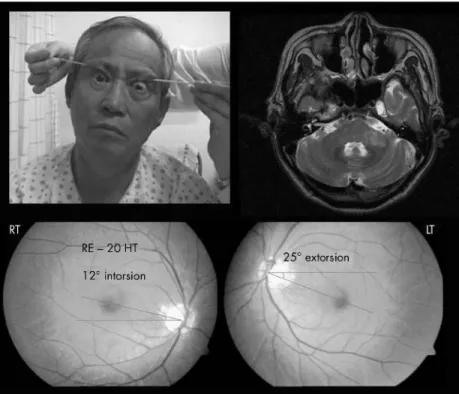
Fundus photography showed 25˚extorsion of the left eye and 12˚intorsion of the right eye.
He had a skew deviation with a right hypertropia of 20 prism diopters in primary gaze (fig 1). Magnetic resonance imaging (MRI) including diffusion images showed acute infarcts in the left middle cerebellar peduncle and the left lateral inferior pontine tegmentum (fig 1).
The second patient was a 58 year old woman with type 2 diabetes mellitus and hypertension who developed severe vertigo, hearing loss, tinnitus on the left side, dysarthria, and imbalance. She had bilateral gaze evoked nystagmus with a horizontal- rotatory component. There was left peripheral facial weakness and numbness, dysmetria of the left limbs, and gait ataxia. Pure tone audiometry showed a 65 dB sensorineural hearing loss on the left side. Fundus photo- graphy showed 14˚extorsion of the left eye and 3˚extorsion of the right eye. Prism testing showed a skew deviation with a right hypertropia of 6 diopters in the primary position. Subjective visual vertical with bino- cular viewing was tilted 13˚to the left (that is, counterclockwise from the patient’s point of view). Caloric response was absent on the left side. MRI showed new infarcts in the left middle cerebellar peduncle, left lateral infer- ior pontine tegmentum, and anterior inferior cerebellum, possibly including the flocculus.
Two months later the subjective visual vertical was normal. Fundus photography
The patient described in this letter consented to his details being published doi: 10.1136/jnnp.2005.067322 Competing interests: none declared
Figure 1 Tonic ocular tilt reaction in patient 1. Note sustained head tilt and concurrent vertical divergence of the eyes (skew deviation). T2 weighted axial magnetic resonance imaging of the brain showed acute infarcts in middle cerebellar peduncle and lateral inferior pontine tegmentum.
There is conjugate leftward torsion of the eyes (that is, counterclockwise from the patient’s point of view): a 25˚extorsion of the left eye and a 12˚intorsion of the right. HT, hypertropia; LT, left; RE, right eye; RT, right. Patient consent was obtained for publication of this figure.
1742 PostScript
www.jnnp.com
group.bmj.com on December 21, 2016 - Published by
http://jnnp.bmj.com/
Downloaded from
now showed 1˚of extorsion of the left eye, indicating that the left eye had been extorted by 13˚at the first examination (that is, by 14˚ minus 1˚) and 9˚of extorsion of the right eye, indicating that at the first examination the right eye had in fact been intorted by 6˚(that is, by 3˚minus 9˚).
Comment
Most earlier reports of AICA infarction have focused on the brain stem or cerebellar findings. Recently, there have been several reports describing the clinical importance of inner ear symptoms, vertigo and/or sudden deafness.2 3However, a detailed description of OTR as a sign of AICA infarction has not been reported previously.
OTR, a sign of vestibular dysfunction in the roll plane, is characterised by a triad of conjugate ocular torsion, skew deviation, and heal tilt. It results from destructive or irritative lesions of central or peripheral graviceptive vestibular pathways. Although head tilt is a common component of OTR, skew deviation with conjugate ocular torsion often occurs without head tilt as in our patient. Thus the pathophysiology of a partial OTR (that is, skew deviation and conjugate ocular torsion without head tilt) is the same as that of a complete OTR, and skew deviation with conjugate ocular torsion is sufficient for the diagnosis of OTR.
In addition to lesions of the central and peripheral vestibular pathways conveying grav- iceptive signals, lesions of the cerebellum may also result in OTR. Skew deviation is commonly seen with cerebellar infarction. Mossman and Halmagyi described two patients with cerebel- lar stroke, presumably in the territory of the posterior inferior cerebellar artery, who had tonic conjugate ocular torsion without asso- ciated head tilt.4These investigators speculated that interruption of nodular inhibitory projec- tions to graviceptive neurones in the ipsile- sional vestibular nuclei may have accounted for the contraversive conjugate ocular torsion.4
Sensorineural hearing loss and canal paresis to caloric stimulation on the left side clearly indicated involvement of the peripheral audio- vestibular system. Ipsiversive OTR with acute peripheral vestibular lesions was described in a previous report.5Considering the direction of OTR and known vascular anatomy of the AICA, damage to the inner ear or the root entry zone of the eighth nerve probably accounts for the ipsilesional OTR with AICA infarction.
In conclusion, this is the first report of well documented OTR with AICA infarction. The ipsiversive OTR in these patients probably resulted from infarction of the inner ear or the root entry zone of the eighth nerve.
Acknowledgements
This study was supported by grants of the Oriental Medicine R&D Project (03-PJ9-PG6-SO02-0001), Ministry of Health and Welfare, Republic of Korea.
H Lee Department of Neurology, Keimyung University School of Medicine, 194 Dongsan dong, Daegu, 700- 712, South Korea S-Y Lee Department of Ophthalmology, Keimyung University School of Medicine H Lee, S-R Lee Brain Research Institute, Keimyung University School of Medicine B-R Park Department of Physiology, Medicine and Hanbang Brain Disease Research Centre, Wonkwang University School of Medicine, Iksan, South Korea
R W Baloh Department of Neurology, UCLA School of Medicine, Los Angeles, California, USA Correspondence to: Dr Hyung Lee; [email protected] doi: 10.1136/jnnp.2005.069104
References
1 Oas JG, Baloh RW. Vertigo and the anterior inferior cerebellar artery syndrome. Neurology 1992;42:2274–9.
2 Lee H, Whitman GT, Lim JG, et al. Bilateral sudden deafness as a prodrome of anterior inferior cerebellar artery infarction. Arch Neurol 2001;58:1287–9.
3 Lee H, Sohn Il, Jung DK, et al. Sudden deafness and anterior inferior cerebellar artery infarction.
Stroke 2002;33:2807–12.
4 Mossman S, Halmagyi M. Partial ocular tilt reaction due to unilateral cerebellar lesion.
Neurology 1997;49:491.
5 Safran AB, Viber D, Issoua D, et al. Skew deviation following vestibular neuritis.
Am J Opthalmol 1994;118:238–45.
Video assessment of rTMS for Tourette syndrome
In a recent study, subthreshold 1 Hz repeti- tive transcranial magnetic stimulation (rTMS) over left motor or premotor cortex failed to improve tics in patients with Gilles de la Tourette syndrome (GTS) as determined by self assessment scores.1 However, video ratings of this study had not been analysed.
Here, we present the results of blinded analysis of the video of GTS patients who participated in the previous study. We show that rTMS has a placebo effect and confirm that low intensity motor or premotor rTMS does not have a specific effect on tics in GTS.
In a placebo controlled cross-over study of 16 patients with GTS, subthreshold 1 Hz rTMS (2400 stimuli delivered on 2 consecu- tive days) were applied under three condi- tions in random order: left motor, left premotor, and left motor sham stimulation.
Videotapes were recorded before and after each rTMS intervention in eight patients. One of the authors (AHS) who did not know the patients and was blinded to the treatment conditions, rated the video recordings. Data were analysed using two different rating scales, the Modified Rush Video Scale
(MRVS)2 and an ‘‘Adapted Yale’’ Video Scale (AYVS) which was developed for this study. With the MRVS, the following five tic domains are rated from 0 to 4 according to severity: number of body areas involved with tics, motor tic severity, phonic tic severity, frequency of motor tics, and frequency of phonic tics. The sum of the five domain scores provides a total tic impairment score (0–20). As the MRVS does not consistently score the complexity, intensity, and inter- ference of tics, we devised an additional scale using the categories of the Yale Global Tic Severity Scale (YGTSS).3This new scale, the AYVS, rated the following five domains from 0 to 5 according to severity: number of different tics, frequency of tics, intensity of tics, complexity of tics, and interference of tics. Each domain was rated separately for motor and vocal tics. The sum of the five domains gave a total motor tic score and a total vocal tic score; these scores combined yielded the total tic impairment score (0–50).
Tics were partially, albeit non-significantly, suppressed following each of the three inter- ventions (table 1 and fig 1, two factorial repeated measures ANOVA with the factors time and rTMS). However, the effect of active rTMS did not exceed the effect observed following sham stimulation. rTMS effects in this study were variable and tic scores showed a regression towards the mean, that is patients with high scores at baseline tended to have lower scores after the rTMS intervention, and vice versa (univariate ANOVA with the difference in video score before and after rTMS as dependent factor and baseline scores as covariant). In other words, the changes of tic severity that we observed most likely reflect the waxing and waning course of tics rather than an intrinsic rTMS effect.
The results of this video assessment are in keeping with patients’ self assessment based on the Motor tic, Obsessions and compul- sions, Vocal tic Evaluation Survey (MOVES),4 a self rating scale that patients completed before and after rTMS.1 Neither motor and vocal tic subscales nor obsession and compul- sion subscales were changed by rTMS, which indicates that rTMS as used in the present study is not an effective treatment for tics or obsessions/compulsions in GTS patients.
However, because ADHD symptoms were not assessed, we cannot exclude the fact that rTMS as used in the present study might have an effect on ADHD.
There was good correlation between the MRVS and the AYVS (r = 0.69; p,0.01, Spearman’s correlation). The AYVS thus Competing interests: none declared
Patient consent was obtained for publication of figure 1
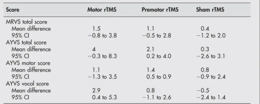
Table 1 Mean differences (before and after rTMS) and confidence intervals of clinical scores
Score Motor rTMS Premotor rTMS Sham rTMS
MRVS total score
Mean difference 1.5 1.1 0.4
95% CI 20.8 to 3.8 20.5 to 2.8 21.2 to 2.0
AYVS total score
Mean difference 4 2.1 0.3
95% CI 20.3 to 8.3 0.2 to 4.0 22.6 to 3.1
AYVS motor score
Mean difference 1.1 1.4 0.8
95% CI 21.3 to 3.5 0.5 to 0.9 20.9 to 2.4
AYVS vocal score
Mean difference 2.9 0.8 20.5
95% CI 0.4 to 5.3 21.1 to 2.6 22.4 to 1.4
AYVS, Adapted Yale Video Scale; CI, confidence interval; MRVS, Modified Rush Video Scale.
PostScript 1743
www.jnnp.com group.bmj.com
on December 21, 2016 - Published by http://jnnp.bmj.com/
Downloaded from