—
171—
ISSN: 1225-8598 (Print), 2288-3371 (Online)서 론
근육은 에너지 대사 및 운동능력에 있어서 매우 중요한 기관이다(Braun and Gautel, 2011). 근육분화(myogenesis)는 근원전구세포인 근육위성세포(muscle satellite cell, MSCs)가 수축성을 가지는 다핵성의 근섬유로 형성되는 과정을 말하
며 paired box protein 3 (Pax3), paired box protein 7 (Pax7), myocyte enhancer factor 2 (MEF2), myogenic regulatory fac- tors (MRFs) 등과 같은 다양한 전사인자들에 의해 조절된다 (Lee et al., 2013). Pax3, Pax7은 세포의 생존, 재생, 분열 및 근육 전구 세포의 분화에 관여하며 특히 Pax3는 초기 발생 과정에서 근육 전구세포의 분화에 영향을 미친다(Relaix et
al., 2005). MyoD, Myf5, MRF4, myogenin을 통틀어 MRFs
라 부르며 이 중 MyoD와 Myf5는 근원세포의 분열, MRF4, myogenin과 MEF2는 근관세포로의 분화에 영향을 미친다제브라피쉬 근육성장에서의 carnosic acid의 효과
김정환∙진덕희∙김영대1∙진형주*
강릉원주대학교 해양분자생명공학과, 1국립수산과학원 동해수산연구소
Effects of Carnosic Acid on Muscle Growth in Zebrafish (Danio rerio)
by Jeong Hwan Kim, Deuk-Hee Jin, Young-Dae Kim1and Hyung-Joo Jin* (Department of Marine Molecular Biotechnology, Gangneung-Wonju National University, Gangneung 210-702, Korea; 1Aguaculture Division, East Sea Regional Fisheries Research Institute, Gangneung 210-860, Korea)ABSTRACT Myogenesis is the formation process of multinucleated myofiber with a contractile capac- ity from muscle satellite cell (MSCs) during life. This process is tightly controlled by several transcrip- tion factors such as Pax3 and Pax7 (paired box protein 3 and 7), MEF2C (myocyte enhancer factor 2) and MRFs (myogenic regulatory factors) etc. On the contrary, myostatin (MSTN) is a transforming growth factor-β
β superfamily, which functions as a negative regulator of skeletal muscle development andgrowth. Carnosic acid (CA) is a major phenolic component in rosemary (Rosmarinus officinalis) and have been reported various biological activities such as anticancer, antioxidant, antimicrobial and ther- apeutic agents for amnesia, dementia, alzheimer’s disease. This study was confirmed to effects of CA on muscle cell line and muscle tissue alteration of zebrafish by intramuscular injection or feeding meth- ods. 10
μμM CA showed a non-cytotoxic on myoblast and a complete inhibition effect against myostatinactivity on luciferase assay. In intramuscular injection experiment, the total protein and triglyceride amount of 10
μμM/kg of CA injected group increased by 11% and decreased by 13% compared to theseof the no injected group. In histology analysis of muscle tissues by hematoxylin/eosin staining, the number of muscle fiber of 10
μμM/kg of CA injected group decreased by 29% and fiber area increased40% compared to these of no injected group. In feeding experiment, the total protein and triglyceride amount no significance difference compared to these of the normal feeding group. In histology analy- sis, the number of muscle fiber of 1% CA fed group decreased by 35% and fiber area increased 56%
compared to these of normal fed group. We identified that CA have an effect on hypertrophy of muscle fiber in adult zebrafish and the results of this study are considered as the basic data that can reveal the mechanisms of muscle formation via gene and protein level analysis.
Key words : Carnosic acid, zebrafish, muscle, hypertrophy, hyperplasia
*Corresponding author: Hyung-Joo Jin Tel: 82-33-640-2349 Fax: 82-33-640-2340, E-mail: [email protected]
Accepted: July 30, 2014
http://www.fishkorea.or.kr
(Relaix et al., 2005). 한편 근육의 분화와 분열을 억제하는 인 자인 myostatin은 TGF-β (transforming growth factor β) super-
family에 속하는 전사인자로서 자연적인 돌연변이로 인한 myostatin 유전자의 결손이 나타난 Belgian blue와 piedomon- tese종은 근육의 양이 정상개체보다 두 배 많은 대신 지방 의 함량은 적은 것으로 보고되어 있다(McPherron and Lee, 2005). 또한 유전자 조작 기법을 통해 마우스의 myostatin 유전자를 결손시킨 결과 정상개체에 비해 근섬유의 과비대 및 과 형성에 의한 근육양의 증가가 보고되었다(Fries et al., 1997; Kambadur et al., 1997; McPherron and Lee, 2005). 어류 의 myostatin 구조 및 기능은 포유류와 유사하지만 조직 특 이적 발현 정도 및 유전자 동형은 차이를 보인다(Ostbye etal., 2007). 이러한 이유로 다양한 어류에서 유전자 knock-out
또는 knock-down 기법을 통해 myostatin에 관한 연구가 진 행되고 있다. 최근 RANi 기법을 통한 myostatin 유전자의 knock-out된 zebrafish의 경우 정상개체에 비해 근섬유의 수 가 증가 및 MRFs의 발현 증가가 보고되었다(Amali et al., 2004). 또한 myostatin의 대표적인 antagonist로 알려진 myo- statin prodomain의 과 발현 및 우성음성 돌연변이(dominant negative mutant) 제브라피쉬의 경우 근섬유의 과비대로 인한 근육양의 증가가 나타났다(Xu et al., 2003; Fuentes et al., 2013). 어류의 성장 및 근육의 발생에 있어서 myostatin 역 할의 중요성을 나타내지만 아직까지 myostatin 억제에 관한 분자적 메커니즘이나 신호 경로에 대한 연구는 미흡한 실 정이다.로즈마리는(Rosmarinus officinalis) 지중해 지역이 원산지 로 강한 향과 다량의 방향족 화합물을 함유하고 있는 특성 때문에 향신료 및 식품 방부제 원료, 민간 요법으로 많이 이 용되고 있다(Benzie and Wachtel-Galor, 2011). 특히 항암, 항 균, 항비만, 항산화 효과가 보고되어 있는 페놀 화합물 계열 인 carnosic acid (CA), carnosol, rosmarinic acid, ursolic acid 등과 다양한 생리활성물질들을 함유하고 있다(Yesil-Celo- ktas et al., 2010; Benezie and Wachtel-Galor, 2011; Lopez- Jimenez et al., 2013). 최근 ursolic acid가 IGF 신호경로를 통해 골격근의 위축을 억제할 수 있다는 사실이 마우스 연 구를 통해서 밝혀졌고, 이로 인해 다양한 페놀계 화합물과 근육 성장 및 근육 위축에 관한 연구가 다양하게 진행 중에 있다.
제프라피쉬는 유전자 및 기관형성이 인간과 유사한 특징 을 가지고 있으며, 투명한 배아는 기관형성 과정, 다양한 생 물학적 반응들에 대해서 실시간 관찰이 용이하며 유전자 조 작 및 유지비용의 경제성 등의 장점으로 동물모델로서 높은 가치를 인정받고 있다(Seth et al., 2013). 포유류와는 달리 제 브라피쉬는 한번에 200~300개의 알을 낳으며 발생이 빠른 장점을 이용하여 한번에 많은 수의 천연물 스크리닝에 이용 되고 있으며 이러한 in vivo 수준에서의 스크리닝을 통해 포
유류를 이용한 동물실험에서 경우의 수를 낮출 수 있는 장 점이 있기 때문에 최근에 다양한 연구에서 사용되고 있다 (Leonard and Randall, 2005). 따라서 본 연구에서는 로즈마 리가 포함하고 있는 대표적인 페놀계 화합물인 carnosic acid 가 제브라피쉬의 근육성장에 미치는 영향을 확인해보고자 한다.
재료 및 방법
1. WST assay
L6 (KCBL No. 21458), C2C12 (ACTC No. AC30004) 근육 세포에 대한 carnosic acid (CA, TCI, Japan)의 세포독성을 WST assay (ITBio, Korea)를 통해 확인하였다. 각 세포는 96 well plate에서 well당 1×10⁵cells개씩 DMEM (10% FBS, 1%
penicillin/streptomycin) 배지를 이용하여 5% CO2, 37�C가 유지한 상태에서 배양하였다. 24시간 후 CA를 1, 5, 10, 50, 100 μM/well로 처리하였고 10시간 후 10 μL의 WST reagent 를 첨가하고, 1시간 후 450 nm (Tecan, Durham, NC, USA)에 서 흡광도를 측정하였다.
2. pGL3-(CAGA)
12-luciferase reporter assay
L6 세포에서 pGL3-(CAGA)12-luciferase reporter assay를 이용한 myostatin 활성 억제능을 측정하였다. 세포를 96 well plate에 well당 8×10³cell/200 μL로 24시간 동안 배양한 후 0.1 μg의 pGL-(CAGA)12plasmid와 0.05 μg의 pRL-TK plas- mid (Promega, Madison, USA)를 형질 도입하였다(FuGENE 6 transfection Reagent, Promega, Madison, USA). 24시간 후, 혈청이 제거된 DMEM으로 교체하였고, 9시간 후 3 ng/mL의 재조합 myostatin (R&D systems, USA)과 CA를 1, 5, 10 μM/
well로 각각 처리하였다. 6시간 후 Dual-Glo luciferase assay system (Promega, USA)을 이용하여 microplate luminometer (Berthold, USA)에서 luciferase activity를 측정하였다.
3. 제브라피쉬 사육
제브라피쉬는 생후 3개월된 개체를 구입하여(Seoulaqua, Korea) 사용하였다. 사육 수는 UV 멸균된 수돗물을 이용하 여 1일 1회 환수하였고 수온을 28.5±0.5�C로 유지하였다.
광주기는 14 L:10 D을 유지하여 사육하였으며 먹이는 1일 3회 사료를(Color charasin, 43.8% crude protein, 4.5% crude lipid, 2.75% calcium, 2.62% crude fiber, 11.79% crude ash, 1.8% phosphorus, JAQNO, Korea) 급여하였다.
4. CA 근육 내 주사
근육 내 주사 실험을 위해 각 그룹은 0.7 L 사육조 내에
5마리씩 총 4개의 사육조(n==20)로 구성하여 배양기 내에서 사육하였다. 대조구는 주사를 실시하지 않은 그룹과 corn oil을 주입한 그룹, 실험구는 5 μM/kg과 10 μM/kg의 CA를 주입한 그룹으로 실험을 진행하였다. CA는 corn oil (Sigma, USA)에 녹여 사용하였으며 시료의 주입은 10 μL micosyr- inge (701RN, HAMILTON, USA)에 32 gauge needle을 이용 하여 개체당 1 μL씩 주입하였다. 근육 내 주사의 부위는 등 지느러미를 기준으로 좌, 우 1 mm 내에 근육이 많은 부분에 주입하였으며 매주 그 위치를 바꾸어 주입하였다. 근육 내 주사 회수는 주 1회 주입, 총 4주간 실험하였으며 매주 개체 간 체중 및 체장을 측정하였다.
5. CA 사료
사료를 통한 CA의 효과를 알아보기 위한 실험을 위해 각 그룹은 0.7 L 사육 조 내에 5마리씩 총 4개의 사육 조(n==20) 로 구성하여 배양기 내에서 사육하였다. CA는 ethanol에 녹 여 사료에 0, 1, 5%가 포함되도록 배합하여 급여하였다. 사 료는 1일 3회 급여하였으며 매주 개체간 체중 및 체장을 측 정하였다.
6. 근육 내 단백질 및 중성지방 함량
제브라피쉬 근육 내 단백질 함량은 근조직으로부터 BCA protein assay kit (Pierce, USA)을 이용하여 측정하였으며 중 성지방을 함량은 Triglyceride quantification kit (Biovision, USA)을 이용하여 측정하였다.
7. 근조직 분석
근섬유 분석은 근육조직을 bouin’s 용액에 고정한 후 파라 핀 침투 후 microtome을 이용하여 5 μm 두께로 박편을 제 작하였다. 이후 hematoxylin/eosin 염색을 한 후 Image J 프 로그램을(http://rsbweb.nih.gov/ij) 이용하여 근섬유의 수, 면 적, 지름을 측정하였다.
8. 통계처리
통계처리는 SPSS 20.0 프로그램을 이용하여 one way ANOVA test를 실시하여 Duncan 검정을 통해 개체 무게, 단백질과 중성지방의 함량, 근섬유의 수와 평균 면적을 분 석하였다.
결 과
1. CA 세포 독성 및 myostatin 활성 저해 능
마우스 유래의 근육세포인 C2C12세포에서 CA의 IC50은
41.88 μM, 랫 유래의 근육세포인 L6세포의 IC50은 168.4 μM 로 확인되었다(Fig. 1A, B). Luciferase assay system을 이용 하여 CA의 myostatin 활성 저해 능을 확인해본 결과 10 μM 에서 myostatin 활성을 저해하는 것으로 확인되었다(Fig. 1C).
2. 근육 내 주사에 의해 CA가 제브라피쉬 근육에 미치는 영향
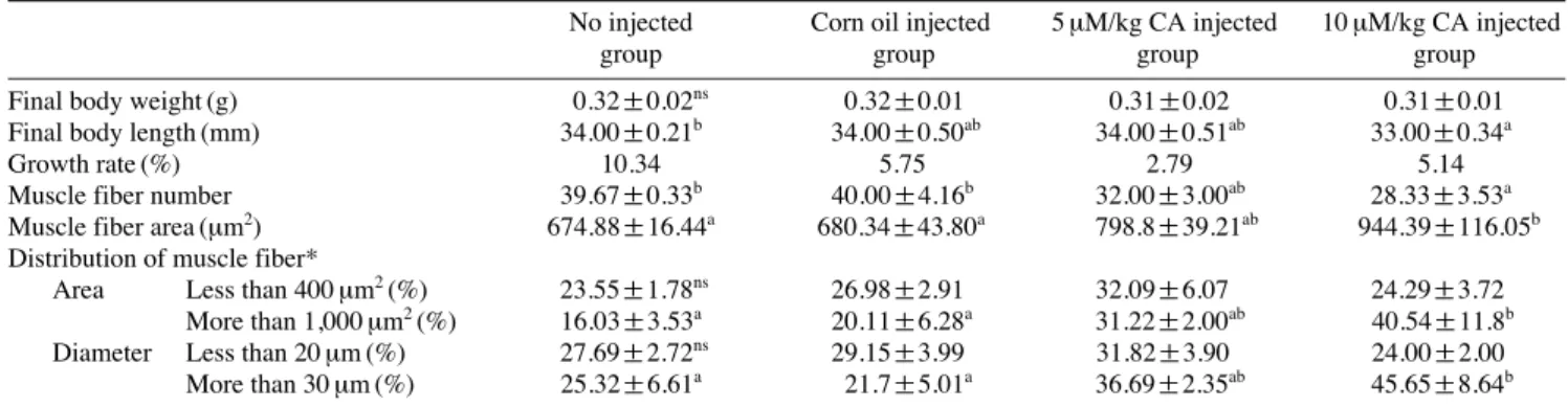
4주 동안 CA를 근육 내 주사를 통해 주입한 결과 제브라 피쉬의 체중은 그룹 간에 유의적인 차이를 보이지 않았지만 10 μM/kg의 CA를 주입한 그룹이 대조구에 비해 체장이 감 소한 것을 확인하였다. 무게에 따른 성장률에서 대조구의 경우 약 10%의 성장률을 보인 반면, 5 μM/kg의 CA를 주입 한 그룹은 3%, 10 μM/kg CA 주입 그룹은 5%의 증가율을 Fig. 1.The cell cytotoxicity and myostatin signal inhibitory activity.
(A) Effect of carnosic acid on L6 cell viability by WST assay (IC50== 168.4 μM). (B) Effect of carnosic acid on C2C12 cell viability by WST assay (IC50==41.88 μM). Data are expressed as percentage of un-treat- ment control (which is set at 100%)±SD (n==3). (C) Myostatin signal inhibitory effect of carnosic acid on L6 by Luciferase assay (IC50== 1.23 μM).
(A) WST assay on L6 cell
(B) WST assay on C2C12 cell
(C) Luciferase assay on L6 cell 150
100
50
0
150
100
50
0
20.0 15.0 10.0 5.0 0.0
Cell viability(%)Cell viability(%)Firefly/Renilla liciferase ratio
No Ethanol 1 5 10 50 100
treatment only
No Ethanol 1 5 10 50 100
treatment only
No Myostatin 1 5 10
myostatin only
Carnosic acid concentration (μM/well)
Carnosic acid concentration (μM/well)
Carnosic acid concentration (μM/well)
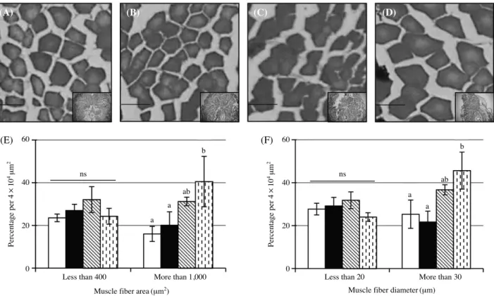
보였다(Table 1). 근육조직 내 단백질과 중성지방의 함량 분 석결과 10 μM/kg CA 주입 그룹에서 대조구에 비해 단백질 함량은 11% 증가하였고, 중성지방의 함량은 13% 감소하였 다(Fig. 2A, B). 근섬유의 크기 및 평균면적 분석을 위한 조 직분석 결과, 대조구에 비해 10 μM/kg CA 주입 그룹이 근섬 유의 수는 29% 감소하였고, 근섬유의 평균 면적은 40% 증 가하였다. 유사하게 1,000 μm
2이상의 근섬유의 분포가 대조 구에 비해 153% 증가하였고 30 μm 크기의 지름을 가지는 근섬유의 분포 또한 80% 증가하였다(Table 1, Fig. 3).
3. 사료를 통한 CA가 제브라피쉬 근육에 미치는 영향
근육 내 주사 실험과 마찬가지로 4주간 사료에 CA를 첨
가하여 급여한 결과 체중 및 체장 모두 대조구와 유의적인 차이를 보이지 않았다. 체중에 따른 성장률은 대조구가 15%
증가한 반면, 1% CA 사료 그룹이 11%, 5% CA 사료 그룹이 4%의 성장률을 보였다(Table 2). 근육 조직 내 단백질과 중 성지방의 함량을 분석해본 결과 그룹 간에 유의적인 차이를 보이지 않았다(Fig. 4A, B). 근육 섬유의 조직 분석 결과, 1%
CA 사료 그룹이 대조구에 비해 근섬유의 면적은 35% 감소 하고 근섬유의 평균 면적은 56% 증가하였다. 유사하게 1,000 μm
2이상 근섬유 및 30 μm 이상의 지름을 가진 근섬유의 분포가 대조구에 비해 각각 136%, 98% 증가하였다(Table 2, Fig. 5).
고 찰
천연물의 경우 합성 화합물에 비교적 경제적이고, 멀티타 깃 효과 및 부작용이 적은 장점으로 인해 여러 분야에서 높 은 관심을 보이고 있다(Harvey, 2008). 생명공학 분야에 있 어서 천연물에 관한 연구는 인간에게 나타날 수 있는 질환 의 치료 목적으로서 사용할 수 있도록 다양한 생리활성을 검증하고 있고 그 결과 전 임상 및 임상단계 수준의 천연물 도 다수 보고되고 있다(Harvey, 2008).
개체성장에 있어서 근육의 발달은 근섬유의 과형성과 과 비대의 메커니즘이 적절히 조절되면서 일어나게 된다. 특히 이러한 메커니즘에 있어서 IGF/Akt/mTOR 신호와 myostatin 신호는 근육을 형성하는 단백질의 합성 및 억제에 있어서 매우 중요한 신호 경로로 알려져 있다(Thomas et al., 2000;
Bonaldo et al., 2013). 특히 myostatin은 노화 또는 근육성이 영양증(DM, muscular dystrophy), 루게릭병(ALS, amyotrophic lateral sclerosis), 만성폐쇄성폐질환(COPD, congestive obstruc- tive pulmonary disease), 만성심부전증 (CHF, chronic heart failure), 후천성 면역결필증후군(ADIS, acquired immune defi-
Table 1.Biometric and morphometric properties of muscle fiber in zebrafish injected with carnosic acid (CA)No injected Corn oil injected 5 μM/kg CA injected 10 μM/kg CA injected
group group group group
Final body weight (g) 0.32±0.02ns 0.32±0.01 0.31±0.02 0.31±0.01
Final body length (mm) 34.00±0.21b 34.00±0.50ab 34.00±0.51ab 33.00±0.34a
Growth rate (%) 10.34 5.75 2.79 5.14
Muscle fiber number 39.67±0.33b 40.00±4.16b 32.00±3.00ab 28.33±3.53a
Muscle fiber area (μm2) 674.88±16.44a 680.34±43.80a 798.8±39.21ab 944.39±116.05b Distribution of muscle fiber*
Area Less than 400 μm2(%) 23.55±1.78ns 26.98±2.91 32.09±6.07 24.29±3.72
More than 1,000 μm2(%) 16.03±3.53a 20.11±6.28a 31.22±2.00ab 40.54±11.8b
Diameter Less than 20 μm (%) 27.69±2.72ns 29.15±3.99 31.82±3.90 24.00±2.00
More than 30 μm (%) 25.32±6.61a 21.7±5.01a 36.69±2.35ab 45.65±8.64b
Values are given as means together with standard deviation, Number and area of muscle fibers within 4.0×104μm2, N==3 Growth rate calculated as [(final weight-initial weight)/initial weight}×100]
nsNo significance, different letters are significantly difference (p⁄0.05, *p⁄0.1)
Fig. 2.Biochemical analysis of muscle fiber in zebrafish injected with CA. (A) Total protein amount, (B) triglyceride amount (black bar), dif- ferent letters are significantly difference (p⁄0.15).
(A) Protein amount
(B) Triglyceride amount 150
100
50
0
150
100
50
0 Relative amount of protein (% of no injected group)Relative amount of triglyceride (% of no injected group)
No Corn oil 5 μM/kg CA 10 μM/kg CA injected group injected group injected group injected group
No Corn oil 5 μM/kg CA 10 μM/kg CA injected group injected group injected group injected group
ciency syndrome), 암악액질(cancer cachexia), 신부전(renal failure), 요독증(uremia), 류마티즘 관절염(RA, rheumatoid arthritis) 등과 같은 다양한 질병에서 과잉 발현되는 것으로 알려져 있다(Bonaldo et al., 2013). 그러므로 천연물을 이용 한 myostatin 신호 억제에 관한 연구가 다양한 질병에 따른 근육 소실을 해결할 수 있는 치료제로 부각되기 시작했다.
하지만 천연물을 이용한 근육성장효과를 어류수준에서 확인한 연구는 매우 미흡하다. 특히 제프라피쉬의 경우 주 로 배아를 이용한 천연물의 독성 평가에 주로 활용되며 성 체의 경우 재조합 단백질 및 유전자 조작 개체를 이용하여 근육 성장 메커니즘에 대한 연구가 대부분이다(Amali et al., 2004; Kim et al., 2011; Fuentes et al., 2013).
본 연구에서 in vitro 수준에서 CA가 myostatin 신호의 억 제능을 확인하였으며 성체 제브라피쉬를 이용한 in vivo 실 험을 통해 로즈마리의 대표적인 페놀화합물인 CA가 제브라 피쉬 근섬유에 영향을 미치는 것을 확인할 수 있었다. 또한 in vitro 실험에서 CA의 효과는 세포에 따라 다른 경향을 나타내었다. 50 μM의 CA는 랫 유래의 근원세포인 L6 세포 에는 아무런 영향을 미치지 않았지만, 마우스 유래의 근원 세포인 C2C12 세포에는 약 60%의 세포 사멸을 확인할 수 있었다. 앞선 연구에 의하면 K-563, MCF-7, Hep-3B, PC-3, DU-145, MDA-MB-231 등과 같은 인간 유래의 다양한 암
세포의 경우 CA가 항 증식효과가 있는 것으로 알려져 있 고(Yesil et al., 2010) 백혈구 세포의 항 증식효과 및 분화 촉진, 항산화효과를 이용한 어류 및 육류의 부패방지, 지방
Fig. 3.Histology analysis of muscle fiber in zebrafish injected with CA. (A~D) histology image of muscle fiber, (E) muscle fiber distribution by average area, (F) muscle fiber distribution by diameter. No injection group (A, white bar), corn oil injected group (B, black bar), 5 μm/kg CA inject- ed group (C, diagonal bar), 10 μm/kg CA injected group (D, dotted bar), scale bar is 50 μm, different letters are significantly difference (p⁄0.1).(A) (B) (C) (D)
(E) (F)
Less than 400 More than 1,000 Muscle fiber area (μm2)
Percentage per 4×104μm2 Percentage per 4×104μm2
Muscle fiber diameter (μm)
Less than 20 More than 30
60
40
20
0
60
40
20
0
Fig. 4.Biochemical analysis of muscle fiber in zebrafish fed with CA. (A) Total protein amount, (B) triglyceride amount (black bar), different letters are significantly difference.
(A) Protein amount
(B) Triglyceride amount 200
150 100 50 0
200 150 100 50 0 Relative amount of protein (% of normal feeding)Relative amount of triglyceride (% of normal feeding)
Normal Ethanol 1% CA 5% CA
fed group fed group fed group fed group
Normal Ethanol 1% CA 5% CA
fed group fed group fed group fed group
ns ns
a
a a a
ab
ab
b b
ns
ns
산화 촉진 및 지방 축적 억제(Frankel et al., 1996; Steiner et al., 2001; Taka-hashi et al., 2009; Wang et al., 2012)와 같은 효과를 볼 때 근섬유에 미치는 CA의 효과는 새로운 발견이 라 할 수 있겠다.
In vivo 수준에서 볼 때 근육 발달에 있어서 포유류의 경 우 근섬유의 과형성이 일어나고 성장기간 동안에 근섬유의 과비대만 일어나는 반면 어류의 경우 발생 초기뿐만 아니라
성장단계에서도 동시 다발적으로 근섬유의 과형성과 과비대 가 일어난다(Kiessling et al., 2006; Rescan, 2008). 또한 어류 의 경우 지름 20 μm 이하의 근섬유의 경우 과형성 메커니즘 에 의해 새롭게 형성된 근섬유로 판단한다(Kiessling et al., 2006; Rescan, 2008). 이러한 점으로 미루어 보아 10 μM/kg 의 CA를 근육 내 주사 그룹과 1% CA가 포함된 사료를 급 식한 제브라피쉬의 경우 근섬유의 과형성이 일어났으며 5%
Fig. 5.Histology analysis of muscle fiber in zebrafish fed with CA. (A~D) histology image of muscle fiber, (E) muscle fiber distribution by aver- age area, (F) muscle fiber distribution by diameter. Normal feed group (A, white bar), ethanol treated with feed group (B, black bar), 1% CA treat- ed with feed group (C, diagonal bar), 5% CA treated with feed group (D, dotted bar), scare bar is 50 μm, different letters are significantly differ- ence (p⁄0.1).
(A) (B) (C) (D)
(E) (F)
Less than 400 More than 1,000 Muscle fiber area (μm2)
Percentage per 4×104μm2 Percentage per 4×104μm2
Muscle fiber diameter (μm)
Less than 20 More than 30
60
40
20
0
80
60
40
20
0
Table 2.Biometric and morphometric properties of muscle fiber in zebrafish fed with carnosic acid (CA)
Normal fed Ethanol fed 1% CA fed 5% CA fed
group group group group
Final body weight (g) 0.37±0.02ns 0.33±0.02 0.34±0.03 0.33±0.02
Final body length (mm) 33.67±0.47ns 33.89±0.63 34.11±0.54 33.56±0.38
Growth rate (%) 15.25 1.42 11.11 3.86
Muscle fiber number 36.00±1.73bc 31.33±2.03b 23.33±1.33a 43.33±3.53c
Muscle fiber area (μm2) 688.04±84.89ab 763.84±35.81b 1070.67±40.51c 511.01±62.69a Distribution of muscle fiber*
Area Less than 400 μm2(%) 21.79±4.66a 21.21±1.02a 10.14±4.12a 38.47±6.24b
More than 1,000 μm2(%) 20.21±8.86a 23.02±7.83a 47.67±6.06b 9.31±5.32a Diameter Less than 20 μm (%) 26.52±3.58b 23.48±0.76ab 11.66±5.61a 39.35±6.19c
More than 30 μm (%) 30.46±8.68b 43.99±5.96b 60.37±3.26c 13.11±4.96a
Values are given as means together with standard deviation, Number and area of muscle fibers within 4.0×104μm2, N==3 Growth rate calculated as [(final weight-initial weight)/initial weight}×100]
nsNo significance, different letters are significantly difference (p⁄0.05, *p⁄0.1)
a a
a
a a
a a a
c
c
ab b
b
b
b b
CA가 포함된 사료를 급식한 그룹의 20 μm이하의 근섬유의 분포가 대조구에 비해 약 48% 증가한 것으로 미루어 보아 CA가 제브라피쉬 근섬유의 과형성 및 과비대에 영향을 미 쳤다고 생각된다. 따라서 본 연구의 자료를 토대로 근육 성 장과 관련된 유전자 및 단백질 분석, 더 나아가 포유류를 이 용한 동물실험을 통해 CA가 근 섬유에 미치는 메커니즘에 관한 연구가 진행되어야 할 것으로 사료된다.
요 약
로즈마리의 주요 성분인 carnosic acid는 carnosol, rosmari- nic acid, ursolic acid 등과 같은 폴리페놀의 한 성분으로 다 양한 생리활성 기능이 보고되어 있다. 본 연구에서는 로즈 마리 유래 폴리페놀인 carnosic acid가 제브라피쉬 근육성장 에 미치는 영향을 근육 내 주사와 사료를 통해서 확인해 보 았다. 근육 내 주사 실험을 통해서 CA는 제브라 피쉬의 근 육 내 단백질 함량을 증가시키고 중성지방의 함량을 감소 시켰다. 또한 조직학적 분석 결과 근섬유의 평균 면적이 커 지는 근섬유의 과비대 효과를 나타내었다. 사료 실험 결과 근육 내 단백질 및 중성지방의 함량에는 영향을 미치지 않 았으며 조직학적 분석 결과 근육 내 주사 실험에서와 마찬 가지로 근 섬유의 과비대를 유도하였다.
사 사
본 논문은 2012년도 강릉원주대학교 학술연구조성비 지 원에 의하여 수행되었습니다.
인 용 문 헌
Amali, A.A., C.J. Lin, Y.H. Chen, W.L. Wang, H.Y. Gong, C.Y.
Lee, Y.L. Ko, J.K. Lu, G.M. Her, T.T. Chen and J.L. Wu.
2004. Up-regulation of muscle-specific transcription factors during embryonic somitogenesis of zebrafish (Danio rerio) by knock-down of myostatin-1. Developmental Dynamics, 229: 847-856.
Benzie, I.F. and S. Wachtel-Galor. 2011. Herbal medicine: Biomol- ecular and clinical aspects. CRC Press.
Biga, P.R. and F.W. Goetz. 2006. Zebrafish and giant danio as mod- els for muscle growth: Determinate vs. indeterminate growth as determined by morphometric analysis. American Journal of Physiology-Regulatory, Integrative and Comparative Phys- iology, 291: R1327-R1337.
Bonaldo, P. and M. Sandri. 2013. Cellular and molecular mecha-
nisms of muscle atrophy. Disease Models & Mechanisms, 6: 25-39.
Braun, T. and M. Gautel. 2011. Transcriptional mechanisms regulat- ing skeletal muscle differentiation, growth and homeosta- sis. Nature Reviews Molecular Cell Biology, 12: 349-361.
Frankel, E.N., S.W. Huang, A. Aeschbach and E. Prior. 1996. Antiox- idant activity of a rosemary extract and its constituents, carnosic acid, carnosol, and rosmarinic acid, in bulk oil and oil-in-water emulsion. J. Agricultural and Food Chemistry, 44: 131-135.
Fries, R., R. Hanset and M. Georges. 1997. A deletion in the bovine myostatin gene causes the double-muscled phenotype in cattle. Nat. Genet., 17: 71.
Fuentes, E.N., K. Pino, C. Navarro, I. Delgado, J.A. Valdés and A.
Molina. 2013. Transient inactivation of myostatin induces muscle hypertrophy and overcompensatory growth in zebra- fish via inactivation of the SMAD signaling pathway. J.
Biotechnol., 168: 295-302.
Harvey, A.L. 2008. Natural products in drug discovery. Drug Dis- Covery Today, 13: 894-901.
Kambadur, R., M. Sharma, T.P. Smith and J.J. Bass. 1997. Mutations in myostatin (GDF8) in double-muscled belgian blue and piedmontese cattle. Genome Res., 7: 910-915.
Kiessling, A., K. Ruohonen, M. Bjørnevik. 2006. Muscle fibre growth and quality in fish. Arch Tierz Dummerstorf, 49: 137-146.
Kim, J.H., S.B. Lee, M.J. Cho, J.Y. Ahn, S.K. Lee, S.Y. Hong, K.B.
Seong and H.J. Jin. 2011. Growth effect of oncorhychus masou by recombinant myostatin prodomain proteins derived from fish. J. Life Science, 21: 1149-1155.
Kunkel, S.D., C.J. Elmore, K.S. Bongers, S.M. Ebert, D.K. Fox, M.C. Dyle, S.A. Bullard and C.M. Adams. 2012. Ursolic acid increases skeletal muscle and brown fat and decreases diet-induced obesity, glucose intolerance and fatty liver disease. PloS One, 7: e39332.
Kunkel, S.D., M. Suneja, S.M. Ebert, K.S. Bongers, D.K. Fox, S.E.
Malmberg, F. Alipour, R.K. Shields. and C.M. Adams. 2011.
mRNA expression signatures of human skeletal muscle atrophy identify a natural compound that increases muscle mass. Cell Metabolism, 13: 627-638.
Lee, E.J., A.R. Bhat, M.R. Kamli, S. Pokharel, T. Chun, Y.H. Lee, Y.H. Nahm, J.H. Nam, S.K. Hong, B. Yang, K.Y. Chung, S.H. Kim and I. Choi. 2013. Transthyretin is a key regulator of myoblast differentiation. PloS One, 8: e63627.
López-Jiménez, A., M. García-Caballero, M.Á. Medina and A.R.
Quesada. 2013. Anti-angiogenic properties of carnosol and carnosic acid, two major dietary compounds from rosemary.
Eur. J. Nutr., 52: 85-95.
McPherron, A.C. and S. Lee. 1997. Double muscling in cattle due to mutations in the myostatin gene. Proceedings of the Na- tional Academy of Sciences, 94: 12457-12461.
Michael, S., P. Irene, G. Judith, L. Joseph, S. Yoav and D. Michael.
2001. Carnosic acid inhibits proliferation and augments dif-
ferentiation of human leukemic cells induced by 1,25-Dihy- droxyvitamin Dsub3 and Retinoic acid. Nutrition and Cancer, 41: 135-144.
Ø