• 검색 결과가 없습니다.

Phagocytic osteoclasts in the alveolar bone of diabetic rats with periodontitis

N/A
N/A
Protected

Academic year: 2021

Share "Phagocytic osteoclasts in the alveolar bone of diabetic rats with periodontitis"

Copied!
7
0
0

로드 중.... (전체 텍스트 보기)

전체 글

(1)

Introduction Introduction

Osteoclasts are tartrate-resistant acid phosphatase (TRAP)- positive multinucleated cells that degrade bone by secreting protons and enzymes. However, the roles of osteoclasts are not limited to bone resorption. For example, osteoclasts are in- volved in coupling bone resorption and formation by stimulation of osteoblast differentiation [1]. Additionally, several studies suggest that osteoclasts have phagocytic activity under physi-

ological conditions. Osteoclasts containing cells are found in various bones, such as tibia, femur, calvaria, and alveolar bone of young animals that show active bone modeling or remodel- ing [2-5]. Cells phagocytosed by osteoclasts have been char- acterized as macrophages, osteoblasts, or osteocytes based on morphological features or immunohistochemical analysis [3,4,6-9]. Apoptotic bone cells have also been identified within osteoclasts in the alveolar bone of normal young rats [5,10]. In another study, rats treated with parathyroid hormone showed Int J Oral Biol 45:92-98, 2020

pISSN: 1226-7155 • eISSN: 2287-6618 https://doi.org/10.11620/IJOB.2020.45.3.92

Phagocytic osteoclasts in the alveolar bone of diabetic rats with periodontitis

Eun-Jung Bak 1 , Ae Ri Kim 1,2,3 , Ji-Hye Kim 4 , and Yun-Jung Yoo 1 *

1 Department of Oral Biology, Yonsei University College of Dentistry, Seoul 03722, Republic of Korea

2 BK21 PLUS Project, Yonsei University College of Dentistry, Seoul 03722, Republic of Korea

3 Department of Applied Life Science, The Graduate School, Yonsei University, Seoul 03722, Republic of Korea

4 Department of Dental Hygiene, Baekseok University, Cheonan 31065, Republic of Korea

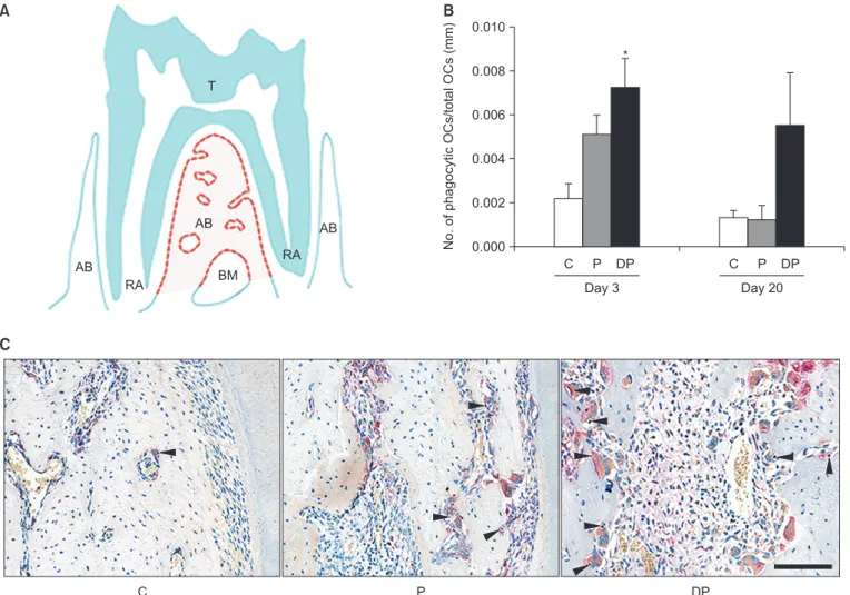
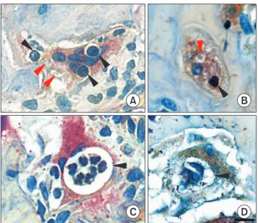
Periodontitis is a bacteria-induced inflammatory disease associated with alveolar bone loss. Osteoclast is a macrophage-lineage cell that exhibits phagocytic activity; however, osteoclast phagocytic activity has not been demonstrated under pathological conditions. Diabetes is a pathological condition that exacerbates alveolar bone loss via periodontitis; therefore, we examined phagocytic osteoclasts in diabetic rats that had periodontitis. The rats were divided into the control (C), periodontitis (P), and diabetes with periodontitis (DP) groups. Diabetes and periodontitis were induced by streptozotocin injection and ligature of the mandibular first molars, respectively. On days 3 and 20 after the ligature, the rats were sacrificed, and osteoclasts containing inclusions were quantified by tartrate-resistant acid phosphatase staining. On day 3, there were more osteoclasts containing inclusions in the DP group than in the C group. Among inclusions, osteocyte-like cells and dense bodies were more frequently observed in the DP group than in the C group. Cytoplasm-like structures were elevated more in the DP group than in the C and P groups. However, no differences were observed on day 20. Interestingly, some osteoclasts were in contact with the osteocytes within the exposed lacunae and contained several inclusions within a large vacuole. Thus, the elevation of phagocytic osteoclasts in rats with diabetes and periodontitis provides insight into the role of osteoclast phagocytic activity under pathological conditions.

Keywords: Periodontitis, Diabetes mellitus, Osteoclasts, Phagocytosis

Received June 3, 2020; Revised August 21, 2020; Accepted August 22, 2020

*Correspondence to: Yun-Jung Yoo, E-mail: [email protected] https://orcid.org/0000-0002-0045-9597 Copyright © The Korean Academy of Oral Biology

CC

This is an open-access article distributed under the terms of the Creative Commons Attribution Non-Commercial License (http://creativecommons.org/licenses/by- nc/4.0/), which permits unrestricted non-commercial use, distribution, and reproduction in any medium, provided the original work is properly cited.

Original Article IJOB

(2)

increased osteoclasts in tibiae, but did not exhibit an increase of osteoclasts with cellular inclusions [3]. These studies pro- vide insight into the role of phagocytic osteoclasts under physiological conditions, but the frequency and significance of phagocytic osteoclasts under pathological conditions remains unknown.

Periodontitis is an inflammatory disease caused by bacteria that is characterized by alteration of the alveolar bone that sur- rounds the roots of teeth [11]. We have previously reported that osteoclast formation and alveolar bone loss is increased in diabetic rats with periodontitis as well as in rats with peri- odontitis compared to control rats [12,13]. Therefore, rats with periodontitis or coincident diabetes and periodontitis provide models for observing osteoclast formation in the alveolar bone under pathological bone resorption. We hypothesized that osteoclast phagocytic activity as well as osteoclast formation would be increased under bone resorption-stimulated condi- tions. In this study, in order to estimate increase of osteoclast phagocytic activity in diabetes with periodontitis, we evaluated osteoclasts with inclusions in rats with streptozotocin (STZ)- induced diabetes with periodontitis.

Materials and Methods Materials and Methods

1. Induction of diabetes and periodontitis

Six-week-old male inbred F344 rats were divided into con- trol (C; n = 6 per time point), periodontitis (P; n = 8 on day 3 and n = 12 on day 20), and diabetes with periodontitis (DP; n = 8 on day 3 and n = 11 on day 20) groups. Diabetes was induced by intravenous administration of STZ (50 mg/kg dissolved in 0.1 M citrate buffer; Sigma-Aldrich, St. Louis, MO, USA) after fast- ing for 16 h. The C group was injected with 0.1 M citrate buffer alone. To confirm induction of diabetes, blood glucose levels were measured using the Accu-Check active system (Roche Diagnostics, Mannheim, Germany) after overnight fasting in accordance with the manufacturer’s instructions. Induction of diabetes was considered successful when fasting glucose levels were higher than 300 mg/dL. One week after admin- istration of STZ or citrate buffer, periodontitis was induced by ligature of the mandibular first molars using dental floss. Rats were sacrificed on days 3 and 20 after ligature. All animal pro- tocols were approved by the Institutional Animal Care and Use Committee of Yonsei University (2014-0393).

2. TRAP staining and quantification

After fixation with 10% neutral buffered formalin for 2 days, mandibles were decalcified with 10% ethylenediaminetet- raacetic acid for 2 months, embedded in paraffin, and cut into serial 4- μm thick sagittal sections. To identify osteoclasts, sections were stained using a TRAP kit (Sigma-Aldrich) ac- cording to the manufacturer’s protocol. Measurement of TRAP-positive multinucleated osteoclasts with cellular inclu- sions was performed in a region of interest (ROI) from the alveolar bone crest to the root apex in the furcation (Fig. 1A).

The number of phagocytic TRAP-positive multinucleated osteoclasts was counted along the bone surface in the ROI using Viewpoint software (Precipoint, Silver Spring, Freising, Germany). The number of phagocytic osteoclasts is presented relative to the total number of osteoclasts and was calculated per mm of bone surface. Based on the criteria of previous studies [5,9], inclusions within osteoclasts were classified into three types: osteocyte-like cells with distinct nuclei and faint basophilic cytoplasm similar to surrounding osteocytes located in lacuna, dense round or ovoid bodies (dense bodies) with nuclei strongly stained by hematoxylin and a few cytoplasm, and cytoplasm-like structures with lightly stained cytoplasm and no nucleus. The number of osteocyte-like cells, dense bodies, and cytoplasm-like structures phagocytosed by TRAP- positive multinucleated osteoclasts in the ROI was counted.

Images were taken using an Olympus microscope system (BX53; Tokyo, Japan). The individual performing the analyses was blinded to the experimental conditions.

3. Statistical analyses

All statistical analyses were performed using a statistical analysis program (SPSS ver. 25; IBM Corp., Armonk, NY, USA).

One-way analysis of variance (ANOVA) followed by Scheff é’s method was used to determine significant differences. p < 0.05 was considered statistically significant. Data are expressed as the means ± standard error of the mean. In the Figs. 1B and 2B, homogeneous subsets are not marked with asterisks.

Results Results

1. Phagocytic osteoclasts are elevated in diabetic rats with periodontitis during early bone loss

Three days after ligature, the number of TRAP-positive mul-

(3)

tinucleated osteoclasts with inclusions in the DP group was higher than the C group (Fig. 1B, F = 5.816, p = 0.012 and 1C).

The P group showed a similarly increased pattern compared to the C group, but there was no statistical difference. Twenty days after ligature, there were no differences among C, P, and DP groups.

2. Cellular inclusions are elevated in phagocytic osteoclasts of diabetic rats with periodontitis

Three types of phagocytosed inclusions were quantified within TRAP-positive multinucleated osteoclasts: osteocyte- like cells, dense bodies, and cytoplasm-like structures (Fig.

2A). On day 3, osteocyte-like cells (F = 5.870, p = 0.012) and dense bodies (F = 11.510, p = 0.001) were higher in the DP group than the C group (Fig. 2B). Cytoplasm-like structures (F = 10.097, p = 0.001) were also higher in the DP group than both the C and P groups. On day 20, there were no significant differences among the three groups.

3. Inclusion morphology in diabetic rats with periodontitis

A large number of osteoclasts in the DP group contained inclusions within clear vacuoles (Fig. 3A-3C). Many inclusions engulfed by osteoclasts were oval or round and stained dark AB

AB

RA BM

RA AB T

A B

C

No. of phagocytic OCs/total OCs (mm)

0.000 0.010

0.008

0.006

0.004

0.002

P DP C P DP

Day 3 Day 20

*

C

C P DP

Fig. 1. Tartrate-resistant acid phosphatase (TRAP)-positive multinucleated osteoclasts containing inclusions. (A) Region of interest for counting TRAP-pos- itive osteoclasts with inclusions in the furcation area of mandibular first molar (red dotted lines in pale pink color). (B) The number of TRAP-positive osteo- clasts with inclusions in the C, P, and DP groups on days 3 and 20 after ligature. The number of phagocytic osteoclasts is presented relative to the total num- ber of osteoclasts and was calculated per mm of bone surface. (C) Representative images of TRAP-positive osteoclasts within in the C, P, and DP groups on day 3. Black arrowheads indicate TRAP-positive osteoclasts with inclusions. Data are represented as the means ± standard error of the mean. Data were evaluated by ANOVA. Scale bar = 100 μm.

AB, alveolar bone; BM, bone marrow; T, tooth; RA, root apex; No, number; OCs, osteoclasts; C, control; P, periodontitis; DP, diabetes with periodontitis.

*p < 0.05 compared with the C group.

(4)

blue, and some inclusions were dense nuclei. Some osteo- clasts were in contact with osteocytes within exposed lacunae (Fig. 3D-3F). This contact was observed under several condi- tions. Cytoplasmic extension of osteoclast was found to be inserted into exposed lacunae and in contact with osteocyte (Fig. 3E). Additionally, some osteocytes were localized partially

within osteoclasts and partially within lacunae (Fig. 3F).

Interestingly, some osteoclasts contained several vacuoles with an individual type of inclusion, such as dense bodies or cytoplasm-like structures (black and red arrowheads in Fig. 4A and 4B, respectively). And, one osteoclast had a large vacuole containing several inclusions with segmented condensed nu-

Osteocyte-like cell 12

10

8

6

4

2

No. of phagocytosed inclusions

0

Osteocyte-like cell Dense body Cytoplasm-like structure

Dense body Cytoplasm-like structure

Osteocyte-like cell

Dense body Cytoplasm-like structure

C P DP

*

*

*

**

A

B

Day 3 Day 20

Fig. 2. Inclusions engulfed by tartrate-resis- tant acid phosphatase (TRAP)-positive mul- tinucleated osteoclasts. (A) Representative images of inclusions within a vacuole on day 3. From left to right, the black arrowheads in- dicate an osteocyte-like cell in the C group, a dense body in the P group, and a cytoplasm- like structure in the C group, respectively. (B) The number of inclusions engulfed by TRAP- positive osteoclasts in the region of interest.

Data are represented as the means ± stan- dard error of the mean. Data were evaluated by ANOVA. Scale bar = 10 μm.

C, control; P, periodontitis; DP, diabetes with periodontitis.

*p < 0.05 compared with the C group. **p <

0.05.

A B C

D E F

Fig. 3. Representative images of tartrate- resistant acid phosphatase-positive multinu- cleated osteoclasts contained inclusions and contacted with osteocytes in the diabetes with periodontitis group. (A-C) Osteoclasts with oval or round dense body within a vacuole on day 3 (black arrowheads). (D) An osteoclast in contact with an osteocyte within the ex- posed lacuna on day 3 (black arrowhead). (E) A cytoplasmic extension of an osteoclast in- serted into the exposed lacuna and in contact with an osteocyte on day 3 (black arrowhead).

(F) An osteoclast with an osteocyte partially

placed within lacuna on day 3 (black arrow-

head). Scale bar = 10 μm.

(5)

clei (Fig. 4C). Moreover, another osteoclast engulfed several osteocyte-like inclusions within a large vacuole (Fig. 4D).

Discussion Discussion

Alveolar bone loss is characteristic of periodontitis, and dia- betes aggravated alveolar bone loss induced by periodontitis [13,14]. In previous report, we have demonstrated that the dis- tance from the cemento-enamel junction to the alveolar bone crest and the number of osteoclasts is markedly increased on days 3 and 20 in rats with periodontitis and diabetic rats with periodontitis compared with control rats [13]. Therefore, in this study, we examined phagocytic osteoclasts in alveolar bone at two time-points that have previously exhibited an elevated presence of osteoclasts. Osteoclasts with inclusions were sig- nificantly elevated on day 3 in the DP group compared to the C group. The differences in the number of phagocytic osteo- clasts and inclusions between the DP group and the C group appeared to be consistent on days 3 and 20, but there was no statistical significance on day 20. The number of phagocytic osteoclasts and inclusions in the DP group tended to decrease on day 20 compared to day 3. In previous report, the number

of osteoclasts in the DP group was increased on day 3 and day 20 than in the C group, but the increase on day 20 was less on day 3 [13]. The results of the number of phagocytic osteo- clasts and inclusions in this study also showed similar trends.

These results suggest elevation of osteoclast phagocytosis in diabetes and periodontitis condition.

Phagocytosis of osteoclasts has been suggested as a way removing exposed osteocytes or apoptotic cells during bone resorption. Suzuki et al. [9] reported that the osteocytes en- gulfed by osteoclasts had features similar to those of osteo- cytes embedded in the lacuna of the bone. Pebp2 aA (isoform of Cbfa1, osteoblast marker)-positive osteocytes are found within osteoclasts in hamster jaw bones around developing tooth germs [8]. Other group has also reported ultrastructural contacts between osteoclasts and osteocytes in calvaria of young rats [4]. In addition, the femoral ultrastructure of young rabbits shows that the ruffled border of osteoclasts extends into lacuna and contacts osteocytes [2]. In this study, there were osteoclasts to be in contact with osteocytes within the exposed lacunae through ruffled borders or partially enclosed osteocytes similar to previous studies. Moreover, DP group showed the increase in inclusion of osteocyte-like cells. These suggest that engulfment of osteocytes by osteoclasts can be increased in diabetes with periodontitis conditions.

It has been suggested that osteoclasts engulf apoptotic cells based on the presence of terminal deoxynucleotidyl trans- ferase dUTP nick end labeling (TUNEL)-positive cells within osteoclasts, gene expression profiles related to the clearance of apoptotic cells, and in vitro evidence of osteoclast phago- cytosis of apoptotic thymocytes [5,10,15]. Other study sug- gests that apoptosis is enhanced in diabetic condition [16].

Consistent with previous report, our unpublished data showed that the number of TUNEL-positive osteocytes in diabetic rats with periodontitis was significantly higher than in control rats and rats with periodontitis on day 3, but not on day 20 (data not shown). Further, in this study, the DP group exhibited more dense bodies and cytoplasm-like structures as well as osteocyte-like cells in osteoclasts than the C group. These re- sults suggest the possibility that elevated osteocyte apoptosis in combination of diabetes and periodontitis may affect osteo- clast phagocytosis. For accurate determination of inclusions within phagocytic osteoclasts, immunohistochemistry using osteocyte or apoptosis markers is required in the future.

Macrophage is a representative phagocyte. We can observe some osteoclasts containing several vacuoles with an indi- vidual inclusion or several inclusions within one large vacuole

A B

C D

Fig. 4. Representative images of tartrate-resistant acid phosphatase-pos-

itive multinucleated osteoclasts containing several vacuoles with individual

inclusions or several inclusions within one vacuole in the diabetes with

periodontitis group. (A, B) Osteoclasts containing several vacuoles with

individual inclusions on day 20 (black arrowheads indicate dense bodies

and red arrowheads indicate cytoplasm-like structures). (C) An osteoclast

containing several segmented condensed nuclei in one large vacuole on

day 20 (black arrowhead). (D) An osteoclast containing several inclusions

within a large vacuole on day 3 (black arrowhead). Scale bar = 10 μm.

(6)

similar to phagocytic characteristic of macrophage [17]. After phagocytosis, professional antigen presenting cells such as macrophages and dendritic cells present fragmented peptides from phagocytosed antigen to T cells [18]. Processes after osteoclast phagocytosis have not been clearly defined. Some studies have shown induction of regulatory T cells to suppress the activation of T cells and the differentiation/activation of os- teoclasts through antigen presentation by osteoclasts [19,20].

These groups suggested that induction of regulatory T cells through antigen presentation by osteoclasts protect bone from autoimmunity and bone loss. It will be interesting to clarify the progresses after osteoclast phagocytosis in diabetes with peri- odontitis.

This is the first report to demonstrate increased phago- cytic activity of osteoclasts in diabetes with periodontitis. Our

results are meaningful to indicate that further study is war- ranted to define the role of osteoclast phagocytic activity in the pathogenesis of diabetes with periodontitis.

Acknowledgments Acknowledgments

This study was supported by the Yonsei University College of Dentistry (6-2018-0029). We thank Dr. Ki-Yeol Kim at Yonsei University College of Dentistry for assistance and advise on statistical analysis.

Conflicts of Interest Conflicts of Interest

No potential conflict of interest relevant to this article was reported.

References References

1. Novack DV, Mbalaviele G. Osteoclasts-Key Players in skeletal health and disease. Microbiol Spectr 2016;4. doi: 10.1128/

microbiolspec.MCHD-0011-2015.

2. Soskolne WA. Phagocytosis of osteocytes by osteoclasts in femora of two week-old rabbits. Cell Tissue Res 1978;195:

557-64. doi: 10.1007/BF00233897.

3. Bargsten G, Stanka P. Light and electron microscopic study on the osteoclastic phagocytosis of cells in the rat. Anat Anz 1985;159:13-20.

4. Elmardi AS, Katchburian MV, Katchburian E. Electron mi- croscopy of developing calvaria reveals images that suggest that osteoclasts engulf and destroy osteocytes during bone resorption. Calcif Tissue Int 1990;46:239-45. doi: 10.1007/

BF02555002.

5. Boabaid F, Cerri PS, Katchburian E. Apoptotic bone cells may be engulfed by osteoclasts during alveolar bone resorption in young rats. Tissue Cell 2001;33:318-25. doi: 10.1054/tice.

2001.0179.

6. Lucht U, Maunsbach AB. Effects of parathyroid hormone on osteoclasts in vivo. An ultrastructural and histochemical study.

Z Zellforsch Mikrosk Anat 1973;141:529-44. doi: 10.1007/

BF00307123.

7. Taniwaki NN, Katchburian E. Ultrastructural and lanthanum tracer examination of rapidly resorbing rat alveolar bone sug- gests that osteoclasts internalize dying bone cells. Cell Tissue Res 1998;293:173-6. doi: 10.1007/s004410051109.

8. Bronckers AL, Sasaguri K, Engelse MA. Transcription and im- munolocalization of Runx2/Cbfa1/Pebp2alphaA in developing

rodent and human craniofacial tissues: further evidence sug- gesting osteoclasts phagocytose osteocytes. Microsc Res Tech 2003;61:540-8. doi: 10.1002/jemt.10377.

9. Suzuki R, Domon T, Wakita M, Akisaka T. The reaction of os- teoclasts when releasing osteocytes from osteocytic lacunae in the bone during bone modeling. Tissue Cell 2003;35:189- 97. doi: 10.1016/s0040-8166(03)00020-x.

10. Cerri PS, Boabaid F, Katchburian E. Combined TUNEL and TRAP methods suggest that apoptotic bone cells are inside vacuoles of alveolar bone osteoclasts in young rats. J Peri- odontal Res 2003;38:223-6. doi: 10.1034/j.1600-0765.2003.

02006.x.

11. Hajishengallis G. Immunomicrobial pathogenesis of periodon- titis: keystones, pathobionts, and host response. Trends Im- munol 2014;35:3-11. doi: 10.1016/j.it.2013.09.001.

12. Kim JH, Lee DE, Cha JH, Bak EJ, Yoo YJ. Receptor activa- tor of nuclear factor- κB ligand and sclerostin expression in osteocytes of alveolar bone in rats with ligature-induced peri- odontitis. J Periodontol 2014;85:e370-8. doi: 10.1902/jop.

2014.140230.

13. Kim JH, Lee DE, Woo GH, Cha JH, Bak EJ, Yoo YJ. Osteo- cytic sclerostin expression in alveolar bone in rats with diabe- tes mellitus and ligature-induced periodontitis. J Periodontol 2015;86:1005-11. doi: 10.1902/jop.2015.150083.

14. Wu YY, Xiao E, Graves DT. Diabetes mellitus related bone metabolism and periodontal disease. Int J Oral Sci 2015;7:63- 72. doi: 10.1038/ijos.2015.2.

15. Harre U, Keppeler H, Ipseiz N, Derer A, Poller K, Aigner M,

(7)

Schett G, Herrmann M, Lauber K. Moonlighting osteoclasts as undertakers of apoptotic cells. Autoimmunity 2012;45:612-9.

doi: 10.3109/08916934.2012.719950.

16. Chen H, Liu W, Wu X, Gou M, Shen J, Wang H. Advanced glycation end products induced IL-6 and VEGF-A production and apoptosis in osteocyte-like MLO-Y4 cells by activating RAGE and ERK1/2, P38 and STAT3 signalling pathways. Int Immunopharmacol 2017;52:143-9. doi: 10.1016/j.intimp.

2017.09.004.

17. Sigola LB, Fuentes AL, Millis LM, Vapenik J, Murira A. Effects of toll-like receptor ligands on RAW 264.7 macrophage mor- phology and zymosan phagocytosis. Tissue Cell 2016;48:389-

96. doi: 10.1016/j.tice.2016.04.002.

18. Trombetta ES, Mellman I. Cell biology of antigen processing in vitro and in vivo. Annu Rev Immunol 2005;23:975-1028. doi:

10.1146/annurev.immunol.22.012703.104538.

19. Buchwald ZS, Kiesel JR, DiPaolo R, Pagadala MS, Aurora R.

Osteoclast activated FoxP3 + CD8 + T-cells suppress bone resorption in vitro. PLoS One 2012;7:e38199. doi: 10.1371/

journal.pone.0038199.

20. Kiesel JR, Buchwald ZS, Aurora R. Cross-presentation by osteoclasts induces FoxP3 in CD8 + T cells. J Immunol 2009;

182:5477-87. doi: 10.4049/jimmunol.0803897.

수치

Fig. 1. Tartrate-resistant acid phosphatase (TRAP)-positive multinucleated osteoclasts containing inclusions
Fig. 2.  Inclusions engulfed by tartrate-resis- tartrate-resis-tant acid phosphatase (TRAP)-positive  mul-tinucleated osteoclasts
Fig. 4.  Representative images of tartrate-resistant acid phosphatase-pos- phosphatase-pos-itive multinucleated osteoclasts containing several vacuoles with individual  inclusions or several inclusions within one vacuole in the diabetes with  periodontitis

참조

관련 문서

At 12 weeks after surgery, bone loss was confirmed and the trabecular bone in the femoral neck was compared between the SHAM and OVX groups using Micro-CT.. One week later,

macrophylla seeds on streptozotocin induced diabetic rats as well the establishment of efficacious role of extract to alleviate the augmented oxidative state associated

The case records of 24 patients in Seoul National University Hospital who had bone grafting of 29 alveolar clefts between 2001 and 2004 were examined. Details were recorded of age,

Purpose: The purpose of this study was to evaluate the optimal diabetes duration for bone regeneration experiments in an alloxan monohydrate (ALX)–induced diabetic rabbit calvarial

Samples of alveolar bone height measured are presented at 0, 4, 8, and 12 weeks of the general diet (GD) control group (E–H) and of the high-salt and -glu- cose diet

Anti-inflammatory, anti-osteoclastic, and antioxidant activities of genistein pro- tect against alveolar bone loss and periodontal tissue degra- dation in a mouse

Although gerbils have different tooth anatomical characteristics from rats and mice, inflammation and prominent alveolar bone loss in the distal areas induced by

The results of our study suggest that alveolar bone loss can be readily detected by volumetric micro-CT analysis as an increase in the distance between periodontal ligaments