INTRODUCTION
More than 700 bacterial strains may be found in oral cav- ity [1]. Oral microbes are composed of proportionally bal- anced beneficial, commensal and pathogenic bacteria and form biofilm as a micro-ecosystem [2]. Once established, the microflora in the biofilm remains relatively stable over time despite regular minor perturbations to the oral envi- ronment, and this stability is termed microbial homeostasis.
Also they co-exist with communication among interspecies
or intraspecies that contribute to ecologic stability [3,4].
This microbial homeostasis can break down on occasion by substantial change of oral environment. Significant param- eters regulating the homeostasis include the host defenses and the composition of the diet. In case of dental caries, the ecological biofilm was changed by the composition of the diet, and the ratio of cariogenic bacteria in the biofilm was increased [2]. Also, when the microbial homeostasis was broken by some major factors, Gram-negative anaer- obes as periodontitis related microbes increase in the accu-
Probiotic effect of feeding Lactococcus lactis and
Streptococcus thermophilus on periodontitis induced rat model
Won-Jae Choi1, Sung-Hoon Lee2*
1Department of Visual Communication Design, College of Arts and Design, Dankook University, Jukjeon, Korea
2Department of Oral Microbiology and Immunology, College of Dentistry, Dankook University, Cheonan, Korea
Dysbiosis of oral microbiome by change of the environments induces pathogen related disease. Especially, Porphyromonas gingivalis is associated with periodontitis. Various studies have showed probiotic efficacy for periodontitis related bacteria. The purpose of this study was to investigate the therapeutic effect of probiotics on periodontitis using periodontitis induced rat model. The ligature- induced periodontitis rats using P. gingivalis were fed the regular diet with or without probiotics for 20 days, and the periodontal status was then analyzed by micro computed tomograpy and levels of inflammatory cytokines. Also, the levels of P. gingivalis and the colonization of probiotics were analyzed by quantitative real-time polymerase chain reaction. The rat that fed on the diet containing Streptococcus thermophilus, Lactococcus lactis, and both probiotics significantly showed higher mass of the alveolar bone compared to the rat that fed on regular diet. Also, the rat that fed on the diet containing probiotics reduced the levels of inflammatory cytokines.
The count of P. gingivalis was reduced in the rat fed containing probiotics. Furthermore, S. thermophilus and L. lactis were detected in the rat containing each probiotic bacterium. This study established that S. thermophilus and L. lactis may be suitable probiotics for therapeutic and preventive periodontitis.
Key Words: Lactococcus lactis, Periodontitis, Probiotics, Streptococcus thermophilus
This is an open-access article distributed under the terms of the Creative Commons Attribution Non-Commercial License (http://creativecommons.org/licenses/by-nc/4.0) which permits unrestricted noncommercial use, distribution, and reproduction in any medium, provided the original work is properly cited.
Original Article
BIOLOGY RESEARCH
Received October 30, 2018; Revised [1] November 17, 2018; [2] December 4, 2018; Accepted December 11, 2018
Corresponding author: Sung-Hoon Lee, Department of Oral Microbiology and Immunology, College of Dentistry, Dankook University, 119 Dandae-ro, Dongnam-gu, Cheonan 31116, Korea.
Tel: +82-41-550-1867, Fax: +82-41-550-1859, E-mail: [email protected]
mulated subgingival biofilm [1,5,6], by which periodontitis is occurred. Therefore, periodontal disease is considered to associate with multi-bacterial infection with anaerobic microbes such as Porphyromonas gingivalis, Tannerella forsythia, and Treponema denticola [5]. The bacterial com- plex interacts with host tissue and cell causing the release of broad array of inflammatory and osteoclastogenic cyto- kines.
Probiotics are beneficial bacteria to host and produce antimicrobial agents as bacteriocin against pathogenic bac- teria [7]. Among probiotics, Lactococcus lactis and Strep- tococcus thermophilus are Gram-positive and facultative anaerobic bacterium [8,9]. These bacteria are widely used in the production of fermented dairy foods. Also, both pro- biotics produce antimicrobial agents against gut pathogenic bacteria and reduce inflammation [10-12]. Furthermore, these probiotics showed antimicrobial activity against peri- odontopathogens and inhibited their virulence factors in vitro study [11]. In order to provide the beneficial effects of probiotics on oral disease such as dental caries and peri- odontitis for the host, probiotics have to colonize on host at the site. Therefore, in this study, the beneficial effects of both probiotics on periodontitis were investigated using periodontitis rat model.
MATERIALS AND METHODS
Bacterial strain and cultivation
P. gingivalis ATCC 33277 was used for induction of peri- odontitis and cultured with brain heart infusion (BHI) broth (BD Biosciences, San Jose, CA, USA) supplemented with hemin (1 μg/mL) and vitamin K (0.2 μg/mL) at 37°C in an anaerobic condition (5% H2, 10% CO2, and 85% N2). Also, L.
lactis HY 449 and S. thermophilus HY 9012 were gratefully donated from Yakult (Korea Yakult Co., Yongin, Korea) and cultivated with BHI broth at 37°C, anaerobically.
Animals and induction of experimental periodontitis
Male Wistar rats (250–300 g) were purchased from ORI- ENT Co. (Seongnam, Korea) and used to induction of peri- odontitis. Also, all experiments using rat were carried out
in separate space with facilities for animal experiments.
The rats were allowed food and water ad libitum and were maintained on a 12 hours light and dark cycle at 24°C with 50% to 60% humidity for 1 week before use. Also, all ani- mals were maintained by according to the guidelines of laboratory animal ethics committee in Dankook University.
The rats were anesthetized with isoflurane gas and the sterile 4-0 (diameter, 0.4 mm) braided silk (Perma-hand silk; Ethicon, Somerville, NJ, USA) was placed around the cervix of the lower second molars and knotted medially.
The rats were randomly allocated into five groups with 5 rats per group. Control group was not ligated with the silk and the others were ligated with P. gingivalis inoculated silk. The grouping of experimental rats was described in more detail in Table 1. In order to induce periodontitis, P.
gingivalis suspension (100 μL, 1×10⁹ colony-forming unit [CFU]/mL) was inoculated into the knotted silk, and the rats were bred for 20 days. Control group (10 rats) was fed the regular diet, and the experimental groups (15 rats per group) were fed the diet containing L. lactis, S. thermophi- lus, or both probiotics (1×10⁷ CFU/g). The diets with mixture regular food and probiotics were made by Korean Yakurt. After displacing the ligatures, gingival crevicular fluid (GCF) and bacteria in subgingiva were collected with four paper points (#40, taper size 0.04 mm). Two paper points were used for enzyme-linked immunosorbent assay (ELISA) analysis to investigate inflammatory cytokines, and the remaining paper points were used for bacterial count.
Analysis of micro-computed tomography
Mandible was subjected by micro-computed tomogra- phy (CT) using an X-ray micro-CT scanner (SkyScan 1172;
Table 1. Groups of experimental rats
Group Treatment
Control None-ligature and regular diet
Regular diet Ligature and regular diet Streptococcus thermophilus diet Ligature and diet containing
S. thermophilus
Lactococcus lactis diet Ligature and diet containing L. lactis
S. thermophilus+L. lactis diet Ligature and diet containing S. thermophilus+L. lactis
SkyScan Co., Kontich, Belgium) after collection at end time point of the experiment. The image intensifier to obtain two-dimensional images of each level, the scanning was performed under the condition of 80 keV, 100 μA, and 16.8 magnification with the spot size of 8 μm. The level of alve- olar bone resorption was analyzed according to the method by Park et al. [13]. Briefly, linear measurements were taken from the cementoenamel junction to the root apex line in the interdental region between the first and second molars.
The ratio of remaining alveolar bone between first and second molar in the left mandible was compared among each group. The three-dimension (3D) area of alveolar bone was measured by program under development which can calculate 3D area from 3D image of micro-CT data and confocal laser microscopy data.
Bacterial count with real-time polymerase chain reaction
For count of P. gingivalis or probiotics in subgingiva of the ligature site of the rats, quantitative real-time poly- merase chain reaction (PCR) was performed using specific primer of each bacterium. First, the standard curve was generated using from various bacterial count (1×10³ to 1
×107) and Cycle threshold (Ct) of amplified DNA of each bacterium. Each bacterial DNA was extracted from various amount of the bacteria by G-spin Genomic DNA extrac- tion kit (iNtRON Biotech., Seongnam, Korea) after counting the bacteria with bacterial counting chamber (Marienfeld- Superior, Lauda-Königshofen, Germany). DNA was mixed with TB Green premix Ex Taq GC (Takara Bio Inc., Kyoto, Japan), 0.4 μM of each primer pair in 20 μL final volume.
The mixture was carried out real-time PCR with ABI prism 7500 real-time PCR system (Applied Biosystems, Foster City, CA, USA). The condition of the PCR was 40 cycles at denaturing 94°C for 10 seconds, annealing at 60°C for 10 seconds, and extension at 72°C for 33 seconds. The se- quence of the used primers was as follows: 5’-CTC GTG GTC ACA AGC AGT AG-3’ and 5’-GGA ATG ACG GTT TCA ATC GTG-3’ for L. lactis gene; 3’-TCA CTA TGC TCA GAA TAC AAA TC-3’ and 5’-ACC CAT ACA AAG ATG GAA GTA G-5’ for S. thermophilus gene; 5’-TGC AAC TTG CCT TAC AGA GGG-3’ and 5’-ACT CGT ATC GCC CGT TAT TC-3’
for P. gingivalis gene. The bacterial level was calculated using Ct level from the standard curve. The PCR products were investigated for each specific amplification product using a dissociation curve of amplification.
Enzyme-linked immunosorbent assay
The collected GCF using paper point from the rat were eluted by Dulbecco’s phosphate-buffered saline to inves- tigate inflammatory cytokines, and the levels of interleukin (IL)-1β and tumor necrosis factor (TNF)-α was measured by ELISA kit (BD Biosciences) according to manufacturer’s protocol.
Statistical analysis
All results were expressed as mean±standard deviation and analyzed by Kruskal–Wallis non-parametric analysis and Mann–Whitney non-parametric analysis using SPSS ver. 24 (IBM Corp., Armonk, NY, USA). p-value less than 0.05 were considered statistically significant.
RESULTS
Micro-CT analysis
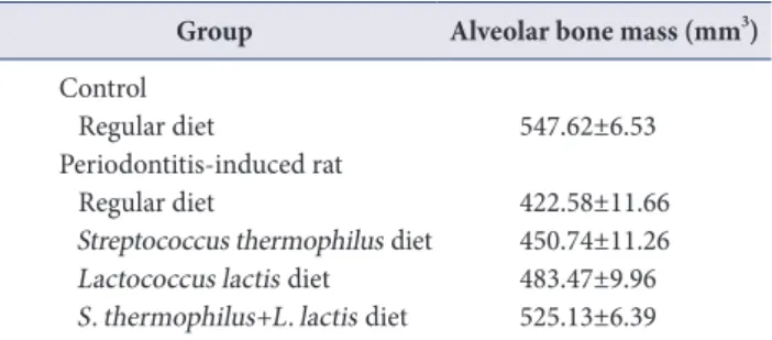
When the alveolar bone was investigated the periodonti- tis-induced rat after feeding various diets by micro-CT, the area of alveolar bone in the rat fed regular diet was 422.58
±11.66 mm3. Compared with the control group (547.62
±6.53 mm3), it is 125.04 mm3 lower area. As shown Table 2, the rat fed the diet containing S. thermophilus, L. lactis,
Table 2. Alveolar bone mass of periodontitis induced rat fed various diet
Group Alveolar bone mass (mm3) Control
Regular diet 547.62±6.53
Periodontitis-induced rat
Regular diet 422.58±11.66
Streptococcus thermophilus diet 450.74±11.26 Lactococcus lactis diet 483.47±9.96 S. thermophilus+L. lactis diet 525.13±6.39 Values are presented as mean±standard deviation.
and both probiotics significantly showed higher area of the alveolar bone (450.74±11.26, 483.47±9.96, and 525.13±
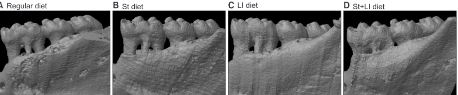
6.39 mm3, respectively). When 3D image of alveolar bone was generated, the interdental space reduced in the rat fed the diet containing probiotics compared to the rat fed regular diet (Fig. 1).
Investigation of cytokine levels
In ligation-induced periodontitis rat model, the rat fed the regular diet showed the highest level of IL-1β and IL-6 expression. Also, the rat fed the diet containing S. ther- mophilus or L. lactis significantly exhibited higher level
of IL-1β and IL-6 expression compared to control group (non-periodontitis rat) (p<0.05) (Fig. 2). However, com- paring the rat fed the regular diet, the levels of IL-1β and IL-6 expression was measured less in the rat fed the diet containing S. thermophilus or L. lactis. Interestingly, the rat fed the diet containing both probiotics did not show sig- nificant difference with control group.
Analysis of bacteria levels
Periodontitis-induced rat fed various diets in the pres- ence or the absence of probiotics was investigated the lev- els of P. gingivalis, S. thermophilus, and L. lactis by quan-
Fig. 1. Micro-computed tomography (CT) image of ligature-induced periodontitis rat. The ligature-induced periodontitis rats were fed the diet with Streptococcus thermophilus (St diet; B), Lactococcus lactis (Ll diet; C), and S. thermophilus+L. lactis (St+Ll diet; D) or without probiotics (A), and the ligature site of the rat was analyzed by micro-CT.
Regular diet
A BSt diet CLI diet DSt+LI diet
Fig. 2. Investigation of inflammatory cytokine production. The ligature-induced periodontitis rats were fed the diet with or without Streptococ- cus thermophilus (St diet), Lactococcus lactis (Ll diet), and S. thermophilus+L. lactis (St+Ll diet) probiotics, and the gingival crevicular fluid was collected with paper point. The levels of interleukin-1β (IL-1β; A) and interleukin-6 (IL-6; B) were measured by enzyme-linked immunosorbent assay kit. A letter (a) represents significant difference compared to non-ligature control group (p<0.05), and a letter (b) expresses significant differ- ence compared to the rat fed regular diet (p<0.05).
Control Regular diet
St diet LI diet St+LI diet 180
160 140 120 100 80 60 40 20
IL-1( pg/mL)
0
A
Control Regular diet
St diet LI diet St+LI diet 900
800 700 600 500 400 300 200 100
IL-6(pg/mL)
0
B
a
b
a b
b
a
a b
a
a b
b
titative real-time PCR. As shown Fig. 3, control group was not detected the bacteria, and the levels of P. gingivalis in the rat fed diet containing each probiotics were significant- ly reduced (p<0.05). P. gingivalis was detected the lowest level in the rat fed the diet containing both probiotics.
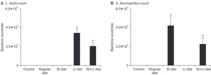
Investigation of probiotics colonization
Probiotics colonization is important to keep beneficial effects on oral disease. Therefore, probiotics colonization in the ligation site of the rat was investigated. As shown Fig. 4, each probiotics was detected in the group of the diet contain each probiotic bacterium. Especially, S. thermoph- ilus and L. lactis were detected together with similar levels in the rat fed the diet containing both probiotics. This data indicates that S. thermophilus and L. lactis can co-exist in oral cavity.
DISCUSSION
Dysbiosis of oral microbiome by change of the envi- ronments induces the disease, such as dental caries and periodontitis [14,15], and epidemiological studies to dental patients suggests that P. gingivalis, T. forsythia, and T. den- ticola are related with periodontitis [1], and Streptococcus mutans is a cariogenic bacterium [16]. Therefore, studies have been performed to efficiently remove these bacteria.
Probiotics have been used to therapy and prevent hu- man diseases [7]. Recently, probiotics have also been tried to apply oral diseases [17], and the antimicrobial activity of various probiotics against oral bacteria [18,19]. Previous studies showed that S. thermophilus and L. lactis have the antimicrobial activity against periodontopathogens and S.
Control Regular diet
St diet LI diet St+LI diet
Bacteriacount/site
2.0 105
0 1.5 105
1.0 105
0.5 105
a b
b b
Fig. 3. Count of Porphyromonas gingivalis on the periodontitis site.
The ligature-induced periodontitis rats were fed the diet with or without Streptococcus thermophilus (St diet), Lactococcus lactis (Ll diet), and S. thermophilus+L. lactis (St+Ll diet), and the bacteria was collected with paper point. DNA from the paper point was extracted by a bacterial DNA extraction kit, and the levels of P. gingivalis was measured by quantitative real-time polymerase chain reaction. A letter (a) represents significant difference compared to non-ligature control group (p<0.05), and a letter (b) expresses significant difference compared to the rat fed regular diet (p<0.05).
A L. lactis count
Control Regular diet
St diet LI diet St+LI diet 6.0 104
Bacteriacount/site
0 4.0 104
2.0 104
B S. thermophilus count
Control Regular diet
St diet LI diet St+LI diet 6.0 104
Bacteriacount/site
0 4.0 104
2.0 104 a
a
a
a
Fig. 4. Colonization of probiotics on the periodontitis site. The rats were fed the diet with or without Streptococcus thermophilus (St diet), Lacto- coccus lactis (Ll diet), and S. thermophilus+L. lactis (St+Ll diet), and the bacteria was collected with paper point. DNA from the paper point was extracted by a bacterial DNA extraction kit, and the L. lactis (A) and S . thermophilus (B) were measured by quantitative real-time polymerase chain reaction. A letter (a) represents significant difference compared to non-ligature control group (p<0.05).
mutans and did not have aciduricity [8,11,16]. Therefore, both probiotics are considered candidate probiotics for dental caries and periodontitis. Next, efficacy of both pro- biotics for periodontitis was investigated using ligation- induced periodontitis animal model.
Ligature-induced periodontitis rat model by placing the wire inoculated P. gingivalis showed high levels of cytokine expression and reduction of alveolar bone mass. Therefore, the rat can be investigated effects of probiotics. When the ligature-induced periodontitis rats were fed the diet in the presence or the absence of S. thermophilus, L. lactis, and both probiotics, the mass of mandibular alveolar bone showed higher levels in the rat fed the diet containing S.
thermophilus, L. lactis, and both probiotics compared to the rat fed the regular diet. Also, the inhibitory effect on bone resorption exhibited to be large in order of the diet containing S. thermophilus, L. lactis, and both probiotics.
Next, we investigated cytokine expression of the rat in var- ious conditions. Similar to micro-CT data, the expression of the inflammatory cytokines, which was highly induced in the rats fed regular diet compared to control group, were lower in the rats fed the diet containing probiotics. These results indicated that the diet containing probiotics showed a definite therapeutic effect on periodontitis.
Next, to investigate whether the inhibitory effects of probiotics on periodontitis was by P. gingivalis elimination or by reduction of inflammation, the count of P. gingivalis on the site was examined by real-time PCR. Various stud- ies have suggested that P. gingivalis was detected greater frequency and at higher levels at the periodontitis sites, and the certain periodontitis indicators are correlated with the presence or levels of P. gingivalis [20-22]. P. gingivalis was detected lower levels in the rats fed containing probiotics compared to the rat fed the regular diet. Therefore, the in- hibitory effect of S. thermophilus and L. lactic on induction of periodontitis may be caused by elimination of P. gingi- valis.
Finally, probiotics colonization is important to keep ben- eficial effects on infectious disease [23]. Therefore, the rats of each group were investigated and measured probiotics levels by quantitative real-time PCR using specific prim- ers for S. thermophilus and L. lactis. The primers did not
show non-specific reaction for any oral bacteria (data not shown). S. thermophilus and L. lactis were detected in the rats fed the diets containing each probiotic bacteria. Inter- estingly, S. thermophilus and L. lactis were detected in the rat fed the diet containing both probiotics. Most bacteria compete interspecies to survive and to occupy favorite place. On the basis of the results, S. thermophilus and L.
lactis may not only compete with each other but coexist, and their competition, exclusion, and displacement act only against P. gingivalis.
Most probiotics have aciduricity (acid tolerance) which can induce dental caries [24,25]. The acid tolerance of probiotics can be an advantage for intestinal disease, but it can be a disadvantage for oral diseases. Therefore, the application of probiotics for prevention and treatment of periodontitis are careful because of dental caries. However, S. thermophilus and L. lactis did not have aciduricity [11,16]
and show the antimicrobial activity against P. gingivalis.
S. thermophilus and L. lactis reduced cytokine expression and alveolar bone loss and showed the antimicrobial activ- ity against P. gingivalis on the ligature site. Furthermore, these probiotics colonized the oral cavity of the rat model.
On the basis of these results, S. thermophilus and L. lactis may be suitable probiotics for therapeutic and preventive periodontitis.
ACKNOWLEDGEMENTS
The present research was conducted by the research fund of Dankook University in 2018.
CONFLICTS OF INTEREST
The authors declare that they have no competing inter- ests.
ORCID
Won-Jae Choi
https://orcid.org/0000-0003-4477-3171 Sung-Hoon Lee
https://orcid.org/0000-0002-8852-0419
REFERENCES
1. Socransky SS, Haffajee AD, Cugini MA, Smith C, Kent RL Jr. Microbial complexes in subgingival plaque. J Clin Peri- odontol 1998;25:134-144. doi: 10.1111/j.1600-051X.1998.
tb02419.x.
2. Marsh PD. Microbial ecology of dental plaque and its sig- nificance in health and disease. Adv Dent Res 1994;8:263- 271. doi: 10.1177/08959374940080022001.
3. Marsh PD. The commensal microbiota and the develop- ment of human disease-an introduction. J Oral Microbiol 2015;7:29128. doi: 10.3402/jom.v7.29128.
4. Marsh PD, Do T, Beighton D, Devine DA. Influence of sa- liva on the oral microbiota. Periodontol 2000 2016;70:80- 92. doi: 10.1111/prd.12098.
5. Socransky SS, Haffajee AD. Periodontal microbial ecology.
Periodontol 2000 2005;38:135-187. doi: 10.1111/j.1600- 0757.2005.00107.x.
6. Socransky SS, Haffajee AD. Dental biofilms: difficult therapeutic targets. Periodontol 2000 2002;28:12-55. doi:
10.1034/j.1600-0757.2002.280102.x.
7. Sanders ME. Probiotics: considerations for human health.
Nutr Rev 2003;61:91-99. doi: 10.1301/nr.2003.marr.91- 99.
8. Lee SH, Baek DH. Effects of Streptococcus thermophilus on volatile sulfur compounds produced by Porphyromo- nas gingivalis. Arch Oral Biol 2014;59:1205-1210. doi:
10.1016/j.archoralbio.2014.07.006.
9. Oh S, Kim SH, Ko Y, Sim JH, Kim KS, Lee SH, Park S, Kim YJ. Effect of bacteriocin produced by Lactococcus sp. HY 449 on skin-inflammatory bacteria. Food Chem Toxicol 2006;44:552-559. doi: 10.1016/j.fct.2005.08.030.
10. Ali D, Lacroix C, Thuault D, Bourgeois CM, Simard RE.
Characterization of diacetin B, a bacteriocin from Lacto- coccus lactis subsp. lactis bv. diacetylactis UL720. Can J Microbiol 1995;41:832-841. doi: 10.1139/m95-114.
11. Shin HS, Baek DH, Lee SH. Inhibitory effect of Lactococ- cus lactis on the bioactivity of periodontopathogens.
J Gen Appl Microbiol 2018;64:55-61. doi: 10.2323/
jgam.2017.06.003.
12. Tong Z, Zhou L, Li J, Kuang R, Lin Y, Ni L. An in vitro inves- tigation of Lactococcus lactis antagonizing cariogenic bac- terium Streptococcus mutans. Arch Oral Biol 2012;57:376- 382. doi: 10.1016/j.archoralbio.2011.10.003.
13. Park CH, Abramson ZR, Taba M Jr, Jin Q, Chang J, Kre- ider JM, Goldstein SA, Giannobile WV. Three-dimen- sional micro-computed tomographic imaging of alveolar bone in experimental bone loss or repair. J Periodontol
2007;78:273-281. doi: 10.1902/jop.2007.060252.
14. Jiao Y, Hasegawa M, Inohara N. The role of oral pathobi- onts in dysbiosis during periodontitis development. J Dent Res 2014;93:539-546. doi: 10.1177/0022034514528212.
15. Hoare A, Marsh PD, Diaz PI. Ecological therapeutic op- portunities for oral diseases. Microbiol Spectr 2017;5. doi:
10.1128/microbiolspec.BAD-0006-2016.
16. Kim YJ, Lee SH. Inhibitory effect of Lactococcus lactis HY 449 on cariogenic biofilm. J Microbiol Biotechnol 2016;26:1829-1835. doi: 10.4014/jmb.1604.04008.
17. Han X, Zhang J, Tan Y, Zhou G. Probiotics: a non-con- ventional therapy for oral lichen planus. Arch Oral Biol 2017;81:90-96. doi: 10.1016/j.archoralbio.2017.04.026.
18. Kim SK, Lee SJ, Baek YJ, Park YH. Isolation of bacteriocin- producing Lactococcus sp. HY 449 and its antimicro- bial characteristics. Korean J Appl Microbiol Biotechnol 1994;22:259-265.
19. Snel J, Marco ML, Kingma F, Noordman WM, Rademaker J, Kleerebezem M. Competitive selection of lactic acid bac- teria that persist in the human oral cavity. Appl Environ Microbiol 2011;77:8445-8450. doi: 10.1128/AEM.06043- 11.
20. Grossi SG, Zambon JJ, Ho AW, Koch G, Dunford RG, Machtei EE, Norderyd OM, Genco RJ. Assessment of risk for periodontal disease. I. Risk indicators for attach- ment loss. J Periodontol 1994;65:260-267. doi: 10.1902/
jop.1994.65.3.260.
21. Haffajee AD, Socransky SS, Smith C, Dibart S. Relation of baseline microbial parameters to future periodontal at- tachment loss. J Clin Periodontol 1991;18:744-750. doi:
10.1111/j.1600-051X.1991.tb00066.x.
22. Moore WE, Moore LH, Ranney RR, Smibert RM, Burmeis- ter JA, Schenkein HA. The microflora of periodontal sites showing active destructive progression. J Clin Periodon- tol 1991;18:729-739. doi: 10.1111/j.1600-051X.1991.
tb00064.x.
23. Sanders ME. Impact of probiotics on colonizing microbiota of the gut. J Clin Gastroenterol 2011;45 Suppl:S115-S119.
doi: 10.1097/MCG.0b013e318227414a.
24. Hassanzadazar H, Ehsani A, Mardani K, Hesari J. Investiga- tion of antibacterial, acid and bile tolerance properties of lactobacilli isolated from Koozeh cheese. Vet Res Forum 2012;3:181-185.
25. Chan ES, Lee PP, Ravindra P, Krishnaiah K, Voo WP. A standard quantitative method to measure acid tolerance of probiotic cells. Appl Microbiol Biotechnol 2010;86:385- 391. doi: 10.1007/s00253-009-2384-y.