•접수 : 2014년 4월 21일 •수정접수 : 2014년 4월 27일 •채택 : 2014년 4월 28일
✽
공동교신저자 : 이영준, 대구한의대학교 한의과대학 예방의학교실, 경북 경산시 한의대로 1, 712-715 전화 : 053-819-1296, 팩스 : 053-819-1576, 전자우편 : [email protected]
구세광, 대구한의대학교 한의과대학 해부학교실, 경북 경산시 한의대로 1, 712-715 전화 : 053-819-1549, 팩스 : 053-819-1576, 전자우편 : [email protected]
#
The two first authors contributed equally to this work
Journal of Society of Preventive Korean Medicine 2014;18(1):125-138Synergic Effects of Mixed Formula Consisted of Polycan and Calcium-gluconate on the Experimental Periodontitis
and Alveolar Bone Loss in Rats
Won-Ho Lee
1)#, Kyung Hu Kim
1)#, Su Jin Kang
2),3), Young Joon Lee
2),3)*& Sae Kwang Ku
1),3)*1)Department of Anatomy and Histology, College of Korean Medicine, Daegu Haany University, Gyeongsan
2)Department of Preventive Medicine, College of Korean Medicine, Daegu Haany University, Gyeongsan
3)The Medical Research Center for Globalization of Herbal Formulation, Daegu Haany University, Gyeongsan
Abstract
Objective : Polycan, exopolymers purified from Aureobasidium pullulans SM-2001 and calcium gluconate have been showed favorable inhibitory effects on the periodontitis and related alveolar bone losses through anti- oxidant and anti-inflammatory activities, respectively. In the present study, we intended to observe the possible synergic effects of mixed formula consisted of Polycan and calcium gluconate on ligation-induced experi- mental periodontitis and related alveolar bone losses in rats, and to select the fittest compositions for further developing as effective agents to ameliorate periodontal diseases.
Method : Experiments were conducted as two separated two tests – first is synergic effects of Polycan and calcium gluconate 1:1, 1:9 and 9:1 mixtures, and second is 1:99, 2:98, 4:96, 8:92 and 1:9 mixtures. Experi- mental periodontal diseases were induced by ligature placed around the cervix of upper left incisior teeth of rats. One day after ligation placements, 200mg/kg of each single or mixed formulas of Polycan or/and calcium gluconate were orally administered for 10 days. The changes on the alveolar bone loss index and maxillary bone mineral density (BMD) were observed for detecting alveolar bone losses, and for anti-infla- mmatory effects, myeloperoxidase (MPO) activities and proinflammatory cytokine (tumor necrosis factor; TNF- α) contents were also evaluated in gingival tissues around ligature placed incisior teeth. The results of mixtures were compared with those of singe Polycan and calcium gluconate treated rat.
Results : Each single or mixed formulas of Polycan or/and calcium gluconate favorably and significantly
inhibited the inflammatory changes. The inhibitory effects of mixed formula consisted of Polycan and calcium
gluconate 1:9 showed against periodontitis and related alveolar bone losses as compared with those of
each Polycan and calcium gluconate single formula (p<0.05). In second experiment, Polycan and calcium
gluconate 2:98, 4:96, 8:92 and 1:9 mixed formulas also showed significant increased anti-inflammatory
and inhibitory effects against alveolar bone losses as compared with those of each single formula. Among
them, Polycan and calcium gluconate 2:98 showed the highest efficacy against to ligation-induced experi-
mental periodontitis and related alveolar bone losses.
Conclusion : The results obtained in this study suggest that appropriated mixtures of Polycan and calcium gluconate showed synergic inhibitory effects against ligation-induced experimental periodontitis and related alveolar bone losses in rats. Moreover, Polycan and calcium gluconate 2:98 showed the highest efficacies in this experiment, suggesting the fittest composition for further developing as effective agents to ameli- orate periodontal diseases.
Key words : Polycan, Calcium gluconate, Synergic effects, Periodontitis, Alveolar bone loss, Rat
I. Introduction
Periodontitis, a relevant cause of tooth loss in adults
1), is a chronic inflammatory disease that is characterized by localized bone resorption
2),3). Recently, involvement of nitric oxide activities, oxidative stresses, in the pathogenesis of perio- dontitis has been revealed
4), and many antioxidants showed favorable effects on periodontitis and related alveolar bone losses
5),6). The induction of periodontal disease by ligature placement is widely used in animal studies, and periodontitis and alveolar bone losses were induced as like humans in this model
7),8). Natural products are gaining space and importance in the pharmaceutical industry as well as inspiring the search for new potential sources of bioactive molecules
9). Herbs, medicinal plants and crude drug substances are considered to be a potential source of antioxidants to combat various diseases including periodontitis and related alveolar bone losses
10).
Polycan is purified exopolymers from Aureo- basidium pullulans SM-2001, and comprises mostly β-1,3/1,6-glucan and other organic materials, such as amino acids, mono- or di-unsaturated fatty acids (linoleic and linolenic acids), and fibrous polysaccharide
11). Recently, we found that Polycan has anti-osteoporotic
12),13); it inhibited bone losses and accelerated the bone formation, and fracture healing promoting effects
14)with anti-inflamma- tory effects on xylene-induced acute inflammation
15)and formalin-induced chronic inflammation
16). It also showed favorable prevention or therapeutic
effects on cisplatin-induced kidney damages
17)and on the ligation-induced experimental perio- dontitis (EPD) and related alveolar bone losses
6)through anti-oxidant and anti-inflammatory mechanisms. Calcium salts (calcium dobestilate, calcium hydroxide, calcium pentosan polysulfate, calcium gluconate) have been showed anti-infla- mmatory activities
18),19). Among them, calcium gluconate have been used for treatment of injuries from direct contact with hydrofluoric acid
20). It markedly reduced proinflammatory cytokines, interleukin-6 and tumor necrosis factor-α in chemical burns in rats
21). Indeed, calcium glu- conate enhanced anti-inflammatory activities of non-steroid anti-inflammatory drugs
22), and it mitigate the ligation-induced EPD and related alveolar bone losses
5)and collagen-induced rheu- matoid arthritis
23)through anti-oxidant and anti- inflammatory mechanisms. It therefore, considered that appropriate mixtures of Polycan and calcium gluconate also might be showed synergic effects on periodontitis and related alveolar bone losses.
In the present study, we intended to observe
the possible synergic effects of mixed formula
consisted of Polycan and calcium gluconate on
ligation-induced EPD and related alveolar bone
losses in rats, and to select the fittest compo-
sitions for further developing as effective agents
to ameliorate periodontal diseases. Changes on
the alveolar bone loss index and maxillary bone
mineral density (BMD) were observed for detecting
alveolar bone losses, and for anti-inflammatory
effects, myeloperoxidase (MPO) activities and
proinflammatory cytokine (tumor necrosis factor;
on the Experimental Periodontitis and Alveolar Bone Loss in Rats
TNF-α) contents were also evaluated in gingival tissues around ligature placed incisior teeth in this experiment.
II. Materials and Methods
1. Animals and husbandry
Total one hundred twenty eight healthy male Sprague-Dawley (Slc:SD) rats (6-wk old upon receipt; SLC, Shizuoka, Japan; Body weight ranged in 170~190g), were used after acclimatization for 10 days. Animals were allocated four per polycarbonate cage in a temperature (20-25°C) and humidity (50-55%) controlled room. Light : dark cycle was 12hr : 12hr, and standard rodent chow (Samyang, Korea) and water were supplied free to access. All animals were treated according to the international regulations of the usage and welfare of laboratory animals, and approved by the Institutional Animal Care and Use Committee in Daegu Haany University (Gyeongsan, Gyeongbuk, Korea) prior to animal experiment. Experiments were conducted as two separated two tests – first is synergic effects of Polycan and calcium gluconate 1:1, 1:9 and 9:1 mixtures, and second is 1:99, 2:98, 4:96, 8:92 and 1:9 mixtures. In first experiment, 8 rats per group, total seven groups – intact and EPD control, Polycan and calcium gluconate single formula, Polycan and calcium gluconate 1:1, 1:9 and 9:1 mixed formulas treated groups were selected base on the body weights after 10 days of acclimatization, and also eight rats per group, total 9 groups – intact and EPD control, Polycan and calcium gluconate single formula, Polycan and calcium gluconate 1:99, 2:98, 4:96; 8:92 and 10:90 mixed formulas treated groups were used in second experiment, respectively.
2. Preparations and administration of test materials
Appropriated amounts of Polycan (purified exo- polymers from Aureobasidium pullulans SM-2001, contains 14% of β-1,3/1,6-glucan, 18% of β-1,4 -glucan, 8% of α-(1,4)-(1,6)-glucan, 37.7% of glucose, 0.8% of galactose, 1,5% of mannose, 3.1% of protein (3.1%) and 7.2% of ashin dried materials), and calcium gluconate (CAS No. 299- 28-5; purity: 98%) were kindly supplied by Aribio Inc. (Seoul, Korea) as brownish and off white powders, respectively. Both materials well dissolved in distilled water, up to 40mg/ml concentration, at least. Appropriate amounts of Polycan or calcium gluconate were dissolved in distilled water as vehicle, and to prepare administration solutions of the mixed formulas consisted of Polycan and calcium gluconate, appropriated amounts of Polycan and calcium gluconate were directly dissolved into distilled water, respectively. One day after ligation placements, 200mg/kg of each single or mixed formulas of Polycan and calcium gluconate were orally administered, once a day for 10 days, in a volume of 5ml/kg, respectively. In intact and EPD controls, same volume of distilled water was orally administered, instead of test substances, once a day for 10 continuous days from 24hrs after ligation placement.
3. Induction of EPD
EPD were induced by sterilized nylon (3-0)
thread ligature placed around the cervix of upper
left incisior teeth of rats, under anesthetized with
25mg/kg intraperitoneal injection of Zoletile mixture
(Zoletile 50; Virbac Lab., Paris, France) according
to previous methods
5),6)with some modifications.
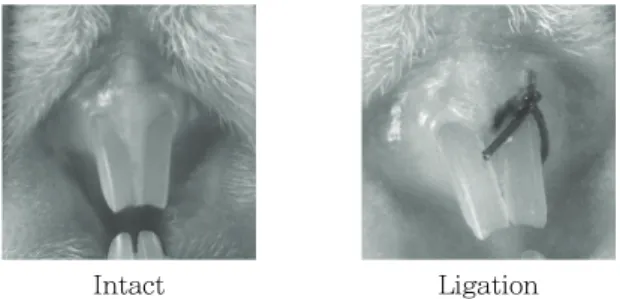
Intact Ligation
Figure 1. Representative images of experimental periodontal disease inducement
The ligature was knotted on the buccal side of the tooth, resulting in subgingival position pala- tinally and in supragingival position buccally. In intact vehicle control rats, only cervix of upper left incisior teeth was identified, instead of liga- tion placement in this experiment (Fig 1).
4. Measurements of alveolar bone loss
The animals were sacrificed at 10 days after administration (on 11 days after ligature place- ment), and maxillary bone contain ligature place- ment site (second molar) were excised and then, the horizontal alveolar bone loss, the distance between the cusp tip and the alveolar bone, was measured using a modification of the methods of Crawford et al
24)as described by Samejima et al
3). Measurements were made along the axis of root of the upper left incisior teeth, as mm/rats
25),26).
5. Measurements of maxillary BMD
At Sacrifice, the upper incisor teeth located maxillary regions were separated from surrounding connective tissues, muscles and any debris, and then dried at 120℃ for 8hrs in high temperature dry oven (LDO-080N, Daihan Labtech Co., Korea).
BMD of upper incisor teeth located maxillary regions (5×5mm) was measured by dual-energy x-ray absorptionmetry (Norland pDEXA; Fort Atkinson, WI, USA) after end of 10 days continuous
oral administration of test substances or vehicle.
6. Measurement of MPO activity
Gingival tissues around ligature placement were collected at 11 days after EPD induction to deter- mine MPO activity as a measurement of neutrophil accumulation. A spectrophotometric assay was utilized to measure MPO activity, as described previously
27). The buccal gingival tissues surrounding the left incisior teeth were removed and stored at -70℃. The material was suspended in 0.5%
hexadecyltrimethyl-ammonium bromide (Gibco,
Carlsbad, CA, USA) in 50mM potassium phosphate
buffer, pH 6.0, to solubilize MPO. After homo-
genized in an ice bath (15s), the samples were
freeze-thawed twice. Additional buffer was added
to the test tube to reach 400μl of buffer per 15
mg of tissue for 12 min. After centrifuging at
1000×g for 12 min, 0.1 ml of the supernatant was
added to 2 ml phosphate buffer (50 mM, pH 6.0),
containing 0.167 mg/ml o-dianisidine dihydrochloride
(Sigma-Aldrich, St. Louise, MO, USA), distillated
water and 0.0005% hydrogen peroxide to give a
final volume of 2.1 ml per tube. The absorbance
was measured by spectrophotometer (OPTIZEN
POP, Mecasys, Daejeon, Korea) at 460 nm. One
unit of activity was defined as that degrading 1
μmol of peroxide/min at 25℃. Results are expressed
as MPO units/ml.
on the Experimental Periodontitis and Alveolar Bone Loss in Rats
7. Detection of TNF-α contents in rat maxillary gingival tissue
The buccal gingival tissue from the area sur- rounding the ligature placement was collected at 11 days after EPD induction. The tissue collected was homogenized and processed as described by Safieh-Garabedian et al
28)as described by Botelho et al
26). The detection of TNF-α concentrations was determined by enzyme linked immunosorbent assay as described previously by Cunha et al
29). Micro titer plates were coated overnight at 4℃
with antibody against rat TNF-α (10 μg/ml). After blocking the plates, the samples and standard at various dilutions were added in duplicate and incubated at 4℃ for 24 hrs. The plates were washed three times with buffer. After washing the plates, 100 ml of biotinylated sheep polyclonal anti-rat TNF-α (diluted 1/1000 with assay buffer 1% BSA; Abcam, Cambridge, UK), was added to the wells. After further incubation at room tem- perature for 1 hr, the plates were washed and 100 μl of avidin-HRP (Abcam, Cambridge, UK) diluted 1:5000 were added. The color reagent o-phenylenediamine (100 μl; Sigma-Aldrich, St.
Louise, MO, USA) was added 15 min later and the plates were incubated in the dark at 37 ℃ for 20 min. The enzyme reaction was stopped with H
2SO
4and absorbance was measured using a microplate reader (Tecan, Männedorf, Switzer- land) at 490 nm.
8. Statistical analyses
All data were expressed as mean ± standard deviation (SD) of eight rats. Multiple comparison tests for different dose groups were conducted.
Variance homogeneity was examined using the Levene test
30). If the Levene test indicated no significant deviations from variance homogeneity, the obtain data were analyzed by one way ANOVA test followed by least-significant differences
multi-comparison (LSD) test to determine which pairs of group comparison were significantly different. In case of significant deviations from variance homogeneity was observed at Levene test, a non-parametric comparison test, Kruskal- Wallis H test was conducted. When a significant difference is observed in the Kruskal-Wallis H test, the Mann-Whitney U (MW) test was conducted to determine the specific pairs of group comparison, which are significantly different. Statistical analyses were conducted using SPSS for Windows (Release 14.0K, IBM SPSS Inc., Armonk, NY, USA)
31), and p-values < 0.05 were considered significantly different.
III. Results
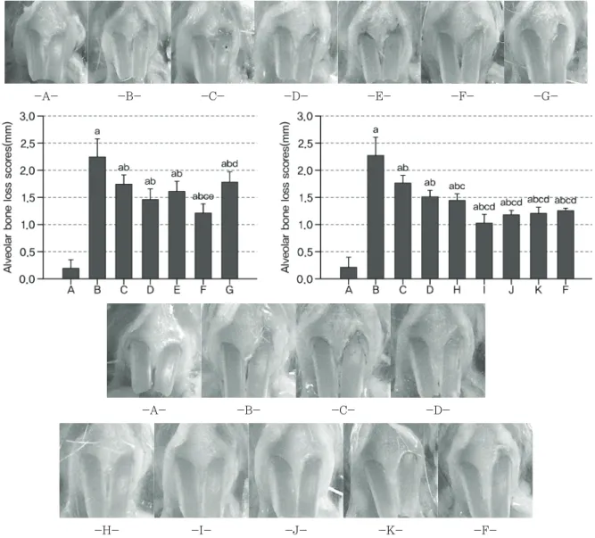
1. Changes on the alveolar bone loss scores
Significant (p<0.01) increases of exposured teeth
roots areas, the alveolar bone loss scores were
detected in EPD control as compared with intact
control in the both first (2.06 mm) and second
(2.06 mm) experiment. However, significant (p<0.01)
decreases of alveolar bone loss scores were detec-
ted in all Polycan (-0.49 mm in first, -0.50 mm
in second) and calcium gluconate (-0.77 mm in
first, -0.75 mm in second) single or mixed formula
(-0.63 mm in 1:1, -1.03 mm in 1:9, -0.45 mm
in 9:1, -0.82 mm in 1:99, -1.25 mm in 2:98,
-1.09 mm in 4:96, -1.06 mm in 8:92, -1.01 mm
in 10:90 mixed formula) treated rats as compared
with EPD control, respectively. Especially, Polycan
and calcium gluconate 1:9 mixed formula treated
rats showed significant (p<0.01 or p<0.05) dec-
reases of bone loss scores as compared with
each Polycan (-0.54 mm) and calcium gluconate
(-0.26 mm) single formula treated rats in first
experiment, respectively. Moreover, Polycan and
calcium gluconate 2:98, 4:96, 8:92 and 10:90
-A- -B- -C- -D- -E- -F- -G-
-A- -B- -C- -D-
-H- -I- -J- -K- -F-
Figure 2. Representative gross images of ligation placed regions and changes on the alveolar bone loss scores after 10 days oral treatment of single or mixed formulas of Polycan and calcium gluconate in EPD rats A: Intact control, B: EPD control, C: Polycan single formula treated rats, D: Calcium gluconate single formula treated rats, E: Polycan and calcium gluconate 1:1 mixture treated rats, F: Polycan and calcium gluconate 1:9 mixture treated rats, G: Polycan and calcium gluconate 9:1 mixture treated rats, H: Polycan and calcium gluconate 1:99 mixture treated rats, I: Polycan and calcium gluconate 2:98 mixture treated rats, J: Polycan and calcium gluconate 4:96 mixture treated rats, K: Polycan and calcium gluconate 8:92 mixture treated rats.
a
p<0.01 as compared with intact control by LSD test,
bp<0.01 as compared with EPD control by LSD test,
cp<0.01 as compared with Polycan single formula treated rats by LSD test,
dp<0.01 and
ep<0.05 as compared with calcium gluconate single formula treated rats by LSD test
mixed formula treated rats also showed significant (p<0.01) decreases of bone loss scores as com- pared with each Polycan (-0.74 mm in 2:98, -0.58 mm in 4:96, -0.55 mm in 8:92, -0.51 mm in 10:90 mixed formula) and calcium gluconate (-0.50 mm in 2:98, -0.34 mm in 4:96, -0.30 mm in 8:92, -0.26 mm in 10:90 mixed formula) single formula
treated rats in second experiment, in that orders, respectively(Fig 2).
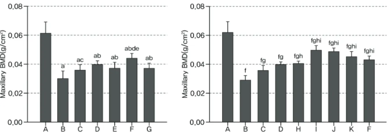
2. Effects on the maxillary BMD
Significant (p<0.01) decreases of maxillary BMD
around ligation placed were detected in EPD control
on the Experimental Periodontitis and Alveolar Bone Loss in Rats
Figure 3. Changes on the maxillary BMD after 10 days oral treatment of single or mixed formulas of Polycan and calcium gluconate in EPD rats
A: Intact control, B: EPD control, C: Polycan single formula treated rats, D: Calcium gluconate single formula treated rats, E: Polycan and calcium gluconate 1:1 mixture treated rats, F: Polycan and calcium gluconate 1:9 mixture treated rats, G: Polycan and calcium gluconate 9:1 mixture treated rats, H: Polycan and calcium gluconate 1:99 mixture treated rats, I: Polycan and calcium gluconate 2:98 mixture treated rats, J: Polycan and calcium gluconate 4:96 mixture treated rats, K: Polycan and calcium gluconate 8:92 mixture treated rats.
a
p<0.01 as compared with in tact control by LSD test,
bp<0.01 and
cp<0.05 as compared with EPD control by LSD test,
dp<0.01 as compared with Polycan single formula treated rats by LSD test,
ep<0.01 as compared with calcium gluconate single formula treated rats by LSD test,
fp<0.01 as compared with intact control by MW test,
g
p<0.01 as compared with EPD control by MW test,
hp<0.01 as compared with Polycan single formula treated rats by MW test,
ip<0.01 as compared with calcium gluconate single formula treated rats by MW test as compared with intact control in the both first
(-0.031 g/cm
2) and second (-0.033 g/cm
2) expe- riment. However, significant (p<0.01 or p<0.05) increases of maxillary BMD were detected in all Polycan (0.006 g/cm
2in first, 0.007 g/cm
2in second) and calcium gluconate (0.010 g/cm
2in first, 0.011 g/cm
2in second) single or mixed formula (0.007 g/cm
2in 1:1, 0.014 g/cm
2in 1:9, 0.007 g/cm2 in 9:1, 0.011 g/cm
2in 1:99, 0.021 g/cm
2in 2:98, 0.019 g/cm
2in 4:96, 0.016 g/cm
2in 8:92, 0.014 g/cm
2in 10:90 mixed formula) treated rats as compared with EPD control, respectively. Espe- cially, Polycan and calcium gluconate 1:9 mixed formula treated rats showed significant (p<0.01) increases of maxillary BMD as compared with each Polycan (0.008 g/cm
2) and calcium gluconate (0.004 g/cm
2) single formula treated rats in first experiment, respectively. Moreover, Polycan and calcium gluconate 2:98, 4:96, 8:92 and 10:90 mixed formula treated rats also showed significant (p<0.01) increases of maxillary BMD as compared
with each Polycan (0.014 g/cm
2in 2:98, 0.013 g/cm
2in 4:96, 0.010 g/cm
2in 8:92, 0.008 g/cm
2in 10:90 mixed formula) and calcium gluconate (0.010 g/cm
2in 2:98, 0.009 g/cm
2in 4:96, 0.006 g/cm
2in 8:92, 0.003 g/cm
2in 10:90 mixed formula) single formula treated rats in second experi- ment, in that orders, respectively(Fig 3).
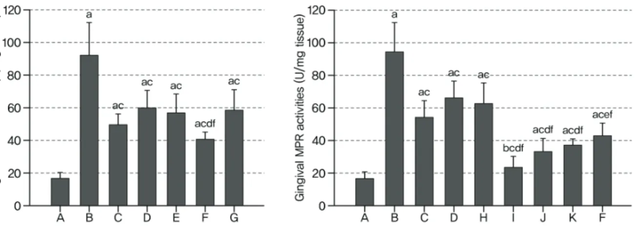
3. Changes on the gingival MPO activities
Significant (p<0.01) increases of MPO activities
around ligation placed gingival tissues were detected
in EPD control as compared with intact control
in the both first (74.68 U/mg) and second (76.92
U/mg) experiment. However, significant (p<0.01)
decreases of gingival MPO activities were detected
in all Polycan (-43.12 U/mg in first, -39.55 U/mg
in second) and calcium gluconate (-31.85 U/mg
in first, -27.84 U/mg in second) single or mixed
Figure 4. Changes on the gingival MPO activities after 10 days oral treatment of single or mixed formulas of Polycan and calcium gluconate in EPD rats
A: Intact control, B: EPD control, C: Polycan single formula treated rats, D: Calcium gluconate single formula treated rats, E: Polycan and calcium gluconate 1:1 mixture treated rats, F: Polycan and calcium gluconate 1:9 mixture treated rats, G: Polycan and calcium gluconate 9:1 mixture treated rats, H: Polycan and calcium gluconate 1:99 mixture treated rats, I: Polycan and calcium gluconate 2:98 mixture treated rats, J: Polycan and calcium gluconate 4:96 mixture treated rats, K: Polycan and calcium gluconate 8:92 mixture treated rats.
a
p<0.01 and
bp<0.05 as compared with intact control by MW test,
cp<0.01 as compared with EPD control by MW test,
dp<0.01 and
ep<0.05 as compared with Polycan single formula treated rats by MW test,
fp<0.01 as compared with calcium gluconate single formula treated rats by MW test
formula (-34.92 U/mg in 1:1, -51.27 U/mg in 1:9, -33.46 U/mg in 9:1, -31.46 U/mg in 1:99, -69.96 U/mg in 2:98, -60.84 U/mg in 4:96, -56.70 U/mg in 8:92, -51.11 U/mg in 10:90 mixed formula) treated rats as compared with EPD control, res- pectively. Especially, Polycan and calcium glu- conate 1:9 mixed formula treated rats showed significant (p<0.01) decreases of gingival MPO activities as compared with each Polycan (-9.14 U/mg) and calcium gluconate (-19.42 U/mg) single formula treated rats in first experiment, respec- tively. Moreover, Polycan and calcium gluconate 2:98, 4:96, 8:92 and 10:90 mixed formula treated rats also showed significant (p<0.01 or p<0.05) decreases of gingival MPO activities as compared with each Polycan (-30.41 U/mg in 2:98, -21.29 U/mg in 4:96, -17.15 U/mg in 8:92, -11.56 U/mg in 10:90 mixed formula) and calcium gluconate (-42.12 U/mg in 2:98, -33.01 U/mg in 4:96, -28.86 U/mg in 8:92, -23.27 U/mg in 10:90 mixed formula) single formula treated rats in second experiment, in that orders, respectively(Fig 4).
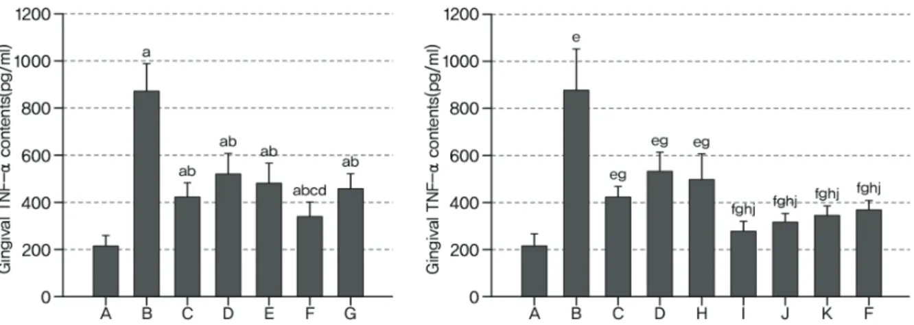
4. Effects on the gingival TNF-α contents
Significant (p<0.01) increases of TNF-α contents
around ligation placed gingival tissues were detected
in EPD control as compared with intact control
in the both first (655.75 ng/ml) and second (660.25
ng/ml) experiment. However, significant (p<0.01)
decreases of gingival TNF-α contents were detec-
ted in all Polycan (-449.38 ng/ml in first, -452.00
ng/ml in second) and calcium gluconate (-350.75
ng/ml in first, -346.00 ng/ml in second) single
or mixed formula (-391.88 ng/ml in 1:1, -534.13
ng/ml in 1:9, -414.50 ng/ml in 9:1, -379.75 ng/ml
in 1:99, -601.00 ng/ml in 2:98, -562.25 ng/ml in
4:96, -533.63 ng/ml in 8:92, -512.00 ng/ml in 10:90
mixed formula) treated rats as compared with EPD
control, respectively. Especially, Polycan and cal-
cium gluconate 1:9 mixed formula treated rats
showed significant (p<0.01 or p<0.05) decreases
of gingival TNF-α contents as compared with
each Polycan (-84.75 ng/ml) and calcium gluconate
on the Experimental Periodontitis and Alveolar Bone Loss in Rats
Figure 5. Changes on the gingival TNF-α contents after 10 Days oral treatment of single or mixed formulas of Polycan and calcium gluconate in EPD rats
A: Intact control, B: EPD control, C: Polycan single formula treated rats, D: Calcium gluconate single formula treated rats, E: Polycan and calcium gluconate 1:1 mixture treated rats, F: Polycan and calcium gluconate 1:9 mixture treated rats, G: Polycan and calcium gluconate 9:1 mixture treated rats, H: Polycan and calcium gluconate 1:99 mixture treated rats, I: Polycan and calcium gluconate 2:98 mixture treated rats, J: Polycan and calcium gluconate 4:96 mixture treated rats, K: Polycan and calcium gluconate 8:92 mixture treated rats.
a