† 주 저자 (e-mail: [email protected]) call: 070-5117-0035
Vol. 47, No. 3, September 2021, 247-254 http://dx.doi.org/10.15230/SCSK.2021.47.3.247
개똥쑥 추출물의 항여드름 효능확인
유 지 영†⋅노 경 백⋅오 세 영⋅정 용 택⋅박 덕 훈⋅정 은 선††
바이오스펙트럼㈜ 생명과학연구소
(2021년 9월 6일 접수, 2021년 9월 27일 수정, 2021년 9월 28일 채택)
Anti-acne Properties of Artemisia annua Extract In Vitro
Jiyoung You†, Kyung-Baeg Roh, Se-young Oh, Yong-Taek Jung, Deokhoon Park, and Eunsun Jung††
Biospectrum Life Science Institute, A-1805, U-TOWER, 767, Sinsu-ro, Suji-gu, Yongin-si, Gyeonggi-do 16827, Korea (Received September 6, 2021; Revised September 27, 2021; Accepted September 28, 2021)
1)
요 약: 여드름은 털피지샘단위(pilosebaceous unit)와 관련된 만성 염증 피부 질환으로, 여드름 병변에서는 피지 과다분비(hyperseborrhea)나 이상분비(dysseborrhea), 염증반응, 그리고 다른 피부상재균들에 비해 증가된 Cutibacterium acnes (C. acnes)로 인한 피부 미생물 균총의 불균형이 관찰된다. 이 연구는 개똥쑥 추출물(Artemisia annua extract: AAE)의 항여드름 효과를 확인한 것으로, 피부 미생물 균총의 균형유지와 관련된 항균효과와 염증반응의 억제, 과도한 피지 분비의 감소 측면에서 실험을 진행하였다. C. acnes와 Staphylococcus epidermidis (S. epidermidis)를 AAE와 공동배양 하였을 때, S. epidermidis의 성장률은 저해되지 않았지만 C. acnes의 성장률 은 저해된 것을 확인하였다. 또한 AAE를 처리하여 배양한 C. acnes 배양배지를 세포에 처리하였을 때, 인터루킨 -1β(IL-1β), 종양괴사인자-α(TNF-α)와 인터루킨-6(IL-6) 같은 사이토카인 분비의 감소와 TLR2 활성 억제도 확인하였다. 마지막으로 피지세포에 AAE를 처리한 결과, 팔미트산에 의해 유도된 피지형성을 감소시키는 것을 확인하였다. 이 결과들은 AAE가 다양한 타깃을 지닌 천연추출물로써 여드름의 주요 원인들인 C. acnes의 선택적 성장저해와 C. acnes로부터 유도되는 염증반응을 억제할 수 있으며, 과도한 피지형성을 감소시켜 결과적으로 여드름을 완화시키는 물질로 사용될 수 있다는 것을 제시한다.
Abstract: Acne vulgaris is a chronic inflammatory skin disease related to pilosebaceous unit. In acne lesions, hyperseborrhea, dysseborrhea, inflammatory event, and an imbalance in skin microflora, particularly an increase in Cutibacterium acnes (C.
acnes) colonization comparing to other bacteria, have been observed. The objective of this study was to evaluate anti-acne effects of Artemisia annua extract (AAE) on antibacterial activity related to preservation of the balance in skin microbiome, inhibition of inflammation, and reduction of excessive sebum production. When C. acnes and Staphylococcus epidermidis (S. epidermidis) were co-cultured in the presence of AAE, the reduction of C. acnes growth by AAE was greater than that of S. epidermidis. In addition, when C. acnes was cultured in a medium containing AAE (C. acnes AAE), levels of cytokines such as interleukin-1β (IL-1β), tumor necrosis factor-α (TNF-α) and IL-6 and toll-like receptors-2 activity were decreased in comparison with C. acnes cultured in a medium without AAE (C. acnes CM). Moreover, AAE significantly inhibited excessive sebum production induced by palmitic acid. These results suggest that AAE, as a natural extract with various targets, can inhibit selective growth of C. acnes and inflammatory reactions derived from C. acnes, which are the main causes of acne, and consequently can be used as a substance to alleviate acne by reducing excessive sebum formation.
Keywords: Artemisia annua, acne vulgaris, microbiome, THP-1, sebocytes
1. Introduction
Skin is the most external part. Problems of skin with changes in appearance can cause aesthetic and psychological stress[1,2]. In particular, westernized eating habits, lifestyle, and fine dust are causing many skin problems[3,4]. Acne vulgaris is increasing as an immune response to exposure to harmful factors. Acne is a chronic inflammatory skin disease involving pilosebaceous units. Acne lesions show hypersecretion or abnormal sebum secretion and inflammatory reaction. In addition, there is an imbalance of skin microbial flora due to increase of Cutibacterium acnes (C. acnes)[5,6].
Microbiome is a compound word of microbiota and genome. Dysbiosis of microorganisms is closely related to disease. Skin microbiota refer to the microbial community of millions of different microorganisms such as bacteria, viruses, and fungi, that inhabit the human skin[7]. Major bacteria that make up the microbiome in the skin are Staphylococcus epidermidis (S. epidermidis) and C. acnes. They are called resident skin flora[8]. Under certain conditions, if the balance of these skin flora is disturbed, skin diseases such as acne, seborrheic dermatitis and atopic dermatitis can occur[3].
Therefore, recovery from dysbiosis is an important part in the prevention and treatment of acne related to changes in cutaneous microbiota[9]. Several reports have shown that one of the causes of acne is C. acnes[10,11]. Overgrowth of C.
acnes is related to the development of acne. It is also a problem if the ratio of S. epidermidis to C. acnes change. In healthy skin, S. epidermidis can inhibit the growth of C. acnes by fermenting glycerol, a natural component of triglycerides in sebum[6]. It can also inhibit C. acnes-mediated inflammatory response by producing succinic acid[12]. However, according to recent studies, the relative ratio of C. acnes in healthy skin is not significantly different from that in acne skin. Instead, it has been reported that when the diversity of skin flora decreases and the proportion of C. acnes phylotypes changes, acne will occur[13]. Inhibiting the growth of all skin commensal microorganisms with indiscriminate use of antibiotics can cause destruction of the ecosystem of symbiotic microorganisms, making acne worse. Thus, it is not an effective method to eliminate the ultimate cause of acne even
if it is treated. Therefore, it is important to find anti-acne substances that selectively act on C. acnes in alleviating or treating acne.
C. acnes, the main causative agent of acne, can activate nuclear factor kappa B (NF-κB) by binding to Toll like receptor2 (TLR2) of keratinocytes and sebocytes, causing an inflammatory response[14]. It can also induce an inflammatory response by forming colonies and secreting various metabolites that degrade host tissues such as lipase and protease[15]. If this response persists, a biofilm will be formed to show antibiotic resistance, leading to a difficulty in treating acne.
Sebaceous glands that secrete sebum exist in high density on the face. They are connected to the isthmus of hair follicles. They are also called sebaceous gland units.
Sebocytes can differentiate during movement from the outside to the inside part of sebaceous glands. Finally, the nucleus and the membrane of differentiated sebocytes are decomposed and sebum is secreted[16].
Artemisia annua (A. annua) is an annual plant belonging to the genus Asteraceae. It is mainly distributed in Japan, China, and Taiwan. It has been widely used for medicinal purposes.
It is used for reducing fever, managing heat-related symptoms, and treating malaria[17]. Artemisinin, the most well-known component of A. annua has been found to be effective in anti-allergic action by suppressing skin hypersensitivity reactions[18]. In addition, flavonoids contained in A. annua extract (AAE) have been used in anticancer treatment as antioxidants. A. annua oil is known to have antibacterial effects. It has been used to relieve inflammatory reactions [19,20].
The objective of this study was to determine anti-acne properties of AAE through its antibacterial effect related to maintaining the balance of skin flora, inhibition of inflammatory response to virulence factor secreted by C.
acnes, and reduction of excessive sebum production.
2. Experimental
2.1. Preparation of Extracts from A. annua
The whole plant of A. annua was purchased from Angang Agrochemicals (Korea). To prepare an ethanol extract, the
plant was extracted with 70% (v/v) ethanol at 80 ℃ for 3 h.
After filtration (No.2 Qualitive Filter paper, 5 µm, Advantec, Japan), the filtrate was concentrated using a rotary vacuum evaporator (EYELA, Japan) and then lyophilized (yield 10.43%).
2.2. Microbial Strains and Growth Condition
C. acnes ATCC 6919T and S. epidermidis ATCC 14990T were obtained from American Type Culture Collection (ATCC, USA) and cultured in tryptic soy agar (TSA, Difco Laboratories, USA) for 48 h at 37 ℃ under anaerobic condition.
2.3. Co-Culture Assays
C. acnes ATCC 6919T and S. epidermidis ATCC 14990T were cultured in tryptic soy broth (TSB, Difco Laboratories, USA) and the turbidity of suspension was adjusted 2 × 10⁷ colony-forming units (CFU)/mL and 2 × 10⁵ CFU/mL respectively. C. acnes ATCC 6919T and S. epidermidis ATCC 14990T were co-incubated in 96 well culture plate. Each well was inoculated with 10 µL of bacteria suspension. Final concentrations of C. acnes ATCC 6919T and S. epidermidis ATCC 14990T in each well were 1 × 10⁶ CFU/mL and 1 × 10⁴ CFU/mL, respectively. Microplates were incubated at 37 ℃ for 3 h under an anaerobic condition. After 3 h of incubation, each well was inoculated with AAE extract (dissolved in DMSO at a concentration of 1,000 µg/mL). These microplates were incubated at 37 ℃ for 21 h under an anaerobic condition. After incubation, bacteria were diluted with TSB media and then inoculated onto selective agar plates containing 5 µg/mL of furazolidone (Sigma-aldrich, USA) and 5 µg/mL ampicillin (Sigma-aldrich, USA) for C. acnes ATCC 6919T and S. epidermidis ATCC 14990T, respectively.
According to a previous study, furazolidone on selective agar plates can completely inhibit the growth of S. epidermidis, but not the growth of C. acnes[21]. CFUs of C. acnes and S.
epidermidis were counted after incubation at 37 ℃ for 5 days.
2.4. Cell Culture
THP-1 cells (ATCC® TIB-202™, ATCC, USA), a human monocytic leukemia cell line were maintained in Roswell Park
Memorial Institute (RPMI) 1640 Medium (Welgene, Korea) supplemented with 10% fetal bovine serum (FBS, Gibco, USA) and 1% penicillin/streptomycin (Thermo Fisher Scientific, USA) at 37 ℃ under 5% CO2. Human sebocytes were purchased from Celprogen (USA) and cultured in Human Sebocyte Complete Growth Media (Celprogen, USA) at 37 ℃ in a 5% CO2 incubator. TLR2-HEK293 (HEK-Blue™ hTLR2 cells, InvivoGen, USA) were co-transfected into human embryonic kidney cells (HEK293) with human TLR2 and an NF-κB-inducible secreted alkaline phosphatase (SEAP) reporter gene. TLR2-HEK293 cells were cultured in Dulbecco’s modified Eagle’s medium (DMEM, Welgene, Korea) with 4 mM L-glutamine, 4500 mg/L glucose, and sodium pyruvate supplemented with 10% fetal bovine serum (FBS, Gibco) at 37 ℃ in a 5% CO2 incubator.
2.5 Preparation of Conditioned Medium of C. acnes C. acnes was anaerobically cultured in Reinforced Clostridial Medium (RCM, Difco Laboratories, USA) at 37 ℃ for 48 h.
AAE was used to treat C. acnes cultured in a 96 well microplate. C. acnes was adjusted to 1 × 10⁷ CFU/mL in RCM. AAE was dissolved in distilled water at concentrations of 10, 50, 100, 500, 1,000, and 2,000 µg/mL for treatment.
The final concentration of C. acnes in each well was 1 × 10⁶ CFU/mL. The microplate was incubated at 37 ℃ for 18 h. After collecting the conditioned media, the supernatant was filtrated using a 0.22 micron pore-sized filter (EMD Millipore, USA).
2.6. Enzyme-linked Immunosorbent Assay (ELISA) Proinflammatory cytokines were detected using an ELISA kit (R&D Systems, USA). THP-1 cells were cultured in the presence of C. acnes CM (C. acnes cultured in a medium without AAE) or C. acnes AAE (C. acnes was cultured in a medium containing AAE) diluted to one hundredth in media for 24 h. After conditioned medium was collected, secreted proinflammatory cytokines including interleukin-1β (IL-1β), tumor necrosis factor-α (TNF-α) and IL-6, were analyzed.
Results were determined using a linear standard curve.
2.7. TLR2/NF-κB/SEAP Activity
TLR2-HEK293 cells were cultured in serum-free DMEM
with C. acnes CM or C. acnes AAE diluted to one hundredth in media. After 6 h, conditioned medium was collected and reacted with QUANTI-Blue™ solution (InvivoGen, USA) and then incubated at 37 ℃ in a 5% CO2 incubator. SEAP activity was determined by measuring the optical density (OD) at 620 nm.
2.8. Measurement of Cell viability
Cell viability of THP-1 cells and TLR2-HEK293 cell were detected by using a 3-(4,5-dimethyl-2-thiazoyl)-2,5-diphenyl- 2H-tetrazolium bromide (MTT, (Sigma-aldrich, USA) and WST-1 (Sigma-aldrich, USA) assay respectively. THP-1 was cultured in the presence of C. acnes CM or C. acnes AAE diluted to one hundredth in media for 24 h. 0.1 mg/mL MTT solution was treated to cultured medium and incubated for 1 h at 37 ℃ under 5% CO2. After dissolving the generated formazan in DMSO, optical density was measured at 570 nm by using a spectrophotometer (Power Wave, Bio-Tek Inc., USA). TLR2-HEK293 also cultured in the presence of C.
acnes CM or C. acnes AAE diluted to one hundredth in media for 6 h. 0.1 mg/mL WST-1 solution was added and incubated for 1 h at 37 ℃ under 5% CO2. The absorbance was measured on an ELISA plate reader (Power Wave, Bio-Tek Inc., USA) at 450 nm.
2.9. Measurement of Sebum Accumulation
Human sebocytes were differentiated with 100 µM palmitic acid (Sigma-aldrich, USA) with or without AAE for 48 h.
Treated cells were washed with PBS and fixed with 4%
formaldehyde at room temperature for 10 min. After washing, cells were stained with 10 µg/mL Nile red (Sigma-aldrich, USA) for 15 min. Nuclei were stained with Hoechst 33342 (Thermo Fisher Scientific, USA). Fluorescence was detected at excitation and emission wavelengths of 550 nm and 630 nm, respectively.
2.10. Statistical Analysis
Statistical significance of data was determined with student’s t-test. All results are expressed as mean ± standard deviation (N = 3). A p value of less than 0.05 was considered statistically significant.
3. Results and Discussion
3.1. Selective Antimicrobial Activity of A. annua Extract C. acnes and S. epidermidis are the main symbiotic bacteria in the pilosebaceous unit. They are the most common symbiotic bacteria on the skin. S. epidermidis uses glycerol, naturally produced in the skin to produce short chain fatty acids (SCFAs) and inhibit the growth of C. acnes to maintain the balance of skin flora. However, under certain conditions, host characteristics such as stress and hormonal disorder can lead to abnormal overgrowth of C. acnes and cause an imbalance of the skin flora[6]. Therefore, the use of antibiotics in acne lesions caused by the imbalance of skin flora can indiscriminately inhibit the growth of symbiotic microorganisms, further disrupting the balance of commensal microorganisms and consequently worsening the acne.
To evaluate the effect of AAE as an antibacterial activity against C. acnes, S. epidermidis (1 × 10⁴ CFU/mL) and C.
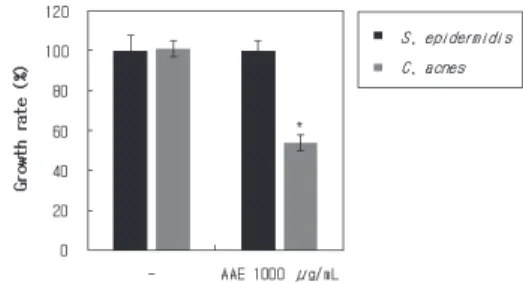
acnes (1 × 10⁶ CFU/mL) were co-cultured in culture media in the presence or absence of AAE. Under co-culture conditions growths of S. epidermidis and C. acnes did not affect each other. However, AAE effectively suppressed the growth of C.
acnes, but not the growth of S. epidermidis (Figure 1). These results suggest that AAE could selectively inhibit the growth of C. acnes known to aggravate acne vulgaris.
3.2. Anti-inflammatory Activity of A. annua Extract C. acnes can secrete virulence factors such as co-hemolytic
Figure 1. A. annua extract inhibits the growth of C. acnes selectively. S. epidermidis (1 × 10⁴ CFU/mL) and C. acnes (1
× 106 CFU/mL) were co-cultured in the presence or absence of AAE under anaerobic conditions for two days at 37 ℃. Data are expressed as mean ± SD, *p < 0.05 compared to the control.
Christie-Atkins-Munch-Peterson (CAMP) factors, lipase, sialidases, hyaluronate lyase, and porphyrins that can be detected by TLRs as pattern recognition receptors. When C. acnes activates TLR2, NF-κB signal is increased, which upregulates IKK phosphorylation and induces IκB release from cytosolic NF-κ B. Finally, translocated NF-κB can stimulate transcription of pro-inflammatory cytokines in the nucleus[22].
To explore the inhibitory effect of AAE on the secretion of virulence factors from C. acnes, we cultured C. acnes in the presence or absence of AAE. The cultured medium was then collected. TLR2-HEK293 cells were cultured with C. acnes CM to measure TLR2 signal activation. Results showed that C. acnes CM induced TLR2 activation, whereas C. acnes AAE at 2,000 µg/mL inhibited TLR2 activation up to 50%
(Figure 2A). No cytotoxicity was observed at all concentrations tested (Figure 2B).
To determine whether AAE could inhibit the inflammatory response caused by virulence factors secreted by C. acnes, THP-1 was cultured with C. acnes CM or C. acnes AAE.
Secreted proinflammatory cytokines were then measured. C.
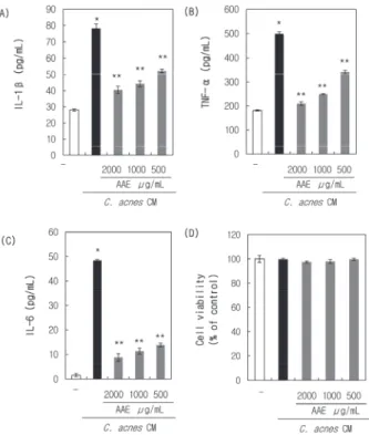
acnes CM stimulated the secretion of IL-1β (Figure 3A), TNF-α (Figure 3B), and IL-6 (Figure 3C), whereas C. acnes AAE effectively inhibited the production of these cytokines
from THP-1. No cytotoxicity was observed at all concentrations tested (Figure 3D). These results suggest that AAE can effectively reduce virulence factors secreted from C. acnes and inflammatory responses following stimulation of virulence factors.
3.3. Reduction of Excessive Sebum Production by A. annua Extract
Excessive sebum production is the main cause and consequence of acne lesions. Sebum is composed of several lipid components such as triglycerides (TG), fatty acids, wax esters, and squalene. It acts to protect the skin by inhibiting Figure 3. A. annua extract inhibits the expression of pro- inflammatory cytokines in THP-1 cells induced by C. acnes culture medium. THP-1 cells were cultured in the presence of C.
acnes CM with or without AAE diluted to one hundredth in media. Secreted proinflammatory cytokines such as (A) IL-1β, (B) TNF-α and (C) IL-6 from THP-1 cells were then measured by ELISA. (D) Cell viability of THP-1 was detection by MTT assay. Values are expressed as mean ± SD, *p < 0.001 versus C.
acnes CM untreated control. **p < 0.01 versus C. acnes CM treated control.
Figure 2. A. annua extract regulates C. acnes CM-induced TLR2 signaling pathways. (A) TLR2-HEK293 cells were cultured in the presence of C. acnes CM with or without AAE diluted to one hundredth in media. TLR2 signal activation was then measured using a Quanti-blue solution. (B) Cell viability of TLR2-HEK293 was detection by WST-1 assay. Data are expressed as mean ± SD, *p < 0.001 versus C. acnes CM untreated control;
**p < 0.01 versus C. acnes CM treated control.
moisture evaporation and bacterial infection. These lipids are involved in functions and differentiation of sebocytes as well as immune response and proliferation of keratinocytes. Under excessive sebum production condition, the proportion of these lipids can be altered which increases sebogenesis, inflammation, and comedogenesis, finally leading to acne[23].
In addition, the increase in sebum can promote an overgrowth of C. acnes and the formation of biofilm, which not only intensifies the inflammatory response, but also makes it difficult to treat acne[15]. Therefore, suppressing the excessive secretion of sebum is considered to be the main goal to effectively suppressing the development of acne.
In this experiment, palmitic acid was used as a factor to promote sebum production. Palmitic acid is a saturated fatty acid that is also used as extracellular free fatty acids (FFAs).
It is known to be involved in lipogenesis through SREBP. It can increase lipid contents and TG levels in sebocytes, the formation of comedone, and inflammatory response through TLR2[24,25].
To determine the effect of AAE on sebum production,
sebocytes were cultured in the presence of palmitic acid with or without AAE. Accumulated sebum droplets were then measured by Nile red staining. The fluorescence intensity of the droplet was calculated using Image J. As a result, it was confirmed that AAE decreased sebum production induced by palmitic acid in a dose-dependent manner. 10 µg/mL, 50 µg/mL and 100 µg/mL AAE reduced sebum production up to 27%, 35% and 60% respectively compared to palmitic acid (Figure 4). Therefore, AAE can alleviate acne by reducing excess sebum production.
4. Conclusion
Results of this study confirmed anti-acne properties of AAE through antibacterial, anti-inflammatory, and suppression of excessive sebum production. AAE selectively inhibited the growth of C. acnes, a major species in acne lesions, without affecting the growth of S. epidermidis in a co-culture condition.
In addition, AAE inhibited TLR2 activity and pro-inflammatory cytokine by reducing secretion of virulence factors from C.
Figure 4. A. annua extract suppresses sebum accumulation induced by palmitic acid in sebocytes. Sebocytes were cultured in DMEM in the presence of palmitic acid with or without AAE. Sebum synthesized in sebocytes was detected by Nile red staining. Scale bars, 100 µm.
Values are expressed as mean ± SD, *p < 0.001 versus palmitic acid untreated control, **p < 0.001 versus palmitic acid treated control.
acnes. AAE also suppressed excessively increased sebum production caused by palmitic acid. These results suggest that AAE as a natural product can be used as an acne alleviation material by inhibiting the growth of C. acnes selectively, suppressing the inflammatory response, and reducing excessive sebum production. Based on the in vitro results, we are planning a clinical trial to confirm the practical efficacy in acne patients.
References
1. A. K. Asadi and A. Usman, The role of psychological stress in skin disease, J. Cutan. Med. Surg., 5(2), 140 (2001).
2. L. Verhulst and A. Goossens, Cosmetic components causing contact urticaria: A review and update, Contact Dermatitis, 75(6), 333 (2016).
3. S. R. Ellis, M. Nguyen, A. R. Vaughn, M. Notay, W. A.
Burney, S. Sandhu, and R. K. Sivamani, The skin and gut microbiome and its role in common dermatologic conditions, Microorganisms, 7(11), 550 (2019).
4. K. E. Kim, D. Cho, and H. J. Park, Air pollution and skin diseases: Adverse effects of airborne particulate matter on various skin diseases, Life Sci., 152, 126 (2016).
5. T. X. Cong, D. Hao, X. Wen, X. H. Li, G. He, and X.
Jiang, From pathogenesis of acne vulgaris to anti-acne agents, Arch. Dermatol. Res., 311(5), 337 (2019).
6. M. Fournière, T. Latire, D. Souak, M. G. J. Feuilloley, and G. Bedoux, Staphylococcus epidermidis and Cutibacterium acnes: Two major sentinels of skin microbiota and the influence of cosmetics, Microorganisms, 8(11), 1752 (2020).
7. A. L. Cogen, V. Nizet, and R. L. Gallo, Skin microbiota:
A source of disease or defence?, Br. J. Dermatol., 158(3), 442 (2008).
8. A. L. Byrd, Y. Belkaid, and J. A. Segre, The human skin microbiome, Nat. R ev. Microbiol., 16(3), 143 (2018).
9. E. A. Grice and J. A. Segre, The skin microbiome, Nat.
Rev. Microbiol., 9(4), 244 (2013).
10. J. McLaughlin, S. Watterson, A. M. Layton, A. J. Bjourson, E. Barnard, and A. McDowell, Propionibacterium acnes and acne vulgaris: New insights from the integration of population genetic, multi-omic, biochemical and host-
microbe studies, Microorganisms, 7(5), 128 (2019).
11. B. Dréno, S. Pécastaings, S. Corvec, S. Veraldi, A. Khammari, and C. Roques, Cutibacterium acnes (Propionibacterium acnes) and acne vulgaris: A brief look at the latest updates, J. Eur. Acad. Dermatol. Venereol., 32(Suppl 2), 5 (2018).
12. Y. Wang, S. Kuo, M. Shu, J. Yu, S. Huang, A. Dai, A.
Two, R. L. Gallo, and C. M. Huang, Staphylococcus epidermidis in the human skin microbiome mediates fermentation to inhibit the growth of Propionibacterium acnes: implications of probiotics in acne vulgaris, Appl.
Microbiol. Biotechnol., 98(1), 411 (2014).
13. A. M. O’Neill and R. L. Gallo, Host-microbiome interactions and recent progress into understanding the biology of acne vulgaris, Microbiome, 6(1), 177 (2018).
14. J. L. Selway, T. Kurczab, T. Kealey, and K. Langlands, Toll-like receptor 2 activation and comedogenesis:
Implications for the pathogenesis of acne, BMC Dermatol., 13, 10 (2013).
15. C. Mayslich, P. A. Grange, and N. Dupin, Cutibacterium acnes as an opportunistic pathogen: an update of its virulence-associated factors, Microorganisms, 9(2), 303 (2021).
16. R. W. Clayton, K. Göbel, C. M. Niessen, R. Paus, M. A.
M. Steensel, and X. Lim, Homeostasis of the sebaceous gland and mechanisms of acne pathogenesis, Br. J.
Dermatol., 181(4), 677 (2019).
17. X. Feng, S. Cao, F. Qiu, and B. Zhang, Traditional application and modern pharmacological research of Artemisia annua L, Pharmacol. Ther., 216, 107650 (2020).
18. Y. Deng, Z. Liu, and Y. Geng, Anti-allergic effect of Artemisia extract in rats, Exp. Ther. Med., 12(2), 1130 (2016).
19. J. F. S. Ferreira, D. L. Luthria, T. Sasaki, and A.
Heyerick, Flavonoids from Artemisia annua L. as antioxidants and their potential synergism with artemisinin against malaria and cancer, Molecules, 15(5), 3135 (2010).
20. W. S. Kim, W. J. Choi, S. W. Lee, W. J. Kim, D. C.
Lee, U. D. Sohn, H. S. Shin, and W. Y. Kim, Anti-inflammatory, antioxidant and antimicrobial effects of artemisinin extracts from Artemisia annua L, Korean J.
Physiol. Pharmacol., 19(1), 21 (2015).
21. Y. Wang, M. S. Kao, J. Yu, S. Huang, S. Marito, R. L.
Gallo, and C. M. Huang, A precision microbiome approach using sucrose for selective augmentation of Staphylococcus epidermidis fermentation against Propionibacterium acnes, Int. J. Mol. Sci., 17(11), 1870 (2016).
22. T. Kawasaki and T. Kawai, Toll-like receptor signaling pathways, Front. Immunol., 5, 461 (2014).
23. M. Ottaviani, E. Camera, and M. Picardo, Lipid mediators in acne, Mediators Inflamm., 2010, 858176 (2010).
24. B. C. Melnik, Linking diet to acne metabolomics, inflammation, and comedogenesis: An update, Clin. Cosmet.
Investig. Dermatol., 8, 371 (2015).
25. C. W. Choi, Y. Kim, J. E. Kim, E. Y. Seo, C. C.
Zouboulis, J. S. Kang, S. W. Youn, and J. H. Chung, Enhancement of lipid content and inflammatory cytokine secretion in SZ95 sebocytes by palmitic acid suggests a potential link between free fatty acids and acne aggravation, Exp. Dermatol., 28(2), 207 (2019).