Cloning and Characterization of Phosphoinositide 3-Kinase γ cDNA from Flounder ( Paralichthys olivaceus )
Tae Hyug Jeong, Joo Yeon Youn, Keunho Ji, Yong Bae Seo and Young Tae Kim*
Department of Microbiology, Pukyong National University, Busan 608-737, Korea Received January 15, 2014 /Revised February 26, 2014 /Accepted March 3, 2014
Phosphoinositide 3-kinase (PI3K) plays a central role in cell signaling and leads to cell proliferation, survival, motility, exocytosis, and cytoskeletal rearrangements, as well as specialized cell responses, superoxide production, and cardiac myocyte growth. PI3K is divided into three classes; type I PI3K is preferentially expressed in leukocytes and activated by βγ subunits of heterotrimeric G-proteins. In this study, the cDNAs encoding the PI3Kγ gene were isolated from a brain cDNA library constructed using the flounder (Paralichthys olivaceus). The sequence of the isolated PI3Kγ was 1341 bp, encoding 447 amino acids. The nucleotide sequence of the PI3Kγ gene was analyzed with that of other species, including Oreochromis niloticus and Danio rerio, and it turned out to be well conserved during evolution. The PI3Kγ gene was subcloned into the expression vector pET-44a(+), and expressed in the E. coli BL21 (DE3) codon plus cell. The resulting protein was expressed as a fusion protein of approx- imately 49 kDa containing a C-terminal six-histidine extension that was derived from the expression vector. The expressed protein was purified to homogeneity by His-tag affinity chromatography and showed enzymatic activity corresponding to PI3Kγ. The binding of wortmannin to PI3Kγ, as detected by anti-wortmannin antisera, closely followed the inhibition of the kinase activities. The results ob- tained from this study will provide a wider base of knowledge on the primary structure and charac- terization of the PI3Kγ at the molecular level.
Key words : Characterization, gene cloning, paralichthys olivaceus, phosphoinositide 3-kinase γ (PI3Kγ)
*Corresponding author
*Tel : +82-51-629-5616, Fax : +82-51-629-5619
*E-mail : [email protected]
This is an Open-Access article distributed under the terms of the Creative Commons Attribution Non-Commercial License (http://creativecommons.org/licenses/by-nc/3.0) which permits unrestricted non-commercial use, distribution, and reproduction in any medium, provided the original work is properly cited.
Journal of Life Science 2014 Vol. 24. No. 4. 343~351 DOI : http://dx.doi.org/10.5352/JLS.2014.24.4.343
Introduction
Phosphoinositides were recognized early as precursors for second messengers in cell surface receptor–coupled signal transduction pathways. Phosphoinositide 3-kinase (PI3K) catalyzes the addition of a phosphate molecule to the three positions of the inositol ring of phosphoinositides (PtdIns), producing four different lipid products: the singly phos- phorylated form PtdIns-3-P, the doubly phosphorylated forms PtdIns-3,4-P2 and PtdIns-3,5-P2, and the triply phos- phorylated form PtdIns-3,4,5-P3 [9].
There are multiple isoforms of PI3K in mammalian cells, and these are subdivided into three main classes on the basis of their structures, in vitro substrate specificity, and mode of regulation [19, 23]. Class I PI3Ks comprise a p110 catalytic subunit and a regulatory adapter subunit. Class II PI3Ks are
large (170-200 kDa) proteins that have a catalytic domain 45-50% homologous to class I PI3Ks. Finally, class III PI3Ks are typified by the yeast protein [8]. Class I PI3Ks have been the major focus of PI3K studies because these isoforms are generally coupled to extracellular stimuli; these PI3Ks are activated by a variety of extracellular stimuli and have been linked to an incredibly diverse set of key cellular functions, including cell cycle progression, cell growth, cell pro- liferation, cell motility, cell differentiation, cell survival and intracellular trafficking [4, 7]. The emerging links between PI3-kinase activity and many human maladies, including al- lergy, inflammation, heart disease, and cancer, has made them the focus of intense study, and inhibitors of these en- zymes are considered potential therapeutic agents.
A class I PI3K is a heterodimeric complex, comprising a p110 catalytic subunit, of which there are four characterized isoforms (α, β, γ, and δ). Class I PI3Ks are subdivided into class IA and IB. Type IA PI3Ks p110α, p110β, and p110δ share 42-58% amino acid sequence identity and are asso- ciated with the p85 family of regulatory subunits; on the other hand type IB PI3K P110γ binds to a p101 adaptor molecule. Whereas class IA PI3Ks are activated by inter- action with tyrosine-phosphorylated molecules, class IB p110
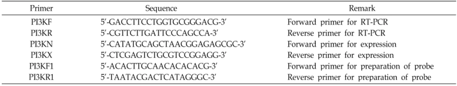
Table 1. Oligonucleotide primers used for this study
Primer Sequence Remark
PI3KF PI3KR PI3KN PI3KX PI3KF1 PI3KR1
5'-GACCTTCCTGGTGCGGGACG-3' 5'-CGTTCTTGATTCCCAGCCA-3' 5'-CATATGCAGCTAACGGAGAGCGC-3' 5'-CTCGAGTCTGCGTCCGGAGG-3' 5'-ACACTTGCAACACACACG-3' 5'-TAATACGACTCATAGGGC-3'
Forward primer for RT-PCR Reverse primer for RT-PCR Forward primer for expression Reverse primer for expression
Forward primer for preparation of probe Reverse primer for preparation of probe γ (PI3Kγ) is activated by engagement of heterotrimeric
GTP-binding protein (G protein)-coupled receptors (GPCR).
PI3Kγ is preferentially expressed in leukocytes [10, 20]; fur- thermore, it is activated by βγ subunits of heterotrimeric G-proteins, which thus link seven transmembrane (7TM) he- lix receptor activation to phosphatidylinositol (3, 4, 5)-trisphosphate production [11, 14]. PI3Kγ controls thymo- cyte survival, as well as the activation of mature T cells, but has no role in the development or function of B cells.
PI3Kγ links GPCR stimulation to the formation of phosphati- dylinositol 3,4,5-triphosphate and the activation of protein kinase B, ribosomal protein S6 kinase, and extracellular sig- nal–regulated kinase 1 and 2 [18, 21]. Thus, PI3Kγ regulates thymocyte development, T cell activation, neutrophil migra- tion, and the oxidative burst. Recent studied in mice lacking functional PI3Kγ showed that PI3Kγ plays a key role as a modulator of inflammation and allergy, as well as in the regulation of cardiac contractility [11, 13, 17].
Elucidation of the structural diversity of PI3Kγ in recent years by molecular cloning of cDNAs and genes from vari- ous species has provided insight into their functions.PI3Kγ cDNA genes have been cloned fromMus musculus [2], Rattus norvegicus [1], Danio rerio [16], and Homo sapiens [22].
Knowledge of the molecular structure of PI3Kγ in marine fishes is extremely limited. In addition, the nature of PI3Kγ in these fish and their roles in the control of the PtdIns sig- naling pathways is still unclear.
The flounder (Paralichthys olivaceus), one of the most evolved teleosts, is a commercially important marine aqua- culture species in Korea and has been the object of studies on various functional genes at the molecular level [5, 6, 15].
The present study focuses on the isolation of cDNA encod- ing the flounder PI3Kγ and characterization of the cloned gene. These data will provide a base of knowledge for the PI3Kγ gene at the molecular level and the functional diver- sity of PI3Kγ.
Materials and Methods
RNA isolation and construction of the flounder cDNA library
Total RNA from flounder (P. olivaceus) brain, liver, and kidney tissues were isolated using a total RNA isolation kit (Promega). The complementary DNA (cDNA) library was constructed using a ZAP-cDNA Synthesis Kit (Stratagene), as described in the manufacturer’s instructions. The result- ing library contained approximately 1×10⁵ clones. The li- brary was then amplified up to 3×10⁹ clones/ml.
Screening PI3Kγ cDNA and DNA sequencing Conserved nucleotide sequences of PI3K among the verte- brate species were determined using the National Center for Biotechnology Information (NCBI) nucleotide and protein sequence database and used for the design of oligonucleo- tide primers for screening PI3K, which were synthesized from GenoTech (Taejeon). PCR was carried out using a pair of the “PI3KF1” and “PI3KR1” primers (Table 1). The probe for screening PI3K was labeled with a digoxigenin (DIG) oli- gonucleotide 3'-end labeling kit (Roche). DIG-labeled probes were quantified and used for the immunoscreening procedure. Approximately 1×10⁵of plaques from the cDNA library was screened with the above probes and several pos- itive plaques were isolated. These plaques were recovered and further confirmed by the second screening. Positive pla- ques were recovered from the second screening and the phagemid containing the insert was excised according to the manufacturer’s instructions (Stratagene).
Comparative sequence analysis of flounder PI3Kγ To examine the molecular evolution of PI3Kγ (AY514674) from Paralichthys olivaceus, the following PI3Kγ sequences were imported from the Swiss-Prot databank / GenBank:
D. rerio (BC164683), O. niloticus (XM003448849), M. musculus (NM008841), B. taurus (NM174796), and H. sapiens (NM 001256045). The nucleotide sequences were analyzed using
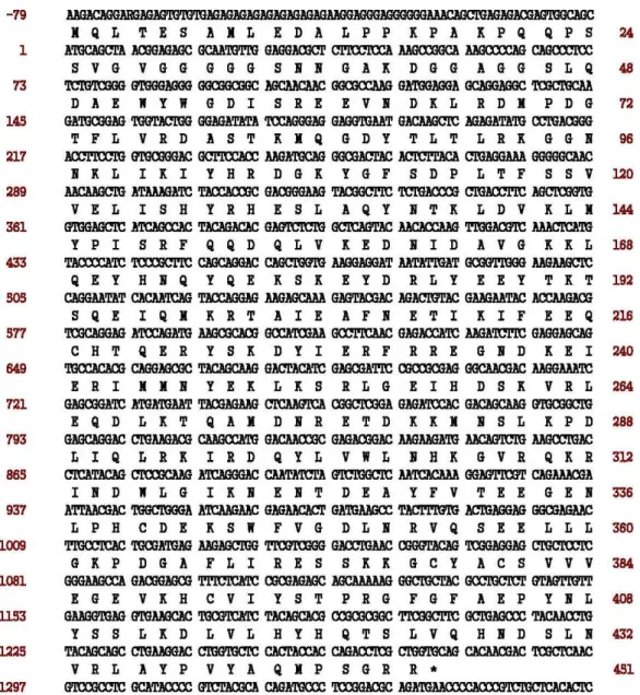
Fig. 1. Nucleotide and deduced amino acid sequences of the cDNA encoding flounder PI3Kγ gene. The nucleotide sequence is numbered to the left and the amino acid to the right.
the BLAST program (http://www.ncbi.nlm.nih.gov/BLAST).
Multiple sequence alignment was conducted using the CLUSTAL W program (http://www.ebi.ac.uk/clustalw), and sequence identities were calculated using GeneDoc (http://www.psc.edu/biomed/genedoc). A phylogenetic tree was constructed by the neighbor-joining (NJ) method using the Treecon program [9] for the amino acid sequences of PI3Kγ fromD. rerio, O. niloticus, M. musculus, B. taurus, and H. sapiens.
Reverse transcription polymerase chain reaction (RT-PCR)
In order to perform RT-PCR, total RNA was isolated from
the brain, liver, and kidney from mature flounder (n=10;
size: 45±10 cm, body weight; 900±300 g; 3 years old). The RT-PCR was performed using Bioneer’s RT-PCR system. The reaction components were set up for Master mix 1 and Master mix 2. Master mix 1 contained template RNA, 50 pmol of primer, and DEPC-water. The sample was incubated for 10 min at 65℃ and cooled down on ice. Master mix 2 consisted of 5X RT-PCR buffer, 2.5 mM dNTP mixture, 100 mM DTT, RNase inhibitor, and MMLV RTase. Mix 1 and mix 2 were added to a 0.2 μl tube. The sample was placed in a thermocycler (GeneAmp PCR system 2,400, Perkin Elmer) and incubated for 1 hr at 42℃ for reverse tran- scription followed by thermocycling. The temperature pro-
file of PI3Kγ was on pre-reaction at 94℃ for 5 min and 30 cycling reaction with 94℃ 40 sec denaturation, 56℃ for 30 sec annealing, 72℃ for 1 min, and finally a 7 min extension at 72℃. After reaction, 15 μl of RT-PCR product was ana- lyzed with 1% agarose gel electrophoresis.
Expression of flounder PI3Kγ gene in Escherichia coli
The PI3Kγ gene was amplified by PCR using a pair of oligonucleotides (Table 1). The PCR product was ligated into the pGEM-T vector and the resulting plasmid was digested withNdeI and XhoI restriction enzymes. Then, the excised fragment was ligated into the pET44-a(+) vector. The result- ing plasmid containingPI3Kγ gene was called pET-44a-PI3K.
The plasmid was transformed into the competent E. coli strain BL21 (DE3) codon plus. Cells harboring a plasmid that contained thePI3Kγ gene were cultured overnight in 10 ml of Luria-Bertani / ampicillin (LB/amp; containing 50 μg/μl ampicillin) broth at 37℃ in a shaking incubator. The cell was induced by adding isopropyl-β-D-thiogalactopyranoside (IPTG) to a final concentration of 1 mM at mid-log growth (OD600=0.5).
Purification of recombinant PI3Kγ proteins The pET-44a(+)-PI3K plasmid contains PI3K-histidine (PI3K-His)-tagged DNA sequences. The PI3K-His fusion pro- tein was eluted using a His Trap Kit (Pharmacia). The pellet from 1 L of induced culture was resuspended in 100 ml of binding buffer containing 5 mM imidazole, 0.5 M NaCl, 50 mM Tris-HCl pH 7.6, 1 mg/ml lysozyme (Sigma-Aldrich), and protease inhibitors (Sigma-Aldrich). The cells were dis- rupted by sonication for 30 sec in VC130 (Sonics and Materials Inc). Cell debris was pelleted by centrifugation at 12,000 rpm in a Sorvall SA-600 rotor for 15 min. The super- natant was filtered through a 0.22 μm pore membrane, di- luted in binding buffer, and then loaded on a His Trap chro- matography column. The supernatant was eluted with three column volumes of 500 mM imidazole, 0.5 M NaCl, and 50 mM Tris-HCl pH 7.6 (elution buffer). Each 3 ml fraction was collected and measured for its protein content on SDS- PAGE.
Enzyme activity assay
PI3Kγ protein activity was measured by the spectrophoto- metric method of Stoyanov et al. [22]. Protein kinase assays using purified PI3Kγ proteins and GST-p110a / p84a protein
were carried out at 30°C. To assay protein phosphorylation, immobilized PI3Kγ was washed twice with kinase buffer without ATP [50 mM Hepes (pH 7.4) / 150 mM, NaCl / 5 mM, EDTA / 5 mM dithiothreitol / 10 mM MgCl2/ 0.01%
Triton X-100] and resuspended in the same buffer (MgCl2
concentrations were varied where indicated). As indicated, TPA (300 nM), BIM (100 nM), wortmannin (100 nM), or lip- osomes were added to the reaction mixture. An equal vol- ume of kinase buffer supplemented with ATP was added to initialize the phosphorylation reaction. Incubation for 20 min at 30°C was followed by denaturation and autoradiog- raphy.
Protein determination
Protein concentration was determined by the Bradford method. The Bradford reagent was from Bio-Rad and bovine serum albumin (BSA) served as a standard protein.
Results and Discussion Nucleotide sequences of flounder PI3Kγ
ThePI3Kγ gene of flounder was isolated using PCR from the flounder brain cDNA library. PCR products were cloned into T vector. Cloned DNA was purified and sequenced with an automatic DNA sequencer using the ABI Prism DNA se- quencing kit.
Fig. 2 shows the nucleotide sequence of the complete cDNA encoding the flounder PI3Kγ gene (GeneBank ac- cession number AY514674) and its deduced amino acid sequence. The sequence of cloned PI3Kγ was analyzed with the NCBI BLAST program. The flounderPI3Kγ gene contains 1,744 bp, including an open reading frame and encoding a 447 amino acid protein. The cDNA consists of 86 bp of a 5'- untranslated region (UTR), 1,341 bp of coding region, and 314 bp of 3'-UTR, followed by a poly (A) sequence. As shown in Fig. 2, the flounder PI3Kγ cDNA clone contains an in-frame termination codon (TGA) at bases 1431-1434.
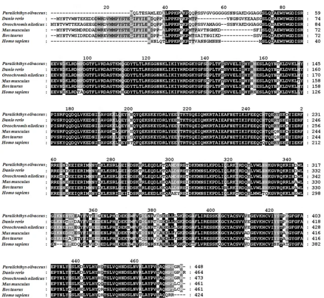
Sequence identity and the phylogenetic tree Fig. 3 shows an alignment of the amino acid sequences of the flounder and other PI3Kγ. The PI3Kγ proteins were compared using the BLAST protein database (NCBI).
The flounder PI3Kγ had a high sequence similarity with other species in its amino acid residues. The deduced flounder amino acid sequence was about 89.6%, 84.7%, 84%, and 74.9% identical with the PI3Kγ of zebrafish (D. rerio),
Fig. 2. Multiple alignment of deduced amino acid sequences of PI3Kγ and other gene. The amino acid sequences are obtained from GeneBank:Paralichthys olivaceus(AY514674),Danio rerio(BC164683)Oreochromis niloticus(XM003448849),Mus musculus (NM008841), Bos taurus(NM174796), Homo sapiens(NM001256045). The amino acid are shaded in different colors of grey, which indicate the degree of consensus between the different sequences. “-“non-conserved amino acids.
mouse (M. musculus), Norway rat (R. norvegicus), and human (H. sapiens), respectively.
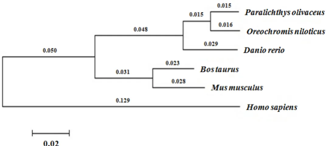
A molecular phylogenetic tree was constructed to analyze the evolutionary relationships of the PI3Kγ protein (Fig. 3).
It shows the evolutionary divergence of thePI3Kγ genes of the zebrafish, flounder, mouse, Norway rat, and human. The flounder PI3Kγ protein was more closely related to the ze- brafish PI3Kγ than to the human one, as reflected in the se- quence identity (89.6% vs. 74.9%).
Tissue distribution of PI3Kγ
In order to determine the expression of thePI3Kγ gene, total RNA was isolated from flounder brain, liver, and kid- ney tissues using a Trizol reagent and the quality of isolated
RNAs was confirmed by formaldehyde RNA gel electro- phoresis. Specific primers PI3KF and PI3KR were synthe- sized on the basis of the consensus sequence of PI3K and used for the detection of PI3Kγ mRNA with RT-PCR. The products (10 μl) of RT-PCR were analyzed with 1% agarose gel electrophoresis. As shown in Fig. 4, an approximately 750 bp DNA fragment was amplified from all total RNAs extracted from the brain, liver, and kidney tissues (Fig. 4).
The resulting RT-PCR patterns provided evidence for the ex- pression of PI3Kγ in tissues from the brain, liver, and kidney, suggesting that the flounder PI3Kγ mRNA has a wide tissue distribution.
Fig. 3. A molecular phylogenetic tree of PI3Kγ based on the NJ method. Numbers at nodes indicate levels of bootstrap support based on 1,000 replicated datasets. Bar, 0.02 substitutions per amino acids position.
Fig. 4. Pattern of the PI3K expression detected by RT-PCR. Lane M, molecular maker; lane B, total RNA template for RT-PCR isolated from flounder brain; lane L, liver; lane K, kidney.
Expression of flounder PI3Kγ inE. coli
In order to subclone for the construction of expression vector ofPI3Kγ gene, a pair of primers was designed based on known PI3Kγ sequences. The resulting PCR fragment of about 1.7 kb was eluted and ligated into the pGEM T-vector.
Then, the flounderPI3Kγ gene was subcloned into the pro- karyotic expression vector, pET-44a(+), which allows ex- pression of recombinant protein with a C-terminal fusion His-tag. The resulting pET-44a-PI3Kγ plasmid (Fig. 5A) was transformed into the E. coli BL21 (DE3) codon plus strain and recombinant protein were expressed by the addition of IPTG. The expression patterns of the PI3Kγ proteins were analyzed using 12% SDS-PAGE (Fig. 5B). The cloned PI3Kγ protein was strongly expressed with IPTG induction. The optimum induction time was approximately 1 hr after IPTG induction. The molecular weight of the PI3Kγ fusion protein is approximately 49 kDa, while the predicted PI3Kγ protein is approximately 46 kDa, corresponding to a C-terminal fu-
sion tag (3 kDa).
Western blot analysis
In order to perform western blot, the induced cells were harvested by centrifugation at 0, 1, 3, and 6 hr. Proteins were electrophoretically transferred from an SDS-PAGE gel to ni- trocellulose membrane, probed with goat antiserum against the 6-His tag, and incubated with alkaline phosphatase cou- pled with the goat antibody against goat IgG. The nitro- cellulose membrane developed using NBT / BCIP. As shown in Fig. 5C, western blot was analyzed and confirmed.
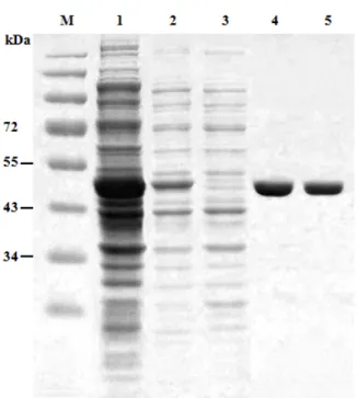
Purification of the PI3Kγ protein
The expression and purification of the recombinant PI3Kγ protein was analyzed by 12% SDS-PAGE. The optimal in- duction of a recombinant PI3Kγ protein was achieved at 9 hr after induction. The recombinant PI3Kγ protein was puri- fied using an affinity chromatography. Affinity chromatog- raphy was applied for the single-step purification in order to separate a particular protein using a specific interaction with a ligand that specifically binds to a target protein from the cellular total proteins. Using this technique, the PI3Kγ protein was purified to homogeneity and the purified pro- tein was shown to be enzymatically active. The molecular mass of the purified protein was 49 kDa, which represents the value calculated from the gene sequence (Fig. 6).
Enzyme activity of PI3Kγ
In view of the potent inhibition of serpentine receptor- mediated PtdIns (3, 4, 5) P3 production and cell responses by wortmannin, the inactivation mechanism of PI3Kγ by this substance was investigated. When GST-p110a / p85a and
A
B C
Fig. 5. Construction of recombinant pET-44a-PI3K plasmid and analysis of expressed proteins using SDS-PAGE and Western blotting.
(A) To express the PI3Kγ gene, the pET-44a-PI3Kγ plasmid was constructed by PCR using a set of primers, PI3KN and PI3KX. These primers, were generated the PI3Kγ sequence bearing both the N- and C-terminal ends of the flounder PI3Kγ coding sequence flanked byNde IandXho Isite, respectively. (B) The expressed proteins were analyzed by 10% SDSPAGE.
Lane M, standard protein molecular weight markers; lane 1, proteins from uninduced cell extracts (control); lanes 2, proteins from induced cell extracts 0 hr after IPTG induction; lanes 3, proteins from induced cell extracts 1 hr after IPTG induction;
lanes 4, proteins from induced cell extracts 3 hr after IPTG induction; lane 5, proteins from induced cell extracts 5 hr after IPTG induction; lane 6, proteins from induced cell extracts 7 hr after IPTG induction; lane 7, proteins from induced cell extracts 9 hr after IPTG induction; lane 8, proteins from induced cell extracts 18 hr after IPTG induction. (C) Western blot analysis of expressed proteins. Lanes 1-8, proteins used the same order as loaded (B).
Table 2. Purification of recombinant flounder PI3Kγ from E.coliBL21(DE3) codon plus cells
Purification step Protein (mg) Activity (nmol/min) Yield (%) Specific activity (nmol/min/mg) Crud extract
Purified enzyme 132
5 90.2
23.7 100
26.3 0.68
4.74 PI3Kγ were incubated with increasing concentrations of
wortmannin under identical conditions, the inhibitor dis- played similar IC50values (approx. 2 nM) for both lipid kin- ases, as measured by the formation of [32P] PtdIns3P from PtdIns and [γ32P] ATP. Cell lysates were incubated after IPTG induction and enzyme purification and PI3Kγ activity was measured (Table 2). Covalent binding of wortmannin to PI3Kγ was detected by anti-wortmannin antisera; this oc- curred in parallel with inhibition and was found to be satu- rated at 20 nM (Fig. 7). As the inhibition of PI3Ks by wort-
mannin is mediated by a covalent modification of the cata- lytic subunit, reaction time, pH, buffer composition, and temperature all influence the inhibitor’s potency and might explain the observed differences. In addition, the pro- nounced phosphorylation of PI3Kγ was demonstrated (Fig.
7). The unaltered incorporation of32P confirmed the PI3Kγ- mediated phosphorylation of the protein.
In the present study, we studied the tissue distribution of cloned PI3Kγ. The resulting RT-PCR DNA banding pat- terns provided evidence for the expression of PI3Kγ in tis-
Fig. 6. SDS-PAGE analysis of purified PI3Kγ. Lane M, standard protein molecular weight markers; lane 1, cell lysate;
lanes 2, pellet; lanes 3, Column flow through; and lanes 4-5, purified enzyme fraction mixture.
Fig. 7. Immobilized, recombinant PI3Kγ and GST-110a / p85 PI3K complexes were exposed to the indicated concen- trations of wortmannin as indicated. PI3K activity was assayed by the formation of [32P] PtdIns3-P after wort- mannin incubation. Legend: ○, Concentration-depend- ent inhibition of GST-p110a/ p85a; ●, Results for PI3Kγ.
sues from the brain, kidney, and liver. Recombinant flounder PI3Kγ was efficiently expressed in E. coli. The molecular weight of the expressed PI3K protein turned out to be ap- proximately 49 kDa. This protein may provide a very useful model for the study of the mechanism of PI3Kγ.
Acknowledgment
This work was supported by the Pukyong National University Research Fund in 2011(PK-2011-CD20110666).
References
1. Aksoy, I. A., Aksoy, S., Cantley, L. C., Fruman, D. A., Ramsey, M. J., Roberts, T. M. and Tucker, J. D. 1999. Mouse phosphoinositide 3-kinase p110 Gnen: cloning, structural or- ganization, and localization to chromosome 3 band B.
Biochem Biophys Res Comm262, 438-442.
2. Altruda, F., Bulgarelli-Leva, G., Hirsch, E., Marengo, S., Patrucco, E., Rocchi, M., Tolosano, E. and Wymann, M. P.
2000. Analysis of the murine phosphoinositide 3-kinase gene. Gene256, 69-81.
3. Bourne, H. R., Rickert, P., Servant, G., Wang, F. and Weiner, O. D. 2000. Leukocytes navigate by compass: roles of PI3K and its lipid products. Trends Cell Biol 10, 466-473.
4. Cantrell, D. A. 2001. Phosphoinositide 3-kinase signalling pathways. J Cell Sci 114, 1439-1445.
5. Cho, J. J., Lee, J. H., Kim, S.-K., Choi, T.-J. and Kim, Y. T.
1999. Complementary DNA encoding nm23/NDP kinase gene from the Korean tiger sharkScyliorhinus torazame.Mar Biltechnol 1, 131-136.
6. Cho, J. J. and Kim, Y. T. 2002. Sharks and cancer: a potential source of antiangiogenic factors and tumor treatments.Mar Biotech4, 521-525.
7. Coelho, C. M. and Leevers, S. J. 2000. Do growth and cell division rates determine cell size in multicellular organism.
J Cell Sci 113, 2927-2934.
8. Ellson, C., Hawkins, P. and Stephens, L. 2002. Roles of PI3Ks in leukocyte chemotaxis and phagocytosis. Curr Opin Cell Biol 14, 203-213.
9. Foster, F. M., Fry, M. J., Siemon, M. and Traer, A. C. J. 2003.
phosphoinositide (PI) 3-kinase family.J Cell Sci116, 3037- 3040.
10. Fry, M. J. 2001. Phosphoinositide 3-kinase signalling in breast cancer: how big a role might it play. Breast Cancer Res 3, 304-312.
11. Hawkins, P., Stephens, L. and Suire, S. 2002. Activation of phosphoinositide 3-kinase by ras. Curr Biol12, 1068-1075.
12. Hazeki, O., Katada, T., Kurosu, H., Okada, T., Suzuki, T.
and Takasuga, S. 1998. Activation of PI 3-kinase by G pro- tein βγsubunits. Life Sci 62, 1555-1559.
13. Hirsch, E., Altruda, F., Mantovani, A., Sozzani, S. and Wymann, M. P. 2000. Lipids on the move: phosphoinositide 3-kinases in leukocyte function.Immunol Today21, 260-264.
14. Hirsch, E., Katanaev, V. L., Garlanda, C., Azzolino, O., Pirola, L., Silengo, L., Mantovani, A., Altruda, F. and Wymann, M.
P. 2000. Central role for G protein-coupled phosphoinositide 3-kinase γ in inflammation. Science287, 1049-1053.
15. Kim, D. S. and Kim, Y. T. 1999. Identification of an embry- onic growth factor IGF-II from the central nervous system of the teleost, flounder, and its expressions in adult tissues.
초록:넙치에서 분리된 phosphoinositide 3-kinase γ 유전자의 클로닝 및 특성 연구 정태혁․윤주연․지근호․서용배․김영태*
(부경대학교 미생물학과)
Phosphoinositide 3-kinase (PI3K)는 항산화 제어반응, 심근세포 성장, 및 세포 내 특수반응 뿐만 아니라 세포분
화, 생장, 운동, 식균 및 내항작용, 세포 골격유지에 관여하는 등 세포 신호체계에서 핵심 역할을 하는 효소이다.
PI3K는 세 그룹으로 나누어지며 type I PI3K는 leukocyte에서 우선적으로 발현되고 G-proteins의 βγ subunits에 의해서 활성화 된다. 본 연구에서는 넙치(Paralichthys olivaceus)의 PI3Kγ를 암호화하는 cDNA를 클로닝하였다. 넙
치의 PI3Kγ는 1,341 bp 염기로 구성되는 한 개의 ORF를 가지며 이 단백질은 447 아미노산으로 구성되어있다.
PI3Kγ는 zebrafish의 PI3Kγ와 89.6%, mouse와는 84.7%, Norway rat와는 84%, human의 PI3Kγ와는 74.9%가 아미 노산 상동성을 나타내었다. PI3Kγ유전자의 대장균에서 발현을 위하여 pET-44a(+)-PI3K 재조합 DNA를 구축하여 대장균에서 발현시킨 결과 49 kDa의 재조합 단백질이 과발현 됨을 확인 할 수 있었다. His-tag affinity chroma- tography를 이용하여 PI3Kγ단백질을 순순 분리하였으며 wortmannin을 이용하여 PI3Kγ의 활성을 분석하였다.
J Microbiol Biotech9, 113-118.
16. Montero, J. A., Kilian, B., Chan, J., Bayliss, P. E. and Heiserberg, C. P. 2003. Phosphoinositide 3-kinase is re- quired for process outgrowth and cell polarization of gas- trulating mesendodermal cells. Curr Biol 15, 1279-1289.
17. Naga Prasad, S. V., Rockman, H. A. and Perrino, C. 2003.
Role of phosphoinositide 3-kinase in cardiac function and heart failure. Trends Cardiovasc Med13, 206-212.
18. Pirola, L. and Wymann, M. P. 1998. Structure and function of phosphoinositide 3-kinase. Biochimica et Biophysica Acta 1436, 127-150.
19. Rameh, L. and Cantley, L. 1999. The role of phosphoinosi- tide 3-kinase lipid products in cell function.J Biol Chem274, 8347-8350.
20. Roymans, D. and Slegers, H. 2001. Phosphatidylinositol 3
kinases in tumor progression. Eur J Biochem268, 487-498.
21. Sasaki, T., Jones, R. G., Stanford, W. A., Ohashi, P. S., Suzuki, A. and Penninger, J. M. 2000. Function of PI3Kγ in Thymocyte development, T cell activation, and neu- trophil migration. Science 287, 1040-1046.
22. Stoyanov, B., Volinia, S., Hanck, T., Rubio, I., Loubtchenkov, M., Malek, D., Stoyanova, S., Vanhaesebroeck, B., Dhand, R., Nuernberg, B., Gierschik, P., Seedorf, K., Hsuan, J. J., Waterfield, M. D. and Wetzker, R. 1995. Cloning and charac- terization of a G protein-activated human phosphoinosi- tide-3 kinase. Science269, 690-693.
23. Vanhaesbroeck, B., Leevers, S. J., Panayaton, G. and Waterfield, M. D. 1997. Phosphoinositide 3-kinase: a con- served family of signal transducers.Trends Biochem Sci22, 267-272.