저작자표시-비영리-변경금지 2.0 대한민국 이용자는 아래의 조건을 따르는 경우에 한하여 자유롭게
l 이 저작물을 복제, 배포, 전송, 전시, 공연 및 방송할 수 있습니다. 다음과 같은 조건을 따라야 합니다:
l 귀하는, 이 저작물의 재이용이나 배포의 경우, 이 저작물에 적용된 이용허락조건 을 명확하게 나타내어야 합니다.
l 저작권자로부터 별도의 허가를 받으면 이러한 조건들은 적용되지 않습니다.
저작권법에 따른 이용자의 권리는 위의 내용에 의하여 영향을 받지 않습니다. 이것은 이용허락규약(Legal Code)을 이해하기 쉽게 요약한 것입니다.
Disclaimer
저작자표시. 귀하는 원저작자를 표시하여야 합니다.
비영리. 귀하는 이 저작물을 영리 목적으로 이용할 수 없습니다.
변경금지. 귀하는 이 저작물을 개작, 변형 또는 가공할 수 없습니다.
5-year Outcomes of Fractional Flow Reserve Guided versus Intravascular UltrasoundGuided Percutaneous Coronary Interventionin IntermediateCoronary Artery Disease
최 상 웅
2 0 2 0 년
2
월
2 0 2 0 년 2 월
지 도 교 수 남 창 욱
최 상 웅
의 학 과
계 명 대 학 교 대 학 원 박 사 학 위 논 문
Coronary Artery Disease
5-year Outcomes of Fractional Flow Reserve Guided versus Intravascular
Ultrasound Guided Percutaneous
Coronary Intervention in Intermediate
5-year Outcomes of Fractional Flow Reserve Guided versus Intravascular
Ultrasound Guided Percutaneous Coronary Intervention in Intermediate
Coronary Artery Disease
의 학 과 내 과 학 전 공 계 명 대 학 교 대 학 원
2 0 2 0 년 2 월
이 논 문 을 박 사 학 위 논 문 으 로 제 출 함 지 도 교 수 남 창 욱
최 상 웅
최상웅의 박사학위 논문을 인준함
주 심 허 승 호
부 심 남 창 욱
부 심 조 윤 경
부 심 이 철 현
부 심 김
O웅
계 명 대 학 교 대 학 원
2 0 2 0 년 2 월
Acknowledgement
이 논문을 완성하기까지 바쁘신 가운데서도 많은 가르침을 주시고 신경 써 주신 남창욱 지도교수님과 늘 중심을 잡아주시는 허승호 교수님과 꼼꼼 하고 면밀하게 논문의 심사를 맡아주신 조윤경 교수님, 진심어린 조언을 해 주신 이철현 교수님, 좋은 말씀과 응원을 아끼지 않으신 김웅 교수님께 깊 은 감사를 드립니다. 박사과정을 시작할 수 있게 인도해주시고 해외연수로 인하여 현재는 미국에 계신 윤혁준 교수님께도 깊은 감사의 인사를 드립니 다. 박사학위 논문을 마무리하며 도움과 응원을 해주셨던 동료직원들에게 또한 감사하다는 말씀을 전합니다. 지면으로 미처 언급하지 못했지만, 격려 해 주셨던 모든 분들께도 감사합니다. 항상 저에게 힘이 되어 주시고 든든 한 지원군이 되어 주시는 가족들에게 이 논문을 바칩니다.
2020년 2월
최 상 웅
Table of Contents
1. Introduction ··· 1
2. Materials and Methods ··· 3
3. Results ··· 8
4. Discussion ··· 20
5. Summary ··· 24
References ··· 25
Abstract ··· 30
국문초록 ··· 32
List of Tables
Table 1. Baseline and Angiographic Characteristics ··· 11
Table 2. Quantitative Coronary Analysis Characteristics ··· 12
Table 3. Predictors of the Patient Oriented Clinical Outcome in Intermediate Coronary Lesions ··· 13
Table 4. 5-year Clinical Outcomes According to the Guided Modality · 14
List of Figures
Figure 1. The rate of performing percutaneous coronary intervention (PCI) according to type of guiding device ··· 15
Figure 2.1 5-year patient oriented clinical outcomes according to the type of guiding device ··· 16
Figure 2.2 5-year vessel oriented clinical outcomes according to the type of guiding device ··· 17
Figure 3. Kaplan-Meier survival curves for the freedom from adverse cardiac events during 5 years of follow-up for both groups
··· 18
Figure 4. 5-year patient oriented clinical outcome rate according to the
guided modality and performing PCI ··· 19
1. Introduction
Although coronary angiography (CAG) is the standard technique for guiding percutaneous coronary intervention (PCI), it is well known that a visual angiographic coronary stenosis evaluation is limited in assessing the exact severity of the lesion (1). Especially in intermediate coronary lesions, this limitation is more challenging. Therefore, the fractional flow reserve (FFR) and intravascular ultrasound (IVUS) have been used as additional diagnostic methods for determining whether to perform PCI of intermediate coronary lesions.
FFR is an invasive method to assess myocardial ischemia defined as the ratio of the maximal blood flow in a stenotic artery to the normal maximal flow (2). FFR-guided treatment of intermediate coronary artery disease has been shown to have good clinical outcomes (3,4). Further, IVUS is a catheter- based imaging modality that provides more accurate information about the appearances of lesions and is useful for stent optimization. IVUS is an imaging modality, but the minimal lumen diameter (MLD) and minimal lumen area (MLA) measured by IVUS are known to be correlated with the FFR values (5), and deferring intervention in intermediate coronary lesions based on the IVUS results (MLA ≥ 4.0 mm
2) has shown favorable clinical outcomes (3).
In the outcomes of percutaneous coronary intervention in intermediate
coronary artery disease study (6), we compared FFR-guided PCI with
IVUS-guided PCI in intermediate coronary disease at 1 year. The study
showed favorable clinical outcomes and insignificant differences between
the two groups, but a significantly lower rate of performing PCI in the
FFR-guided group (6). The result from the study helped to make a
clinical decision in patients with intermediate coronary disease. However,
the long-term safety and efficacy of FFR-guided and IVUS-guided PCI strategies in patients with intermediate coronary artery disease have not been validated thus far.
The aim of this study was to investigate whether the favorable
outcomes with the FFR-guided PCI and IVUS-guided PCI in the
previous study persisted for up to 5 years of follow-up.
2. Materials and Methods
2.1. Study Design and Populations:
From August 2006 through June 2008, we reviewed patients with intermediate coronary lesions who underwent CAG and IVUS or FFR to decide whether to perform PCI. Further, a total of 167 patients (177 lesions) were enrolled. Eighty-three lesions were evaluated by FFR and 94 lesions by IVUS.
The patients (aged ≥ 18 years) were included with de novo intermediate coronary lesions defined as having a 40% to 70% diameter stenosis by visual evaluation. The target vessel was a single lesion in the proximal or mid portion of a major epicardial coronary artery larger than 2.5 mm. The patient did not undergo any noninvasive tests to detect any evidence of ischemia. Exclusion criteria were a primary PCI, previous coronary artery bypass surgery, cardiogenic shock, multiple lesions in the same epicardial artery, left main disease, major life-threatening illness, or contraindication to adenosine, aspirin or clopidogrel.
The primary endpoint was the patient oriented clinical outcome
(POCO) at 5 years, defined as a composite of cardiac death, nonfatal
myocardial infarctions (MIs), and any revascularization after the index
procedure. Cardiac death was defined any death due to a proximate
cardiac cause (e.g., MI, low-output failure, or fatal arrhythmia),
unwitnessed death or death of unknown cause, and all procedure-related
deaths (7). The diagnosis of MI was based on either the development of
new pathological Q waves or ST or T changes in ≥ 2 contiguous
electrocardiogram leads and/or a cardiac enzyme level elevation of > 3 times the upper limit of the normal value with symptom (8). Any revascularization included any PCI or bypass surgery for any lesion.
The secondary endpoint was the vessel oriented clinical outcome (VOCO) at 5 years, defined as a composite of cardiac death, target vessel MI, and ischemia-driven target vessel revascularization. Target vessel MI is defined as a MI case with evidence of myocardial necrosis in the vascular territory of a previously treated target vessel. As well as the direct evidence of invasive angiography, electrocardiographic or other imaging evidence such as echocardiography (e.g., a newly developed regional wall motion abnormality or extension of a previous abnormality) could be used to adjudicate the involvement of the target vessel territory (9). Ischemia-driven target vessel revascularization was defined as a PCI of the index infarct-related artery prompted by symptoms with objective evidence of ischemia.
2.2. Procedural Detail:
CAG was performed using the Judkins method via the femoral or
radial artery approach. An antiplatelet drug and intravenous bolus dose
of weight adjusted heparin (100 U/kg) were given before the procedure
according to the current PCI guidelines. Predilation of the lesion was
performed to facilitate the stent passage across the lesion. After the
predilatation, the stent size was determined using a digital cardiac
imaging system or IVUS. All stents were implanted with a nominal to
moderately high pressure using a stent delivery balloon. All implanted
drug-eluting stents (DES) were commercially available. FFR or IVUS
was used according to the operator’s decision. The PCI decision making
cut-off value was 0.8 in the FFR-guided PCI group and that of the MLA was 4.0 mm
2in the IVUS-guided PCI.
2.3. IVUS Protocol and Quantitative Measurements:
All IVUS guidance was performed after the intracoronary administration of 200 ug of nitroglycerin using a conventional IVUS catheter system (Boston Scientific Corp., Natick, Massachusetts, USA).
We used 6 or 7 french guiding catheters and a 0.014 mm guidewire, and the IVUS was advanced distally to the target lesion and retrogradely pulled back to the coronary ostium at a pullback speed of 0.5 mm/sec.
Quantitative IVUS measurements were obtained within the stented segments and at reference segments 5 mm proximal and distal to the stent edge. The qualitative analysis was performed according to the american college of cardiology clinical expert consensus document on standards for acquisition, measurement and reporting of intravascular ultrasound studies (10). The proximal and distal references were the single slices with the largest lumen and smallest plaque cross-sectional area (CSA) within 5 mm proximally and distally. The lesion site was the site with the smallest lumen CSA. The lumen area was measured by tracing the leading edge of the intima before stenting. The post-intervention minimal stent area was determined to be the smallest lumen cross-sectional area within the stent using a visual estimation.
2.4. FFR Protocol and Quantitative Measurements:
FFR is a technique used in coronary catheterization to measure
pressure differences across a coronary artery stenosis defined as the
ratio of the mean distal coronary pressure to mean aortic pressure at maximal hyperemia. To measure the FFR, a pressure wire (Pressure Wire, Radi Medical Systems, Uppsala, Sweden) was advanced through a 6 or 7 french guiding catheter, and after equalization at the exit of the guiding catheter, the pressure sensor was positioned distal to the stenotic lesion. Aortic and intracoronary pressures were continuously recorded and the ratio of the mean intracoronary versus mean aortic pressure was automatically calculated to determine the FFR.
The FFR value was checked after the administration of adenosine to induce maximal hyperemia. Adenosine was administered as a continuous intravenous infusion (140 ug/kg/min in the right and, 80 ug/kg/min in the left coronary artery).
2.5. Quantitative Coronary Analysis (QCA):
CAG was performed in multiple views after the intracoronary injection
of 0.2 mg of nitroglycerin. At least 4 projections of the left coronary
artery and 2 of the right coronary artery were obtained. All coronary
angiograms were analyzed using standard definitions and measurements
by QCA (Quantcor QCA, version 4.0, Pie Medical Imaging, Maastricht,
the Netherlands) by an experienced physician who was blinded to the
type of PCI guidance. The QCA include the MLD and reference vessel
diameter (RVD). The diameter of the stenosis was calculated as the
percentage of the MLD divided by the mean RVD.
2.6. Statistical Analysis:
Data are expressed as the mean ± SD for continuous variables and as percentages for discrete variables. The continuous variables were compared using a Student t test. The categorical variables were compared using Chi-square tests or a Fisher exact test, as appropriate.
All calculated p values were 2-sided and differences were considered to
be statistically significant when the respective p values were <0.05. A
multivariate logistic regression analysis was used to assess the
independent predictors of the POCO and whether to perform a PCI. The
parameters analyzed in the multivariate analysis were selected when the
p value was less than 0.1 in the univariate analysis. All statistical
analyses were performed using SPSS version 21.0 for Windows software
(SPSS Inc., Chicago, Illinois, USA).
3. Results
3.1. Patient Characteristics:
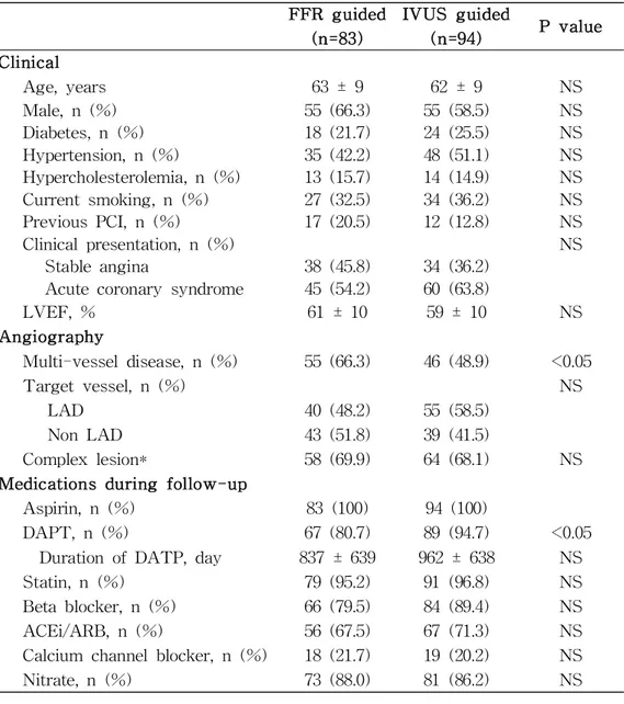
The baseline clinical characteristics are summarized in Table 1. The baseline clinical characteristics were similar between the two groups.
A higher frequency of hypertension in the IVUS-guided group (42.2%
vs. 51.1%, p=0.29) and a previous PCI in the FFR-guided group (20.5%
vs. 12.8%, p=0.22) were observed. However, it was not statistically significant. In both groups, the rate of acute coronary syndrome was higher than that of stable angina. All patients were prescribed aspirin.
The frequency of dual anti-platelet therapy (DAPT) prescriptions was significantly lower in the FFR-guided group than IVUS-guided group (80.7% vs. 94.7, p=0.01). The duration of the DAPT and other medications did not differ between the two groups.
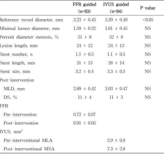
3.2. QCA Characteristics:
Table 1 and 2 shows the coronary angiographic and QCA characteristics
in both groups. Angiographic characteristics were similar between the
two groups except the incidence of multivessel disease and RVD. In the
FFR group, the incidence of multivessel disease was significantly higher
(66.3% vs. 48.9%, p=0.02). The percent diameter stenosis between the two
groups was similar (51 ± 8% vs. 52 ± 8% in the FFR and IVUS groups
,p=0.53). But RVD was larger in the IVUS-guided group (3.23 ± 0.43
mm vs. 3.39 ± 0.49 mm p=0.03). The frequencies of proximal lesions
and middle lesions were similar, and the frequency of complex lesions
was higher than that of simple lesions in both groups. But it was not statistically significant. Other angiographic and QCA characteristics were not different between the 2 groups.
3.3. IVUS and FFR Characteristics:
Table 2 shows the procedural analysis results between the 2 groups.
In this study, a total of 114 (64.4%) DES were inserted in 177 lesions.
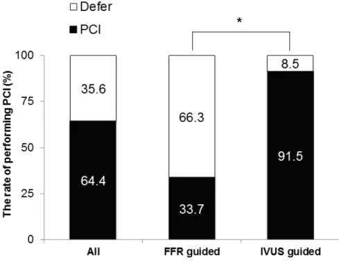
In the FFR-guided group, the incidence of performing PCI was significantly lower than that in the IVUS-guided group (33.7% vs.
91.5%, p<0.001) (Figure 1). The stent number, length and size were similar between the 2 groups. The pre-intervention mean value of the FFR was 0.82 and the post-intervention mean value of the FFR was 0.91. In the IVUS-guided group, the pre-intervention MLA was 2.9 ± 1.0 mm
2and post-intervention minimal stent area was 7.3 ± 2.8 mm
2. The post-intervention MLD and percent diameter stenosis did not differ between the 2 groups (2.89 ± 0.42 mm vs. 3.03 ± 0.47 mm, p=0.19; 11 ± 4% vs. 11 ± 3%, p=0.45).
3.4. Clinical Outcomes:
One hundred fifty-one (90.4%) of 167 patients had a completely
5-year follow-up. The number of patients lost to follow-up was
similarly between the 2 groups (p=0.98). Seven patients in the
FFR-guided group and 9 in the IVUS-guided group were lost to
follow-up due to death. Four cardiac deaths and 3 non-cardiac deaths
occurred in the FFR-guided group. Six cardiac deaths and 3 non-cardiac
deaths occurred in the IVUS-guided group.
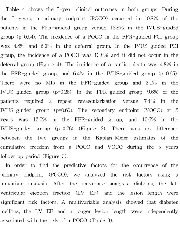
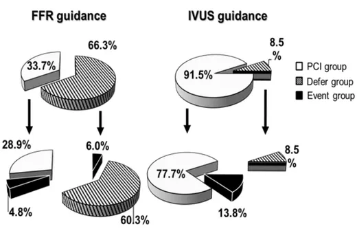
Table 4 shows the 5-year clinical outcomes in both groups. During the 5 years, a primary endpoint (POCO) occurred in 10.8% of the patients in the FFR-guided group versus 13.8% in the IVUS-guided group (p=0.54). The incidence of a POCO in the FFR-guided PCI group was 4.8% and 6.0% in the deferral group. In the IVUS-guided PCI group, the incidence of a POCO was 13.8% and it did not occur in the deferral group (Figure 4). The incidence of a cardiac death was 4.8% in the FFR-guided group, and 6.4% in the IVUS-guided group (p=0.65).
There were no MIs in the FFR-guided group and 2.1% in the IVUS-guided group (p=0.28). In the FFR-guided group, 9.6% of the patients required a repeat revascularization versus 7.4% in the IVUS-guided group (p=0.60). The secondary endpoint (VOCO) at 5 years was 12.0% in the FFR-guided group, and 10.6% in the IVUS-guided group (p=0.76) (Figure 2). There was no difference between the two groups in the Kaplan-Meier estimates of the cumulative freedom from a POCO and VOCO during the 5 years follow-up period (Figure 3).
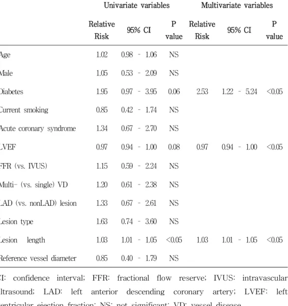
In order to find the predictive factors for the occurrence of the
primary endpoint (POCO), we analyzed the risk factors using a
univariate analysis. After the univariate analysis, diabetes, the left
ventricular ejection fraction (LV EF), and the lesion length were
significant risk factors. A multivariable analysis showed that diabetes
mellitus, the LV EF and a longer lesion length were independently
associated with the risk of a POCO (Table 3).
Table 1. Baseline and Angiographic Characteristics
FFR guided(n=83)
IVUS guided
(n=94) P value
Clinical
Age, years 63 ± 9 62 ± 9 NS
Male, n (%) 55 (66.3) 55 (58.5) NS
Diabetes, n (%) 18 (21.7) 24 (25.5) NS
Hypertension, n (%) 35 (42.2) 48 (51.1) NS
Hypercholesterolemia, n (%) 13 (15.7) 14 (14.9) NS
Current smoking, n (%) 27 (32.5) 34 (36.2) NS
Previous PCI, n (%) 17 (20.5) 12 (12.8) NS
Clinical presentation, n (%) NS
Stable angina 38 (45.8) 34 (36.2)
Acute coronary syndrome 45 (54.2) 60 (63.8)
LVEF, % 61 ± 10 59 ± 10 NS
Angiography
Multi-vessel disease, n (%) 55 (66.3) 46 (48.9) <0.05
Target vessel, n (%) NS
LAD 40 (48.2) 55 (58.5)
Non LAD 43 (51.8) 39 (41.5)
Complex lesion* 58 (69.9) 64 (68.1) NS
Medications during follow-up
Aspirin, n (%) 83 (100) 94 (100)
DAPT, n (%) 67 (80.7) 89 (94.7) <0.05
Duration of DATP, day 837 ± 639 962 ± 638 NS
Statin, n (%) 79 (95.2) 91 (96.8) NS
Beta blocker, n (%) 66 (79.5) 84 (89.4) NS
ACEi/ARB, n (%) 56 (67.5) 67 (71.3) NS
Calcium channel blocker, n (%) 18 (21.7) 19 (20.2) NS
Nitrate, n (%) 73 (88.0) 81 (86.2) NS
* According to the Ameican College of Cardiology/American Heart Association classification, type B2 and C lesions as complex lesions. ACEi:
angiotensin-converting-enzyme inhibitor; ARB: angiotensin II receptor blocker;
DAPT: dual antiplatelet therapy; FFR: fractional flow reserve; IVUS:
intravascular ultrasound; LAD: left anterior descending coronary artery; LVEF:
left ventricular ejection fraction; NS: not significant; PCI: percutaneous coronary intervention.
Table 2. Quantitative Coronary Analysis Characteristics
FFR guided(n=83)
IVUS guided
(n=94) P value
Reference vessel diameter, mm 3.23 ± 0.43 3.39 ± 0.49 <0.05 Minimal lumen diameter, mm 1.59 ± 0.32 1.61 ± 0.45 NS
Percent diameter stenosis, % 51 ± 8 52 ± 8 NS
Lesion length, mm 24 ± 12 24 ± 13 NS
Stent number, n 1.1 ± 0.5 1.1 ± 0.5 NS
Stent length, mm 31 ± 13 28 ± 14 NS
Stent size, mm 3.2 ± 0.4 3.3 ± 0.5 NS
Post-intervention
MLD, mm 2.89 ± 0.42 3.03 ± 0.47 NS
DS, % 11 ± 4 11 ± 3 NS
FFR
Pre-intervention 0.72 ± 0.07
Post-intervention 0.91 ± 0.05 IVUS, mm2
Pre-interventional MLA 2.9 ± 0.9
Post-interventional MSA 7.3 ± 2.8
DS: diameter stenosis; FFR: fractional flow reserve; IVUS: intravascular ultrasound; MLA: minimal lumen diameter; MLD: minimal lumen diameter;
MSA: minimal stent area; NS: not significant.
Table 3. Predictors of the Patient Oriented Clinical Outcome in Intermediate Coronary Lesions
Univariate variables Multivariate variables Relative
Risk 95% CI P
value
Relative
Risk 95% CI P
value
Age 1.02 0.98 – 1.06 NS
Male 1.05 0.53 – 2.09 NS
Diabetes 1.95 0.97 – 3.95 0.06 2.53 1.22 – 5.24 <0.05 Current smoking 0.85 0.42 – 1.74 NS
Acute coronary syndrome 1.34 0.67 – 2.70 NS
LVEF 0.97 0.94 – 1.00 0.08 0.97 0.94 – 1.00 <0.05 FFR (vs. IVUS) 1.15 0.59 – 2.24 NS
Multi- (vs. single) VD 1.20 0.61 – 2.38 NS LAD (vs. nonLAD) lesion 1.33 0.67 – 2.61 NS
Lesion type 1.63 0.74 – 3.60 NS
Lesion length 1.03 1.01 – 1.05 <0.05 1.03 1.01 – 1.05 <0.05 Reference vessel diameter 0.85 0.40 – 1.79 NS
CI: confidence interval; FFR: fractional flow reserve; IVUS: intravascular ultrasound; LAD: left anterior descending coronary artery; LVEF: left ventricular ejection fraction; NS: not significant; VD: vessel disease.
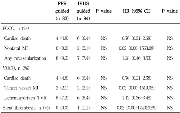
Table 4. 5-year Clinical Outcomes According to the Guided Modality
FFRguided (n=83)
IVUS guided (n=94)
P value HR (95% CI) P value
POCO, n (%)
Cardiac death 4 (4.8) 6 (6.4) NS 0.76 (0.21-2.68) NS Nonfatal MI 0 (0.0) 2 (2.1) NS 0.02 (0.00-1563.08) NS Any revascularization 8 (9.6) 7 (7.4) NS 1.28 (0.46-3.53) NS VOCO, n (%)
Cardiac death 4 (4.8) 6 (6.4) NS 0.76 (0.21-2.68) NS Target vessel MI 2 (2.1) 2 (2.1) NS 0.02 (0.00-1519.35) NS Ischemia-driven TVR 6 (7.2) 6 (6.4) NS 1.12 (0.36-3.48) NS Stent thrombosis, n (%) 0 (0.0) 1 (1.1) NS 0.02 (0.00-174013.89) NS CI: confidence interval; FFR: fractional flow reserve; HR: hazard ratio; IVUS:
intravascular ultrasound; MI: myocardial infarction; NS: not significant; POCO:
patient oriented clinical outcome; TVR: target vessel revascularization; VOCO:
vessel oriented clinical outcome.
Figure 1. The rate of performing PCI according to type of guiding
device. The FFR-guided group had significantly lower rates
of performing PCI than the IVUS-guided group. * The P value
was < 0.001. FFR: fractional flow reserve; IVUS: intravascular
ultrasound; PCI: percutaneous coronary intervention.
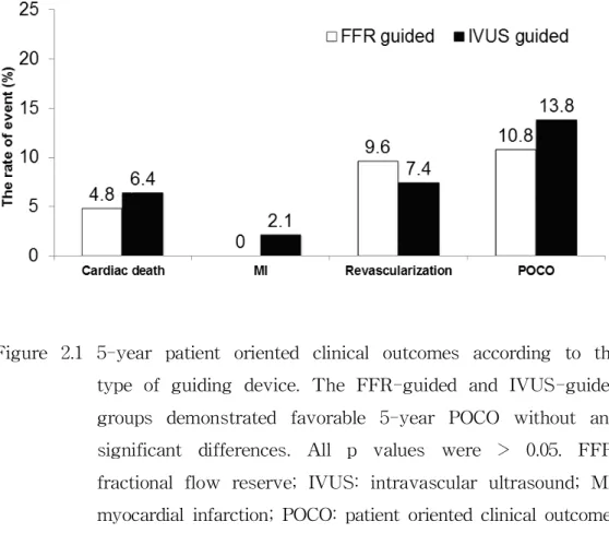
Figure 2.1 5-year patient oriented clinical outcomes according to the type of guiding device. The FFR-guided and IVUS-guided groups demonstrated favorable 5-year POCO without any significant differences. All p values were > 0.05. FFR:
fractional flow reserve; IVUS: intravascular ultrasound; MI:
myocardial infarction; POCO: patient oriented clinical outcome.
Figure 2.2 5-year vessel oriented clinical outcomes according to the type of guiding device. The FFR-guided and IVUS-guided groups demonstrated favorable 5-year VOCO without any significant differences. All P values were > 0.05. FFR: fractional flow reserve; ID-TVR: ischemia driven target vessel revascularization;
IVUS: intravascular ultrasound; MI: myocardial infarction;
TVMI: target vessel myocardial infarction; VOCO: vessel
oriented clinical outcome.
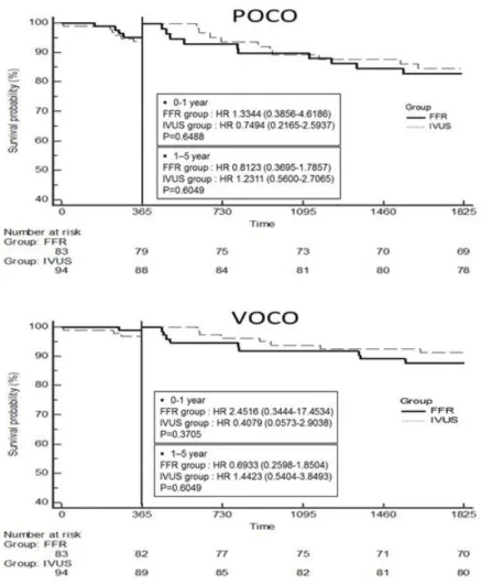
Figure 3. Kaplan-Meier survival curves for the freedom from adverse cardiac events during 5 years of follow-up for both groups.
There was no difference between the two groups in the Kaplan-Meier estimates of the cumulative freedom from a POCO and VOCO. All P value was > 0.05. FFR: fractional flow reserve; HR: hazard ratio; IVUS: intravascular ultrasound;
POCO: patient oriented clinical outcome; VOCO: vessel oriented
clinical outcome.
Figure 4. 5-year patient oriented clinical outcome rate according to the guided modality and performing PCI. There was no difference between the four groups in POCO according to the guided modality and performing PCI. P value was > 0.05. FFR:
fractional flow reserve; IVUS: intravascular ultrasound; POCO:
patient oriented clinical outcome.
4. Discussion
A previous study showed that both an FFR- and IVUS-guided PCI in patients with intermediate coronary lesions were associated with favorable clinical outcomes at 1 year and the FFR-guided PCI reduced the need of revascularization of those lesions as compared to the IVUS-guided group. The 5-year follow-up results showed that the long-term clinical outcome of an FFR- or IVUS-guided PCI in intermediate coronary disease was still favorable and there were no significant differences in any of the clinical outcomes between the two groups.
FFR and IVUS are known to play an important role in the treatment decision of coronary intervention. Furthermore, previous studies have shown good clinical outcomes of PCI guided by these two modalities.
The FAME (Fractional Flow Reserve versus Angiography for
Multivessel Evaluation) study showed that the major adverse cardiac
events were significantly lower in the FFR-guided group than in the
angiography-guided PCI group at 1 year (4). Moreover, at 2 years, an
ongoing favorable outcome was noted with significantly lower rates of
death and myocardial infarction in the FFR-guided group (11). At 5
years of follow up, the long-term safety of the FFR-guided PCI with
multivessel disease was confirmed (12). Also, several meta-analyses
studies have shown that an IVUS-guided PCI is associated with a
significant reduction in the major adverse cardiovascular events
compared to an angiography-guided PCI for both bare metal stents and
DES (13-18). Furthermore, a recent randomized multicenter trial showed
that the use of IVUS resulted in a significantly lower rate of 1-year
coronary lesions (19,20). The result of our study also demonstrated that both FFR and IVUS had a good long-term efficacy in intermediate coronary lesions that were difficult to make a clinical decision. In addition, FFR-guided PCI had good results even with a low stent implantation rate compared with IVUS-guided PCI, suggesting that FFR may be a more efficient modality in intermediate coronary lesions. It can be assumed that optimal medical therapy played an important role during the long-term follow-up period. The Clinical Outcomes Utilizing Revascularization and Aggressive Drug Evaluation (COURAGE) trial (21) showed that the long-term clinical cardiovascular outcomes (death, MIs and hospitalization for acute coronary syndrome) in patients with stable angina did not differ between the optimal medical treatment group and PCI plus optimal medical treatment group. Further, in the ORBITA trial (22) comparing the difference in the exercise time increment between patients who underwent a PCI and placebo procedure with severe (> 70%) single vessel stenosis, there was no difference between the two groups and the importance of medical treatment was emphasized. In this study, aspirin was prescribed in all patients, and 165 (93.2%) patients had a dual antiplatelet treatment. Statin regimens were prescribed in 96.0%, nitrate-based drugs in 87.0%, beta-blockers in 84.7%, ACE inhibitors or angiotensin receptor antagonists in 69.5% and calcium channel blocker in 20.9%. There was no difference except for the dual antiplatelet medications in the comparison of the two groups. In the FFR-guided PCI group, the prescription rate of a dual antiplatelet medication was lower than that in the IVUS-guided PCI group (80.7%
vs. 94.7%, p=0.01). Both groups had a high prescription rate, but the
statistical difference between the two groups was considered to be
related to the lower rate of a stent insertion in the FFR-guided PCI
group.
By a multivariate regression analysis, the occurrence of a POCO was more frequent in patients with diabetes, a lower LV EF, or those with a long lesion. The association of diabetes with coronary artery disease (CAD) is well established. CAD is the main cause of death in diabetic patients, and diabetes is associated with a 2 to 4-fold increased mortality risk from heart disease (23). Our study also showed that diabetes was the strongest predictor of a poor cardiovascular outcome in intermediate coronary disease. The relationship between the LV EF and poor clinical outcomes has been widely studied and the LV EF has proven to be a potent prognostic factor in CAD patients (24,25).
However, in this study, most patients were in the normal range of the LV EF, so it seemed to be a less powerful relative risk of a POCO than in the other studies. Nevertheless, the LV EF must be an important factor in the patients. Also the lesion length is known to be a risk factor of in-stent restenosis (26). Percutaneous intervention of long coronary lesions has been associated with poorer outcomes than that of focal lesions because long lesions often require multiple stent implantation which leads to more extensive vascular injury and has been associated with an increased risk of stent thrombosis and restenosis (27-28). There was no difference between the FFR-guided PCI and IVUS-guided PCI in terms of the occurrence of a POCO. In this study, the prognosis of both modalities was good because of intermediate disease. It is also important to note that the more deferred FFR-guided PCI group had good results.
The present study had several limitations. First, it was not designed
for a follow-up of 5 years. There were several different baseline
characteristics and the choice of FFR and IVUS was in accordance with
the decision of the physician and the choice of medications during the
the two modalities with the same patients. Third, the number of patients
was small. And thus, the statistical power was weak. Therefore, to
compensate for these weaknesses, a larger randomized control head to
head study with a long-term follow-up will be needed to confirm the
results.
5. Summary
During the 5-year follow-up period, both the FFR- and IVUS-guided
PCI for intermediate coronary artery disease were associated with
favorable outcomes. Both FFR and IVUS are useful additional tests for
determining the PCI in patients with intermediate coronary lesions.
References
1. Topol EJ, Nissen SE: The dissociation between clinical and angiographic findings in ischemic heart disease. Circulation 1995; 92: 2333-42.
2. Pijls NH, De Bruyne B, Peels K, Van Der Voort PH, Bonnier HJ, Bartunek JKJJ, et al.: Measurement of fractional flow reserve to assess the functional severity of coronary-artery stenoses. N Engl J Med 1996; 334: 1703-8.
3. Pijls NH, van Schaardenburgh P, Manoharan G, Boersma E, Bech JW, van't Veer M, et al.: Percutaneous coronary intervention of functionally nonsignificant stenosis: 5-year follow-up of the DEFER Study. J Am Coll Cardiol 2007; 49: 2105-11.
4. Tonino PA, De Bruyne B, Pijls NH, Siebert U, Ikeno F, van' t Veer M, et al.: Fractional flow reserve versus angiography for guiding percutaneous coronary intervention. N Engl J Med 2009; 360: 213-24.
5. Briguori C, Anzuini A, Airoldi F, Gimelli G, Nishida T, Adamian M, et al.: Intravascular ultrasound criteria for the assessment of the functional significance of intermediate coronary artery stenoses and comparison with fractional flow reserve. Am J Cardiol 2001; 87:
136-41.
6. Nam CW, Yoon HJ, Cho YK, Park HS, Kim H, Hur SH, et al.:
Outcomes of percutaneous coronary intervention in intermediate coronary
artery disease: fractional flow reserve-guided versus intravascular
ultrasound-guided. J Am Coll Cardiol Intv 2010; 3: 812-7.
7. Cutlip DE, Windecker S, Mehran R, Boam A, Cohen DJ, van Es GA, et al.: Clinical endpoints in coronary stent trials: a case for standardized definitions. Circulation 2007; 115: 2344-51.
8. Thygesen K, Alpert JS, White HD, Jaffe AS, Apple FS, Galvani M, et al.: Universal definition of myocardial infarction. Circulation 2007;
116: 2634-53.
9. Manel S, Angel C, Andres I, Antonio S, Rosa-Ana H-A, Vicente M, et al.: Rationale and design of the EXAMINATION trial: a randomised comparison between everolimus-eluting stents and cobalt-chromium bare-metal stents in ST-elevation myocardial infarction. EuroIntervention 2011; 7: 977-84.
10. Mintz GS, Nissen SE, Anderson WD, Bailey SR, Erbel R, Fitzgerald PJ, et al.: American college of cardiology clinical expert consensus document on standards for acquisition, measurement and reporting of intravascular ultrasound studies (IVUS). A report of the american college of cardiology task force on clinical expert consensus documents.
J Am Coll Cardiol 2001; 37: 1478-92.
11. Pijls NH, Fearon WF, Tonino PAL, Siebert U, Ikeno F, Bornschein
B, et al.: Fractional flow reserve versus angiography for guiding
percutaneous coronary intervention in patients with multivessel
coronary artery disease: 2-year follow-up of the FAME (fractional
flow reserve versus angiography for multivessel evaluation) study. J
12. Van Nunen LX, Zimmermann FM, Tonino PA, Barbato E, Baumbach A, Engstrom T, et al.: Fractional flow reserve versus angiography for guidance of PCI in patients with multivessel coronary artery disease (FAME): 5-year follow-up of a randomised controlled trial.
Lancet 2015; 386: 1853-60.
13. Zhang Y, Farooq V, Garcia-Garcia HM, Bourantas CV, Tian N, Dong S, et al.: Comparison of intravascular ultrasound versus angiography-guided drug-eluting stent implantation: a meta-analysis of one randomised trial and ten observational studies involving 19,619 patients. EuroIntervention 2012; 8: 855-65.
14. Klersy C, Ferlini M, Raisaro A, Scotti V, Balduini A, Curti M, et al.: Use of IVUS guided coronary stenting with drug eluting stent: a systematic review and meta-analysis of randomized controlled clinical trials and high quality observational studies. Int J Cardiol 2013; 170:
54-63.
15. Jang JS, Song YJ, Kang W, Jin HY, Seo JS, Yang TH, et al.:
Intravascular ultrasound-guided implantation of drug-eluting stents to improve outcome: a meta-analysis. J Am Coll Cardiol Intv 2014;
7: 233-43.
16. Ahn JM, Kang SJ, Yoon SH, Park HW, Kang SM, Lee JY, et al.:
Meta-analysis of outcomes after intravascular ultrasound-guided versus
angiography-guided drug-eluting stent implantation in 26,503 patients
enrolled in three randomized trials and 14 observational studies. Am
J Cardiol 2014; 113: 1338-47.
17. Casella G, Klauss V, Ottani F, Siebert U, Sangiorgio P, Bracchetti D: Impact of intravascular ultrasound-guided stenting on long-term clinical outcome: a meta-analysis of available studies comparing intravascular ultrasound-guided and angiographically guided stenting.
Catheterization and cardiovascular interventions 2003; 59: 314-21.
18. Parise H, Maehara A, Stone GW, Leon MB, Mintz GS: Meta-analysis of randomized studies comparing intravascular ultrasound versus angiographic guidance of percutaneous coronary intervention in pre-drug-eluting stent era. Am J Cardiol 2011; 107: 374-82.
19. Kim BK, Shin DH, Hong MK, Park HS, Rha SW, Mintz GS, et al.:
Clinical impact of intravascular ultrasound-guided chronic total occlusion intervention with zotarolimus-eluting versus biolimus-eluting stent implantation: randomized study. Circ Cardiovasc Interv 2015; 8: e002592.
20. Hong SJ, Kim BK, Shin DH, Nam CM, Kim JS, Ko YG, et al.:
Effect of intravascular ultrasound-guided versus angiography-guided everolimus-eluting stent implantation: the IVUS-XPL randomized clinical trial. JAMA 2015; 314: 2155-63.
21. Boden WE, O'Rourke RA, Teo KK, Hartigan PM, Maron DJ, Kostuk WJ, et al.: Optimal medical therapy with or without PCI for stable coronary disease. N Eng J Med 2007; 356: 1503-16.
22. Al-Lamm R, Thompson D, Dehbi HM, Sen S, Tang K, Davies J, et
al.: Percutaneous coronary intervention in stable angina (ORBITA): a
double-blind, randomised controlled trial. Lancet 2018; 391: 31-40.
23. Berry C, Tardif JC, Bourassa MG: Coronary heart disease in patients with diabetes: part II: recent advances in coronary revascularization. J Am Coll Cardiol 2007; 49: 643-56.
24. Holmes DR, Detre KM, Williams DO, Kent KM, King SB, Yeh W, et al.: Long-term outcome of patients with depressed left ventricular function undergoing percutaneous transluminal coronary angioplasty:
the NHLBI PTCA registry. Circulation 1993; 87: 21-9.
25. Germano DS, Giuseppe P, Andrea DA, Annunziata N: Coronary stenting in patients with depressed left ventricular function: acute and long-term results in a selected population. Catheterization and Cardiovascular Interventions 2003; 59: 429-33.
26. Kasaoka S, Tobis JM, Akiyama T, Reimers B, Di Mario C, Wong ND, et al.: Angiographic and intravascular ultrasound predictors of in-stent restenosis. J Am Coll Cardiol 1998; 32: 1630-5.
27. Kastrati A, Elezi S, Dirschinger J, Hadamitzky M, Neumann F-J, Schömig A: Influence of lesion length on restenosis after coronary stent placement. Am J Cardiol 1999; 83: 1617-22.
28. Guagliumi G, Ikejima H, Sirbu V, Bezerra H, Musumeci G, Lortkipanidze
N, et al.: Impact of drug release kinetics on vascular response to
different zotarolimus-eluting stents implanted in patients with long
coronary stenoses: the LongOCT study (optical coherence tomography
in long lesions). J Am Coll Cardiol Intv 2011; 4: 778-85.
5-year Outcomes of Fractional Flow Reserve-Guided versus Intravascular Ultrasound-Guided Percutaneous
Coronary Intervention in Intermediate Coronary Artery Disease
Choi, Sang Woong Department of Internal Medicine
Graduate School Keimyung University
(Supervised by Professor Nam, Chang Wook)
Both fractional flow reserve (FFR)- and intravascular ultrasound
(IVUS)-guided percutaneous coronary intervention (PCI) strategies were
reported to be safe and effective in intermediate coronary lesions for up
to 1 year of follow-up. This study aimed to investigate whether the
favorable clinical outcomes of the lesions persisted over a 5-year
follow-up. One hundred sixty seven patients, with intermediate coronary
lesions evaluated by FFR or IVUS (FFR guided, 83 lesions vs. IVUS
guided, 94 lesions), were included. The primary end-point was the
patient oriented clinical outcome (POCO), defined as a composite of
cardiac death, nonfatal myocardial infarction (MI), and any revascularization.
(VOCO), defined as a composite of cardiac death, target vessel MI and
ischemia-driven target vessel revascularization. The baseline characteristics
were similar except for more multi-vessel disease and a smaller vessel
diameter in the FFR group. The POCO at 5 years was 12.4% (FFR
10.8% vs IVUS 13.8%, p=ns) and VOCO 11.3% (FFR 12.0% vs IVUS
10.6%, p=ns). The Kaplan-Meier survival analysis of the POCO and
VOCO was similar between the two groups. Both the FFR guided and
IVUS guided PCI strategy for intermediate coronary artery disease were
associated with favorable 5-year clinical outcomes.
중간단계의 관상동맥 협착질환에서 분획 혈류 예비력 유도하 경피적 관상 동맥 중재술과 관상동맥 혈관내 초음파
유도하 경피적 관상동맥 중재술의 5년 임상결과 비교
최 상 웅 계명대학교 대학원 의학과 내과학 전공
(지도교수 남 창 욱)