Ⅰ. 서 론
구강 편평세포암(Oral Squamous cell carcinoma)의 발 생율은 전 세계적으로 점점 높아지고 있다. 최근의 보고에 의하면 미국에서 매년 약 36,000례의 새로운 구강 편평세 포암이 발생하고 20,000명 정도가 이로 인해 사망한다1). 구강 편평세포암의 발생과정에 대한 광범위한 연구와 치료 방법의 개선에도 불구하고, 5년 생존율은 약 53%에 불과하 며 이 수치는 25년째 큰 차이가 없다. 구강 편평세포암의 특징은 국소적 침습과 경부 임파절 전이와 원격전이는 드문
편으로 보고되고 있다. 사망하는 환자의 대부분은 국소적 파괴와 경부 임파절 전이로 인한 것이다. 그러므로 국소적 재발을 감소시키거나 저해하는 술식은 구강 편평세포암의 처치 효율의 제고와 생존율 향상에 큰 도움이 될 수 있다2).
종양세포의 침습과 전이는 세포의 구조변화를 매개하는 단백질분자에 의해 세포와 세포사이 그리고 세포와 기질과 의 부착이 조절됨으로써 세포의 탈락과 부착이 반복되는 복 잡한 단계를 거쳐 일어난다. 종양 조직에서의 세포부착분자 감소는 종양세포의 탈분화를 유발하고, 세포 부착과 극성 배열의 소실로 세포와 세포간, 세포와 기저막간의 부착상 표성운∙이광배∙김영실*∙이상화
가톨릭대학교 의과대학 치과학교실 구강악안면외과, *제주대학교 의과대학 병리학교실
구강 편평세포암종에서 E-cadherin과 β -catenin의 발현과 임상병리학적 특징
EXPRESSION OF E-CADHERIN AND
β-CATENIN IN RELATION TO CLINICOPATHOLOGIC FEATURES IN ORAL SQUAMOUS CELL CARCINOMA
Sung-Woon Pyo, Kwang-Bae Lee, Young-Sill Kim * , Sang-Hwa Lee
Department of Oral and Maxillofacial Surgery, College of Medicine, The Catholic University of Korea,
* Department of Pathology, College of Medicine, Cheju National University
Changes in cell adhesion molecules are associated with infiltration and metastatic progression of cancer.
Reduced expression of E-cadherin and β-catenin complex in some carcinomas has been reported. The changes in the expression in oral squamous cell carcinoma (OSCC) is not fully understood and it also remains undetermined whether the expression of these adhesion molecules in metastatic lesions differs from that in the primary lesions.
In the present study, therefore, we immunohistochemically examined the expression of E-cadherin and β- catenin in 45 primary OSCCs and 19 metastatic lymph nodes. We compared the expression of these mole- cules between primary and metastatic lesions and investigated the correlation between the expression and clinicopathologic parameters. The expression of E-cadherin and β-catenin was reduced in 35/45 (78.2%), 14/45 (31.2%) of primary tumors respectively, but 18/19 (94.7%) and 17/19 (89.4%) of lymph nodes showed preserved expression. The reduced expression of the E-cadherin was associated with lymph node metastasis, invasive mode and marginal status but no significant relationship was not found with β- catenin.
In conclusion, the loss of E-cadherin and β-catenin complex function is associated with progression of OSCC and suggest that the expression of this complex will be a supplementary prognostic tool.
Key words: Oral squamous cell carcinoma, E-cadherin, β-catenin, Metastasis, Prognosis
Abstract
실로 침습성을 갖게 되므로 종양에서 세포부착분자의 소실 은 침윤과 전이에 중요한 역할을 하는 것으로 알려져 있다3).
Cadherin은 세포 표면의 당단백으로서 칼슘의존성 접착 물질로 작용하며 태아의 발생과 성인의 조직 구조 유지에 주된 역할을 한다4,5). 세포간 인지, 부착, 신호전달과정에서 cadherin의 역할이 중요하여, E-, N-와 P-cadherin이 암 의 발생과 진전과정에서 미치는 영향에 대한 많은 연구가 있었다6-8). E-cadherin은 주로 상피세포에서 발현되는 세 포부착 단백질로 침습과 종양억제인자로 받아들여지고 있 다. E-cad의 발현 감소는 구강 편평세포암을 포함한 상피 성 악성종양에서 세포 접착성의 감소와 악성도의 증가와 관 련되어 있다. 특히 구강 편평세포암에서 E-cadherin의 down-regulation은 매우 의미 있는 재발과 생존이 관련된 예후 인자이다. 본 연구진의 최근 연구에 의하면 E-cad- herin의 표현감소가 임파절 전이와 재발 양상에 유의한 의 미를 갖고 있다고 밝혀진바 있다9).
Catenin은 α- (102 kDa), β- (88 kDa)와 γ-catenin (82 kDa)의 3종으로 구성된 단백질로, β-와 γ-catenin은 e-cadherin의 세포내 도메인과 직접 결합하여 세포내 골격 물질인 actin을 연결하고 α-catenin은 E-cadherin과 β-, γ-catenin complex를 actin과 연결한다. 그러므로 E-cad- herin의 부착능력은 catenin system의 완전성 여부에 따 라 달려있다고 할 수 있다. 앞에서 언급한 바와 같이 E- cadherin에 의해 유도되는 세포간 부착능력의 유지가 구강 암의 침습 및 전이와 관계가 있음이 밝혀진 바 있으며, 다른 많은 암에서도 catenin의 발현이 저하되었음이 보고되고 있다10-13).
아직까지는 구강암에서의 E-cadherin-catenin system 의 발현 변화 여부가 현재 인정되고 있는 다른 예후 인자와 의 상관관계나 생존율에 끼치는 영향에 관한 연구는 많지 않다14-16).
이에 저자들은 구강 편평세포암으로 진단받고 외과적 절 제술을 받은 환자의 파라핀 포매 조직을 이용하여 면역 조 직화학적 염색방법으로 E-cadherin과 β-catenin의 발현정 도를 관찰하여 이를 구강 편평세포암종의 조직학적 분화도, 침습의 형태, 외과적 절제술의 변연의 침범여부, 임파절 전 이의 유무 및 임상병기와의 상관관계를 조사하여 예후 인자 로서의 유용성을 살펴보고자 한다.
Ⅱ. 연구 재료 및 방법 1. 연구 재료
구강과 구인두부위의 원발성 편평세포암종을 가진 50명 의 환자로부터 50개의 원발성 부위의 종양과 22개의 임파 절 전이 조직을 얻어 통상적인 방법으로 포르말린으로 고정
하고, 파라핀으로 포매된 절편을 이용하였다. 임상 자료와 추적 자료로써 나이, 성별, 흡연, 음주 습관, 종양의 원발 부 위, UICC의 분류에 따른 임상적, 병리학적 TNM 병기, 종 양의 조직 병리학적 단계 (WHO에 따른 분류)등을 조사하 였다.
2. 면역조직화학적 연구
발생된 종양부위와 임파절을 파라핀으로 포매하였다.
4 μm두께의 절편을 얻어 슬라이드에 부착하고 xylene으로 15분 동안 파라핀을 제거하고, 3단계의 에탄올 (100%, 95%, 80%)에 각각 담가 xylene을 제거하고 탈수하였다.
내인성 peroxidase의 작용을 막기 위해 조직 절편을 실온 에서 0.3 % 과산화수소에 30분간 반응시켰다. Tris- buffered saline (TBS, pH 7.6)로 수세한 후 항원 수득률 을 높이기 위해 조직 절편을 citrate buffer (pH 6.0)에 담 아 2100 Retriever (PickCell Laboratories B.V.
Amsterdam, Netherlands)에 120℃에서 8분 동안 배양 하였다. 비특이적 결합은 희석된 normal horse serum에 30분 동안 배양함으로써 배제하였다. 특이적 면역반응의 관찰을 위해 제작한 단일클론항체로 절편을 4℃에서 12시 간동안 배양한 후에 biotinylated anti-mouse 2차항체 (Jackson laboratory, MA, USA)로 30분간 적용 후, 완 충액으로 수세하고 ABC complex로 다시 30분간 배양하였 다. 마지막 발색 과정으로 조직 절편을 2분간 3,3’- diaminobenzidine and hydrogen peroxide (DAB Substrate Kit, Vector, Burlingame, CA. USA)로 발색 한 후, Mayer’s haematoxylin (Fisher Scientific, Pittsburgh, PA. USA)으로 대조염색하고, 탈수한 조직절 편을 Permount (Fisher Scientific, Pittsburgh, PA.
USA)로 밀봉하였다.
3. E-cadherin과 β-catenin의 발현에 대한 결과 판독
정상 구강점막의 E-cadherin과 β-catenin의 발현을 외부 양성 대조군으로 사용하였고, 각 조직 절편의 인접 정상 상 피를 내부 양성 대조군으로 사용하였다. 음성 대조군으로는 일차 항체를 생략하였다. 염색된 조직절편은 임상적 정보가 전혀 없는 병리의사가 반정량적으로 판독하였다. 조직 절편 들은 종양의 침범부위 가장자리에서 세포막이 강하게 염색 된 종양세포의 비율에 따라 분류하였다. 약술하면 score 0:
정상 발현이 5% 미만일 경우, score 1: 5-25%인 경우, score 2: 26-50%인 경우, score 3: 51-75%인 경우 그리 고 score 4: 76-100%인 경우로 세분하였다.
종양세포의 염색결과가 score 4를 보일 때 발현의 보존 (preserved)으로 정의하고, 종양세포가 score 1, 2, 3을 보
일 때 발현의 감소 (reduced)로 정의하였다.
4. 통계 분석
E-cadherin과 β-catenin의 발현정도에 따른 임상, 병리 적 척도와의 연관성은 Chi-Square test를 사용하였고 Fisher’s exact test로 검증하였고, 유의수준은 P값이 0.05이하일 경우로 하였다.
Ⅲ. 연구 결과 1. 연구 대상의 임상병리학적 특징
50명의 원발성 구강 편평세포암종을 가진 환자로부터 50 개의 원발성 종양과 22개의 임파절 전이 조직을 얻어 통상 적인 방법으로 포르말린으로 고정하고, 파라핀으로 포매된 절편을 이용하였다. 그러나 원발부위와 전이된 임파절에서
동시에 염색이 불가능하였던 원발성 부위 5개와 임파절 3 개의 증례는 연구에서 제외하였다. Table 1에서 환자의 임 상병리학적 특징을 정리하였다.
2. E-cadherin과 β-catenin 발현 양상 (Fig. 1-4)
정상 구강점막세포의 세포막에서 E-cadherin과 β- catenin의 발현은 기저층에서 극세포층에 이르기까지 관찰 되었다. 발현이 유지되는 경우에는 세포막에 분명하고 균질 하게 분포된 것을 관찰할 수 있었으나, 발현이 감소된 경우 에는 비균일한 염색소견과 양성 세포와 음성세포가 혼재된 양상을 나타내었다. 특히 종양세포에서는 이들 단백질의 발 현이 세포막 뿐 아니라 세포질 내와 세포핵 내에서도 관찰 되었다. 특히 β-catenin의 세포핵 내에서의 발현은 세포질 내에서 발현과 이에 따른 세포막에서의 발현 감소와 동반하 는 예가 관찰되었다. 종양 조직 내에서 cadherin과 catenin의 발현의 분포는 증례마다 달랐으며 심지어 같은 증례에서도 다른 분포의 형태를 보이는 경우가 있었다. 대 표적인 면역조직화학적 염색 소견은 그림과 같다. 모두 45 건의 증례 중에서 원발 부위에서 E-cadherin과 β-catenin 의 발현 감소는 각각 35예 (78.2%), 14예 (31.2%)이었 고, 임파절 전이 조직 19례 중 E-cadherin과 β-catenin의 발현 감소는 각각 1예 (6.3%), 2예 (10.6%)이었다(Table 2).
3. 임상병리학적 지표와 E-cadherin과 β-catenin의 발현
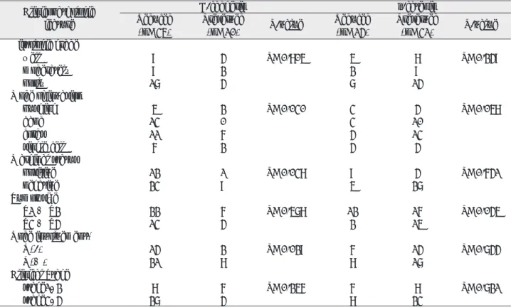
E-cadherin의 발현에 있어서는 암종의 조직학적 분화도, 종양의 국소적 크기 (T-category)와 임상적 진전 상태에 따른 병기 (Stage)에 따른 유의한 차이가 없었다. 그러나 구강암이 주변의 정상 결합조직으로 침윤하는 부위에서 침 습성이 높은 single cell 유형이 pushing 유형이나 band 유 형보다 E-cadherin의 발현감소가 유의성 있게 나타났다 (P=0.030). 또한 수술후 조직 변연의 종양 세포의 존재와 관련된 변연상태(marginal status)에 따른 분류에서도 E- cadherin의 발현 감소군에서 positive가 유의성 있게 높게 나타났다 (P=0.038). 원발 부위에서 E-cadherin의 발현 이 감소된 예에서 주위 임파절로 전이가 많이 나타났다 (P=0.029).
그러나 β-catenin의 발현과 여타 임상병리학적 관계에 있 어서는 종양의 크기가 작을 때 (T1, T2), 상대적으로 β- catenin의 발현이 적은 것으로 밝혀졌으나 (P=0.045), 이 외에 다른 임상적 지표와는 특이할 만한 상관관계가 나타나 지 않았다 (Table 3).
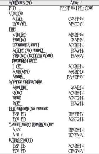
Table 1. Clinicopathologic Characteristics of Patients with Squamous Cell Carcinoma
Patient Data No(%)
Age 65.06 ± 8.5 years
Gender
Male 30(66.7)
Female 15(33.3)
Site
Gingiva 12(26.7)
Tongue 7(15.6)
Floor of Mouth 13(28.9) Palate and tonsil 8(17.8) Lip and buccal mucosa 5(11.1) Histologic grade
Well 13(28.9)
Moderate 11(24.4)
Poorly 21((46.7)
Pattern of invasion
Pushing 7(15.6)
Band 13(28.9)
Cord 17(37.8)
Cell 8(17.8)
Size of primary tumor
T1+T2 28(62.2)
T3+T4 17(37.8)
Lymph node involvement
N(-) 22(48.9)
N(+) 23(51.1)
Clinical stage
S1+S2 13(28.9)
S3+S4 32(71.1)
Table 2. E-cadherin and β -catenin Expression Pattern in Primary Lesion and Metastatic Lymph Node
E-cadherin β -catenin
Frequency % Frequency %
Primary(n=45)
preserved 10 22.2 31 68.8
reduced 35 78.2 14 31.2
Lymph node(n=19)
preserved 1 94.7 17 89.4
reduced 18 6.3 2 10.6
Table 3. Correlation between the Expression of E-cadherin and β-catenin in the Primary Lesion and Clinicopathologic Findings
Clinicopathologic E-cadherin β -catenin
feature Reduced Preserved P-value Reduced Preserved P-value
(n=35) (n=10) (n=14) (n=31)
Histologic grade
well 9 4 P=0.805 5 8 P=0.249
moderately 9 2 2 9
pooly 17 4 7 14
Mode of invasion
pushing 5 2 P=0.030 3 4 P=0.058
band 13 0 3 10
cords 11 6 4 13
single cell 6 2 4 4
Marginal status
positive 12 1 P=0.038 9 4 P=0.641
negative 23 9 5 27
Tumor size
T1 + T2 22 6 P=0.578 12 16 P=0.045
T3 + T4 13 4 2 15
Node involvement*
N(-) 14 2 P=0.029 6 14 P=0.744
N(+) 21 8 8 17
Clinical Stage
stage1, 2 8 6 P=0.256 6 8 P=0.721
stage3, 4 27 4 8 23
( * N(-): No Lymph Node Involvement, N(+): Metastatic Lymph Node Including N1, N2, N3 case.)
Table 4. Association between Expression of E-cadherin and β -catenin in Primary Lesion and Metastatic Lymph Node β -catenin
Primary Lymph node
preserved reduced P-value preserved reduced P-value
Primary(n=45) 31 14 P<0.025
preserved 9 1 χ
2=5.184
reduced 22 13
Lymph node(n=19) 17 2 P�1.0
preserved 1 0 χ
2=0.124
reduced 16 2
4. E-cadherin과 β-catenin의 발현의 상관관계
원발 부위에서 E-cadherin과 β-catenin의 발현은 서로
유의성있게 나타났다 (P<0.025). 그러나 전이된 임파절에 서는 서로의 발현이 서로에게 영향을 미치지 않는 것으로 밝혀졌다 (P�1.0)(Table 4).
Fig. 1. Immunohistochemical staining pattern in primary oral squamous cell carcinoma. (a) Preserved stain- ing of E-cadherin and (b) β-catenin shows distinct and evenly distributed membrane staining, × 100.
Fig. 2. Immunohistochemical staining pattern in primary oral squamous cell carcinoma. (a) Reduced staining of E-cadherin and (b) β-catenin with heterogeneous staining with mixed negative and positive cells, × 100.
Fig. 3. Immunohistochemical staining pattern in metastatic lymph node. (a) Preserved staining of E-cadherin
and (b) β-catenin, × 40.
Ⅳ. 총괄 및 고찰
E-cadherin과 β-catenin은 정상 구강상피조직에서 중요 한 부착 분자이다. α-, β-, 와 γ-의 3종으로 구성된 catenin 은 E-cadherin에 의해 매개되는 세포간 신호전달과 세포간 부착에 매우 중요한 역할을 한다. 정상 부착능력의 변화 또 는 상실은 종양생물학 분야에 매우 결정적인 역할을 한다.
예를 들어 종양 세포의 세포간 부착능력의 변화는 종양 성 장의 형태와 응집에 미세한 수준에서 반영된다. 종양세포의 분리는 주변조직으로 침습이나 장기로의 원격 전이의 초기 단계이며, 이 과정에서 세포 부착능력의 변화가 핵심적인 역할을 하는 것이다. 여러 연구에서 E-cadherin-catenin system이 여러 종류의 종양의 성장 매개와 세포 부착의 유 지에 기여함이 밝혀졌다17-19).
따라서 세포 부착인자의 발현은 종양의 생물학적 행동과 특성을 반영하며, 수술한 환자의 재발과 전이의 위험성을 예측하고 평가하는데 유용할 것으로 개인의 특성에 맞춘 치 료를 제공함에 실제적인 의미가 있을 것으로 사료된다20).
구강 편평세포암에서 경부 전이의 발생은 상대적으로 높 은 편이다21). 본 연구에서는 비록 통계적인 처리는 시행하지 않았으나, T 분류가 임파절 전이의 발생과 가장 관련이 깊 은 것으로 예상되며 이는 다른 연구와 일치한다 (미발표 결 과).
이번 연구에서는 E-cadherin 발현과 임파절 전이와 의미 있는 관계가 있음이 밝혀졌다. Bukholm들에 따르면 유방 암에서 E-cadherin과 β-catenin 중 최소 하나의 면역화학 적 반응도의 감소가 전이와 관계있다고 하였다22). 이 연구에 서는 E-cadherin의 영향은 통계적으로 의미가 있었으나 β- catenin은 의미를 밝힐 수는 없었다. 그러나 아직 어떤 단 백질의 발현이 진단과 전이 또는 종양의 침습 정도에 대한
예측에 유용할 지는 연구가 필요할 것이며, 보다 정확한 통 계학적인 분석을 위해서 구강암 증례수를 확대하여 모집단 을 크게 하는 것이 필요하다고 사료된다.
반면 E-cadherin과 β-catenin의 발현이 서로 연관성을 보이는 것으로 여겨진다. 즉 원발 부위에서 E-cadherin이 보존된 증례에서 β-catenin이 보존된 형태를 보이고, E- cadherin이 감소하면 β-catenin도 따라서 감소하는 것으로 밝혀졌다 (P<0.025). β-catenin은 E-cadherin의 세포질 내 말단에 직접적으로 결합한다17). E-cadherin는 β- catenin과 complex를 이루어 세포내 골격 물질인 actin을 연결하므로, E-cadherin의 발현 능력은 catenin system 의 완전성 여부에 따라 달려있다고 할 수 있을 것이다.
이번 연구에서는 E-cadherin과 β-catenin의 발현이 원발 부위에서보다 임파절에서 증가된 것이 관찰되었다. 이 현상 은 cadherin-catenin 복합체의 발현은 현저하게 변화하고 이 증가는 구강 편평세포암 세포의 전이 과정 중에서 재표 현 되는 것을 의미한다. Tanaka 들에 의하면 원발 부위와 전이 부위의 미세 구조학적인 측면에서 형태학적인 유사함 을 보고하였으나23), 어떤 증례에서는 둘 사이의 부착 복합체 (junctional complex)의 특징이 다르다고 하였다. 그러나 왜 원발 부위와 임파절 전이부위에서 이 cadherin-catenin 복합체의 발현이 차이가 나는 지에 대하여는 밝혀진 바 없 다. 따라서 임파절 전이 과정 중에서 이들이 어떤 기전으로 그 발현이 변화하는 지에 대한 연구가 필요할 것이다. 또한 구강암의 국소적 전이나 임파절 전이가 원발 부위의 세포부 착 인자의 변화같은 특징에 좌우되는 지는 아직 알 수 없다.
그러나 이번 연구에서는 원발 부위와 전이 부위가 독립적인 관계에 있는 것으로 여겨지며, 이는 전이된 구강암 세포가 전이 부위의 환경에 영향을 받는 것으로 사료된다.
E-cadherin의 발현이 저하된 것에 비하여 β-catenin의
Fig. 4. Immunohistochemical staining pattern in metastatic lymph node. (a) Reduced staining of E-cadherin
and (b) β-catenin, × 100.
down regulation은 상대적으로 적은 것으로 보인다. β- catenin은 독립적으로 세포간 부착인자로서와 세포내에서 신호전달물질로 작용한다. E-cadherin에 주도되는 세포간 부착의 조절 역할이외에도 Wnt signalling pathway에서 전사조절인자로 기능하며, 다른 종류의 암의 기시에 관여하 는 Adenomatous polyposis coli (APC) 유전자의 목표이 기도 하다24). 이번 연구에서 특히 β-catenin의 세포핵 내에 서의 발현이 되는 경우 세포질 내에서 발현과 이에 따른 세 포막에서의 발현 감소와 동반하는 예가 관찰되었다. β- catenin이 세포막뿐 아니라 핵에서도 염색이 되는 것에 대 한 이유는 β-catenin이 전사조절물질로 작용하기 때문에 세 포질과 핵 내에서도 발현되는 것으로 유추할 수 있다. 따라 서 구강암에서 β-catenin의 역할과 기능을 규명하기 위하여 다른 pathway에 관여하는 β-catenin에 대한 분석이 요구 된다고 하겠다.
생검된 조직에서 평가된 암종의 침투 형태가 국소적인 전 이와 관련된다고 밝혀진 바 있으며25), 이번 결과에서도 유일 하게 E-cadherin과 β-catenin이 동시에 통계학적인 의미 를 갖는 것으로 관찰된다 (P=0.030, P=0.058).
본 연구를 통해 세포부착 단백질인 E-cadherin과 β- catenin의 발현과 구강 편평세포암의 발생과 전이과정에 기여하는 역할을 밝힘으로, 이를 생물학적인 표지자로 사용 하여, 진단과정에서 치료 방법의 선택에 도움을 줄 수 있을 것이며, 이를 임상적으로 적용할 경우 국소적 치료 효율의 개선을 도모할 수 있으리라 사료된다. 향후 연구를 수행하 여 cadherin-catenin 복합체 간의 관계를 규명하므로써 구 강암 발생 과정 면에서 중요한 정보를 제시하여, 구강 편평 세포암의 발생에 대한 이해를 높일 수 있으며, 새로운 전이 이론을 정립할 수 있을 것으로 사료된다.
Ⅴ. 결 론
원발성 구강 편평세포암종 조직 45예와 전이성 임파절 19례에서 면역 조직화학 염색을 통하여 E-cadherin과 β- catenin 의 발현을 조사하고 임상병리학적 특성과의 상관 관계를 알아보았다. E-cadherin의 발현은 전체 45예 중 35예에서 (78.2%), β-catenin (31.2%)은 14예에서 발현 감소를 보였다. E-cadherin의 발현 감소는 조직학적 분화 정도, 임파절 전이와 연관성을 보였으나, β-catenin은 임상 적 지표와 유의성을 발견할 수 없었다. 이상의 결과로 E- cadherin과 β-catenin complex의 발현정도는 임파절 전이 와 관련성 있는 예후인자가 될 수 있으며, 재발의 고위험군 예측을 가능하게 함으로써 기존의 병리학적 예후인자들에 근거한 위험도 추정을 보완할 수 있어 임상적으로 유용한 지표로 사용될 수 있을 것으로 생각된다.
참고문헌
1. Landis SH, Murray T, Bolden S et al : Cancer statistics, 1999. CA Cancer J Clin 49 : 8, 1999.
2. Smith BD, Haffty BG : Molecular markers as prognostic factors for local recurrence and radioresistance in head and neck squamous cell carcinoma. Radiat Oncol Invest 7 : 125, 1999.
3. Pignatelli M, Vessey CJ : Adhesion molecule ; Novel mole- cular tools in tumor pathology. Hum Pathol 25 : 849, 1994.
4. Takeichi M : The Cadherin, cell-cell adhesion molecules controlling animal morphogenesis. Development 102 : 639, 1988.
5. Takeichi M : Cadherin cell adhesion receptor as a morpho- genetic regulator. Science 251 : 1451, 1991.
6. Shiozaki H, Tahara H, Oka H et al : Expression of immunoreactive E-cadherin adhesion molecules in human cancers. Am J Pathol 139 : 17, 1991.
7. Oka H, Shiozaki H, Tahara H et al : Immuno- histochemical evaluation of E-cadherin adhesion molecule expression in human gastric cancer. Virchows Arch Abt A Pathol Anat 421 : 149, 1992.
8. Schipper JH, Unger A, Jahnke K : E-cadherin as a func- tional marker of the differentiation and invasiveness of squamous cell carcinoma of the head and neck. Clin Otolaryn Allied Sci 19 : 381, 1994.
9. Shin JM, Kim YS, Kim CH et al : Expression of E-cad- herin with correlation to clinicopathologic parameters in oral squamous cell carcinoma. Kor J Oral Maxillofac Surg 31 : 1, 2005.
10. William HK, Sanders DS, Jankowski PA et al : Expression of cadherins and catenins in oral epithelial dys- plasia and squamous cell carcinoma. 27 : 308, 1998.
11. Tanaka N, Odajima T, Ogi K et al : Expression of E-cad- herin, α-catenin and β-catenin in the process of lymph node metastasis in oral squamous cell carcinoma. Br J Cancer 89 : 557, 2003.
12. Chow A, Yuen APW, Lam KY et al : A comparative study of the clinicopathologic significance of E-cadherin and catenins(α, β, γ) expression in the surgical management of oral tongue carcinoma. J Cancer Res Clin Oncol 127 : 59, 2001.
13. Shieh YS, Chang LC, Chiu KC et al : Cadherin and catenin expression in mucoepidermoid carcinoma: correla- tion with histopathologic grade, clinical stage, and patient outcome. J Oral Pathol Med 32 : 297, 2003.
14. Nakanishi Y, Ochiai A, Akimoto S et al : Expression of E- cadherin, α-catenin, β-catenin and plakoglobin in esophageal carcinoma and its prognostic significance.
Oncology 54 : 158, 1997.
15. Bagutti C, Speight PM, Watt FM : Comparison of inte- grin, cadherin and catenin expression in squamous cell carcinoma of the oral cavity. J Pathol 186 : 8, 1988.
16. Lee SJ : Expression of E-Cadherin/ β-Catenin in oral tongue squamous cell carcinoma. J Cathol Med Coll, 25, 2003.
17. Wijnhoven BP, Dinjens WN, Pignatelli M : E-cadherin- catenin cell-cell adhesion complex and human cancer. Br J Surg 87 : 992, 2000.
18. Van Aken E, De Wever O, Correia da Rocha AS et al : Defective E-cadherin/catenin complexes in human cancer.
Virchows Arch 439 : 725, 2001.
19. Behrens J : Cadherins and catenins: role in signal trans-
duction and tumor progression. Cancer Metastasis Rev 18 : 15, 1999.
20. Lim SC, Lee MS : Significance of E-cadherin/β-catenin complex and cyclin D1 in breast cancer. Oncol Rep 9 : 915, 2002.
21. Martis C, Karabouta I, Lazardis N : Incidence of lymph node metastasis in elective (prophylactic) neck dissection for oral carcinoma. J Oral Maxillofac Surg 7 : 182, 1979.
22. Bukholm IK, Nesland JN, Karesen R et al : E-cadherin and α-, β-, and γ-catenin protein expression in relation to metastasis in breast carcinomas. J Pathol 185 : 262,
1998.
23. Tanaka N, Sugihara K, Odajima T et al : Oral squamous cell carcinoma -Electron microscopic and immunohisto- chemical characteristics. Med Electron Microsc 35 : 127, 2002.
24. Ilyas M, Tomlinson IPM : The interactions of APC, E-cad- herin and β-catenin in tumour development and progres- sion. J Pathol 182 : 128, 1996.
25. Yamamoto E, Miyakawa A, Koham G : Mode of invasion and lymph node metastasis in squamous cell carcinoma of the oral cavity. Head Neck 6 : 938, 1984.
저자 연락처