기도상피세포에서 Mentha piperita 발효물에 의한 MUC5AC 억제 효능
지혜진1,2․Katrina Joy Bormate1․임 옥3․정이숙1,2
1아주대학교 약학대학
2아주대학교 약과학연구소
3포천허브아일랜드 천연물과학연구소
Inhibitory Effect of Fermented Mentha piperita on MUC5AC in Lung Epithelial Cells
Hye Jin Jee1,2, Katrina Joy Bormate1, Ok Lim3, and Yi-Sook Jung1,2
1College of Pharmacy and 2Research Institute of Pharmaceutical Sciences and Technology, Ajou University
3Pocheon Herbisland Natural Product Science Research Institute
ABSTRACT Airway mucus hypersecretion is an important clinical manifestation of asthma and can be elicited by a wide variety of endogenous and exogenous stimuli leading to airway constriction and worsening of the disease.
Gene expression for MUC5AC, a principal component of mucin, is known to significantly increase in the epithelial cells of patients with asthma. Therefore, many studies have proposed that regulating the MUC5AC expression in airway epithelial cells could be a target candidate as therapy for asthma treatment. Mentha piperita, also known as peppermint, is a popular medicinal herb due to its various pharmacological uses, and polyphenolic components with strong anti- oxidant activity. Based on these studies, we investigated whether peppermint could improve airway function through its inhibitory effect on phorbol myristate acetate (PMA)-induced expression of MUC5AC in NCI-H292 cells, a human airway epithelial cell line. Peppermint leaves were sourced from Herb Island and fermented with Bacillus subtillis.
The fermented extract of peppermint (FEP) significantly reduced PMA-induced MUC5AC mRNA expression and protein release. FEP also inhibited the phosphorylation of extracellular signal regulated kinases (ERK) and U0126 (ERK in- hibitor) inhibited PMA-induced MUC5AC expression and secretion. FEP also inhibited reactive oxygen species (ROS) production, and Trolox, a ROS scavenging agent, inhibited PMA-induced MUC5AC mRNA expression and protein secretion through the inhibition of ERK activation. These results suggest that FEP can protect the respiratory tract from inflammatory damage by reducing mucus hypersecretion through the ERK signaling pathway and antioxidant activity.
Key words: Mentha piperita, fermented extract of peppermint, MUC5AC, ERK, antioxidant
Received 14 Oct 2020; Revised 19 Feb 2021; Accepted 22 Feb 2021 Corresponding author: Yi-Sook Jung, College of Pharmacy, Ajou University, 206, World cup-ro, Yeongtong-gu, Suwon-si, Gyeonggi 16499, Korea, E-mail: [email protected]
Author information: Hye Jin Jee (Researcher), Katrina Joy Bormate (Graduate student), Yi-Sook Jung (Professor)
서 론
인간의 기도 내부에 존재하는 점액은 호흡기 상피 표면을 덮고 있으며, 점액 섬모 청소기능을 통해 미세분진 입자, 비 산 화학물질, 병원성 미생물 등 인체 외부에서 유입되는 독 성물질과 감염원에 대한 생리적 방어 작용을 수행함으로써 기도 내부를 독성자극으로부터 보호하고 있다(Voynow와 Rubin, 2009). 그러나 천식, 만성 폐색성 폐질환, 낭포성 섬 유증 및 폐렴과 같은 급성 또는 만성 염증성 호흡기 질환에 서는 점액의 분비가 과도하게 일어나고 그 성상이 변화되어
병의 경과를 악화시킬 수 있으므로 점액 과분비를 조절하는 것이 매우 중요하다(Chopra 등, 1991; Voynow와 Rubin, 2009; Ahn 등, 2015). 점액의 성상을 결정하는 여러 성분 중에서 당단백인 뮤신(mucin)은 점액유전자에 의해 생성이 조절되고, 점액의 유동학적, 물리화학적 또는 생화학적 특성 을 결정할 때 가장 큰 역할을 담당한다(Song 등, 2013; Kim 등, 2014).
지금까지 알려진 18가지의 인간 뮤신 중 기도에서 발견되 는 것은 mucin 2(MUC2), mucin 4(MUC4), mucin 5AC (MUC5AC) 및 mucin 5B(MUC5B) 등이 있으며, 그중 MUC 5AC는 주로 기도상피세포 내지 점액분비세포에서 발현된 다(Kim 등, 1989). MUC5AC는 기관지 질환 환자의 증상이 악화될수록 분비가 증가하며, 비정상적인 점액 내지 점액세 포 증식의 원인인 것으로 알려져 있다(Song 등, 2013). 따라 서 점액에 점성을 부여하는 주요 구성요소인 뮤신의 생성 자 체를 조절하거나 혹은 분비를 조절하기 위한 약물학적 접근
은 호흡기 질환의 치료에 있어 매우 중요하다(Liu 등, 2012).
특히 최근에는 우수한 약리효과를 가지면서도 인체에 별다 른 부작용이 없어 안전성이 높은 천연물에 대한 생리활성 연구가 활발히 진행되고 있다.
항염증, 항산화, 항암 효능을 발현하는 것으로 알려진 다 수의 천연물 중에서 호흡기 염증성 질환에서 관찰되는 기도 뮤신의 생성 및 과다분비를 조절할 가능성이 있는 천연물 소 재를 탐색하는 것은 매우 중요하다(Leach와 Page, 2015).
페퍼민트(Mentha piperita)는 물 민트(water mint; Mentha aquatica)와 스피어민트(spearmint; Mentha spicata)가 교 배된 잡종 민트로서 다양한 약리효과를 가진 대표적인 약용 식물 중 하나이다(Savikin 등, 2013). 페퍼민트는 다량의 멘톨을 함유하고 있으며 식물 전체에 존재하는 휘발성 기름 때문에 매우 특징적인 향기가 난다(Mckay와 Blumberg, 2006). 페퍼민트 오일은 동서양에서 전통적으로 암, 감기, 경련, 소화불량 또는 치통 등의 치료에 사용되며 방향제 내 지 방부제로도 사용되었다(Zamin 등, 2013). 특히 페퍼민트 오일은 알레르기성 비염과 호흡기 기관지 병리에서 항알레 르기 효과를 보였으며, 페퍼민트에서 유래된 플라보노이드 인 글리코사이드에 대한 연구에서는 박하가 알레르기성 비 염의 코 증상을 완화시키는 데 도움을 주는 것으로 보고되었 다(Ben-Arye 등, 2011). 또한, 건조된 페퍼민트 잎의 에탄 올 추출물은 염증을 억제하여 육아종을 감소시켰다(Sharafi 등, 2010). 그러나 염증반응에 의한 기도 점액 과분비 과정 에서 페퍼민트가 점액유전자 발현에 어떤 영향을 미치는지 에 관한 연구는 아직 보고된 바가 없다.
점막상피세포에서 점액유전자의 발현과 점액분비를 증가 시키는 물질은 다양하게 존재한다. 그중 하나인 phorbol myristate acetate(PMA)는 protein kinase C(PKC) acti- vator로 호흡상피에서 matrix metalloproteinase-9(MMP9) 을 통해 염증반응을 일으켜 MUC5AC 발현을 증가시킨다고 알려져 있다(Wu 등, 2007). 따라서 본 연구에서는 발효된 페퍼민트 추출물이 인간 기도상피세포에서 PMA에 의해 증 가한 MUC5AC 뮤신의 유전자 발현 및 생성에 대해 어떠한 영향을 나타내는지 탐색함으로써, 후속 연구를 통한 효과적 인 기도 점액 과다생성(분비) 조절 신약의 개발을 위한 단초 를 제시하고자 하였다.
재료 및 방법
페퍼민트 추출 발효물 제조
본 실험에 사용한 페퍼민트는 포천 허브아일랜드(Po- cheon, Korea)로부터 건조된 잎 상태로 제공받아 추출, 발 효하였다. 페퍼민트 전초의 원시료를 증류수에 넣어 100°C 에서 2.5시간 동안 추출, 멸균하여 페퍼민트 추출물을 제조 하였다. 멸균된 페퍼민트 추출물에 Bacillus subtilis를 접종 한 후 온도 25°C, 습도 80%의 환경에서 140 rpm으로 진탕 배양 하면서 5일간 발효하여 페퍼민트 추출 발효물(fer-
mented extract of peppermint, FEP)을 수득하였으며 -20°C에 보관하면서 사용하였다.
인간 기도상피세포(NCI-H292) 배양 및 처치
사람 호흡기 점액상피양 암 세포주(human pulmonary mucoepidermoid carcinoma cell line)인 NCI-H292 세포 (American Type Culture Collection, Manassas, VA, USA)에 100 U/mL penicillin, 100 µg/mL streptomycin, 10% fetal bovine serum(FBS; Gibco, Grand Island, NY, USA)이 포함된 RPMI 1640 배지(Invitrogen, Carlsbad, CA, USA)를 이용하여 5% 이산화탄소, 37°C의 조건에서 배 양하였다. 6-well plate에 0.5×10⁶ cells/well의 밀도로 세 포를 도포하여 배양하였으며, 70~80% 정도 배양이 이루어 지면 세포를 0.5% FBS가 포함된 RPMI 1640 배지로 교체 한 후 24시간 동안 배양하였다. FEP에 의한 MUC5AC 점액 유전자 발현과 단백 생성을 알아보기 위해 NCI-H292 세포 에 각기 다른 농도의 FEP를 전처리한 후 30분 뒤에 10, 20 ng/mL 농도의 PMA를 투여하였다. 대조군은 0.5% FBS가 포함된 RPMI 1640 배지에서 NCI-H292 세포를 단독으로 배양하였다.
DPPH 라디칼 소거능 측정
2,2-Diphenyl-1-picrylhydrazyl(DPPH)을 이용한 전자 공여능(electron donating abilities, EDA)은 각 시료용액 100 µL에 0.25 mM DPPH(Sigma-Aldrich Co., St. Louis, MO, USA) 용액 100 µL를 넣고 교반한 후 실온에서 차광한 상태로 30분간 방치한 다음 517 nm에서 흡광도를 측정하였 다. 전자공여능은 시료용액의 첨가구와 무첨가구의 흡광도 감소율로 나타내었다.
라디칼 소거능(%)=
(
1- 시료 첨가구의 흡광도)
×100시료 무첨가구 흡광도
인간 기도상피세포에서의 MUC5AC 뮤신 생성량 측정 각 약물의 처리기간이 종료된 후, 세포 내 생성되어 저장 된 뮤신을 정량하기 위해 세포 용해용 완충액(20 mM Tris, 0.5% NP-40, 250 mM NaCl, 3 mM EDTA, 3 mM EGTA, protease inhibitor cocktail)을 가하여 세포 내에 존재하는 MUC5AC 뮤신을 추출하여 면역흡착법(enzyme-linked im- munosorbent assay, ELISA)을 이용하여 측정하였다. 뮤신 을 함유한 배양 상층액을 phosphate buffered saline(PBS) 으로 1:10 비율로 희석하고, 희석된 각 sample을 ELISA 전용 96-well plate에 각각 100 µL씩 분포시킨 후 42°C에 서 완전히 건조시켰다. 그 후 Tween 20이 함유된 PBS 용액 (PBS-T) 200 µL/well을 이용하여 각 well 당 3회씩 세척하 였다. 세척 후 PBS-T에 용해된 2% bovine serum albu- min(BSA, Sigma-Aldrich Co.) 용액 200 µL를 각 well에 가하고 다시 1시간 동안 배양하였다. 1시간 후 PBS-T 200 µL로 3회 세척하고 MUC5AC에 대한 monoclonal anti-
body인 mouse anti-MUC5AC clone 45 M1을 2% BSA에 1:200의 비율로 희석한 후, 각 well 당 100 µL씩 첨가하여 1시간 동안 배양하였다. 1시간 후 PBS-T로 3회 세척하고 2차 항체인 horseradish peroxidase-goat anti-mouse IgG conjugate를 2% BSA에 1:3,000의 비율로 희석한 후, 각 well 당 100 µL씩 첨가하고 1시간 동안 배양하였다.
PBS-T로 다시 3회 세척 후 3,3′,5,5′-tetramethylbenzi- dine peroxide(Sigma-Aldrich Co.) 용액 100 µL를 각 well 에 첨가하고 5분 후 1 N H2SO4 50 µL를 첨가하여 반응을 정지시켰다. 450 nm에서 각 well의 흡광도를 측정함으로써 대조군과 약물 처리군에 존재하는 MUC5AC 뮤신을 정량하 였다(Shao 등, 2003; Song 등, 2003; Heo 등, 2007).
Reverse transcription-polymerase chain reaction(RT- PCR) 분석을 이용한 뮤신 발현량 측정
NCI-H292 세포에서 RNA를 추출하기 위해 Trizol re- agent(Ambion, Carlsbad, CA, USA)를 이용하여 RNA를 분리 정제하였고, reverse transcriptase를 포함한 RT re- agent(M-MLV, Beams Biotechnology, Seongnam, Ko- rea)를 첨가하여 37°C에서 1시간 동안 반응시켜 cDNA를 만든 후 cDNA 1 µL를 각 유전자의 sense&antisense pri- mer, Taq polymerase, dNTP와 혼합하여 각 유전자 pri- mer(MUC5AC, β-actin)의 Tm 값에서 PCR을 수행하였다.
MUC5AC mRNA에 대한 PCR은 95°C에서 60초간 변성과 정과 60°C에서 60초간 결합반응, 72°C에서 60초간 연장반 응을 33회 반복한 후 72°C에서 20분간 최종 연장반응을 시행하였다. 증폭된 중합효소연쇄반응의 산물은 1% aga- rose gel(Inclone Biotech, Yongin, Korea)을 EcoDyeTM (SolGent Co., Ltd., Daejeon, Korea)으로 염색한 후 전기 영동을 하여 Gel-Doc 2000 system(Bio-Rad, Hercules, CA, USA)으로 mRNA 발현 정도를 관찰하였다. 실험에 사 용된 primer의 염기배열은 MUC5AC의 경우 sense는 5′
-TCA ACG GAG ACT GCG AGT ACA C-3′, antisense는 5′-CTT GAT GGC CTT GGA GCA-3′이고, β-actin의 경 우 sense는 5′-CAC CAT TGG CAA TGA GCG GTT C- 3′, antisense는 5′-AGG TCT TTG CGG ATG TCC ACG T-3′였다.
Real-time quantitative PCR을 이용한 뮤신 발현량 측정 합성된 cDNA를 AmfiSure qGreen Q-PCR Master Mix (2×)(GenDEPOT, Katy, TX, USA)와 Rotor-gene Q(Qia- gen, Hilden, Germany) 기기를 이용하여 사용법에 맞게 실 험을 진행하였으며, qPCR에 사용된 primer는 RT-PCR과 동일하다. 95°C에서 3분, 95°C에서 5초, 60°C 15초, 72°C 10초의 조건으로 40 cycle을 실행하였다. MUC5AC mRNA 의 상대적 정량은 comparative cycle of the threshold method를 이용하여 얻었고, 대조군으로 β-actin을 사용하 였다.
중합효소연쇄반응의 상대적 정량화
증폭된 중합효소연쇄반응 산물의 정량화를 위해 분광광 도계를 이용하여 농도를 측정한 β-actin을 함께 증폭하여 threshold cycle 수를 얻었고, 이를 기준으로 표준검량선을 작성한 후 검량선의 수식에 의해 검체 내 점액유전자 각각의 산물을 정량화하는 상대적 정량법을 사용하였다. cDNA가 증폭되면 SYBR Green I dye가 결합하여 형광을 발생하는 데, 증폭된 DNA 양이 많을수록 형광이 처음으로 검출되는 연쇄반응 주기의 시점이 빨라짐을 이용하여 threshold cy- cle 수를 구하였다. 점액유전자 MUC5AC의 농도를 β-ac- tin 농도로 나누어 정상화된 점액유전자의 농도를 구하고, 이 값을 정상화된 대조군의 평균 점액유전자 수치로 나누어 PMA 처리 시 정상보다 증가한 점액유전자 MUC5AC mRNA 발현을 상대적으로 정량화하였다.
Western blot을 이용한 ERK 인산화 측정
시약 처리된 세포를 ice-cold PBS로 2회 세척하고 RIPA buffer(pH 7.4, 150 mM NaCl, 20 mM Tris-HCl, 1%
NP40, 1% Na-deoxycholate, 1 mM EDTA, protease in- hibitor 10 µg/mL leupeptin, 10 µg/mL aprotinin)에 넣어 세포를 파쇄하였다. 세포는 glass homogenizer(Thomas Scientific, Swedesboro, NJ, USA)로 1회 더 파쇄하였으 며, 4°C, 14,000 rpm에서 15분간 원심분리를 하여 핵과 세 포 잔해물은 제거하고 상층액을 모아서 immunoblotting에 사용하였다. 단백질은 BCATM protein assay(Pierce, Rock- ford, IL, USA)로 정량하였다. 단백질 시료는 10% sodium dodecyl sulfate-polyacrylamide gel에서 전기영동으로 분 리하고, polyvinylidene difluoride membrane으로 blot- ting 하였다. 5% non-fat dry milk를 넣은 Tris-buffered saline에 blot을 넣고 상온에서 1시간 동안 유지하였고, 1차 항체와 함께 4°C에서 하룻밤 동안 유지하였다. Blot을 Tris- buffered saline으로 씻어낸 후 horseradish peroxidase- conjugated 2차 IgG 항체와 함께 1시간 유지하였다. 결합된 항체는 LAS-1000 Pro(FujiFilm Corporation, Tokyo, Japan)를 사용하여 시각화하고 정량하였다. Extracellular Signal Regulated Kinase(ERK)와 p-ERK antibody는 Cell Signaling(Beverly, MA, USA)에서 구매하였다.
DCF-DA assay를 이용한 항산화능 분석
세포 내 활성산소의 농도변화를 측정하기 위하여 형광 probe는 2′,7′-dichlorofluorescin diacetate(DCF-DA;
Sigma-Aldrich Co.)를 이용하였다. 비형광물질인 DCF- DA는 세포 내 hydrogen peroxide와 관련된 peroxides 존 재 시 ROS에 의해 산화되어 녹색의 형광을 띄게 된다. 따라 서 ROS의 측정은 DCF-DA를 통해 이루어질 수 있다. 2×
105개의 세포를 60 mm culture dish에 접종하여 24시간 동안 배양하였고, FEP와 PMA를 처리 후 1시간 동안 배양하 였다. 세포를 수확하기 전에 10 µM DCF-DA를 배지에 처리
0 0.2 0.4 0.6 0.8 1 1.2 1.4 1.6 1.8 2
CTL 1 2 4 8 12 24
Time of treatment (h)
MUC5AC secretion .
PMA 10 ng/mL
PMA 20 ng/mL *
A
*0 1 2 3 4
CTL VEH U0126 1 3 10 30 100
FEP (μg/mL)
MUC5AC secretion .
B
#
#
#
#
A
B
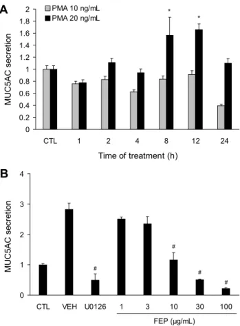
Fig. 1. Effects of FEP on PMA-induced MUC5AC protein secre- tion in NCI-H292. (A) NCI-H292 cells were treated with PMA 10 ng/mL or 20 ng/mL for 1, 2, 4, 8, 12, and 24 h. MUC5AC protein secretion was measured using ELISA. Data are expressed as mean±SEM (n=3). Significant differences are indicated by
*P<0.05 vs CTL. (B) FEP decreased MUC5AC protein secretion in a dose-dependent manner. Cells were pretreated with FEP (1, 3, 10, 30, and 100 μg/mL) or U0126 (20 μM) for 30 min and then stimulated with PMA (20 ng/mL) for 12 h. The total MUC5AC protein secreted in the supernatant was measured us- ing ELISA. Significant differences were indicated by #P<0.05 vs VEH (n=3). CTL, control; VEH, vehicle; PMA, phorbol my- ristate acetate; U0126, MEK/ERK inhibitor; FEP, fermented ex- tract of peppermint.
하여 30분간 배양하였다. 배양한 세포는 PBS로 세척하여 1% trypsin-EDTA 용액을 처리하여 세포를 수확하고, 다시 PBS로 세척하여 공초점 레이저 형광현미경(LSM 510, Carl Zeiss, Oberkochen, Germany)을 이용하여 ×20배율로 관 찰하였으며 excitation 파장은 488 nm(argon laser), emis- sion 파장은 505 nm였다(Okimoto 등, 2000).
통계처리
본 실험 결과는 각 항목에 따라 평균과 표준오차를 구하였 고, 평균치 간의 유의성은 Student’s t-test를 이용한 후 P 값이 0.05 미만일 때 유의한 것으로 판정하였다.
결과 및 고찰
FEP의 MUC5AC 분비 억제 효능 측정
천연물의 생리활성을 극대화할 수 있는 공정으로 많이 알 려진 방법으로 발효공정이 있으며(Kong 등, 2008), 발효공 정에서 주로 사용되는 유산균은 면역증진, 항산화 효능, 항 암효과 등 다양한 효능을 가지고 있다고 보고되었다(Sekine 등, 1985; Lin과 Yen, 1999). 따라서 본 연구에서는 페퍼민 트 물 추출물에 유산균인 Bacillus subtilis를 처리하여 발효 공정을 거친 뒤 FEP를 수득하여 연구를 진행하였다. PMA 는 PKC 및 diacylglyceride(DAG) 활성제로 호흡상피에서 MMP9을 통해 염증반응을 일으켜 MUC5AC 발현을 증가시 킨다고 알려져 있다(Hewson 등, 2004; Wu 등, 2007;
Sikder 등, 2014). MUC5AC는 만성기관지염 및 천식 등의 염증성 호흡기 질환에서 기도의 과도한 점액분비 마커로 널 리 사용된다(Ishinaga 등, 2005). 염증 자극제로 사용되는 PMA에 의해 점액단백질인 MUC5AC가 분비되는 조건을 구하기 위해 NCI-H292 세포에 PMA 10 ng/mL와 20 ng/
mL를 처리한 다음 1, 2, 4, 8, 12, 24시간 후에 세포 외로 분비된 MUC5AC 점액단백질의 생성량을 ELISA assay를 통해 측정하였다. 그 결과 PMA 20 ng/mL의 농도로 처리한 후 8시간과 12시간이 지나고 점액단백질 MUC5AC(각각 1.56±0.3, 1.65±0.09)를 정상군(1±0.05)과 비교했을 때 유의하게 증가하는 것을 관찰할 수 있었다(Fig. 1A). PMA 로 증가한 MUC5AC 단백질에 대한 FEP의 억제 효과를 조 사하기 위해 FEP를 1, 3, 10, 30, 100 µg/mL의 농도로 NCI-H292 세포에 30분 동안 전처리한 후, PMA를 20 ng/mL 농도로 자극하였다. 12시간 후 ELISA를 사용하여 세포 외로 분비된 MUC5AC 점액단백질을 측정하여 수치화 하였다. PMA에 의해 증가한 MUC5AC 점액단백질 분비가 FEP 농도 의존적으로 감소하였으며, FEP 10, 30, 100 µg/
mL 농도에서 통계학적으로 의미 있게 감소하였다(Fig. 1B).
이를 통해 FEP가 NCI-H292세포에서 PMA에 의해 증가한 MUC5AC 분비에 대해 억제 효능이 있음을 확인하였다.
FEP의 MUC5AC mRNA 발현 억제 효능 측정
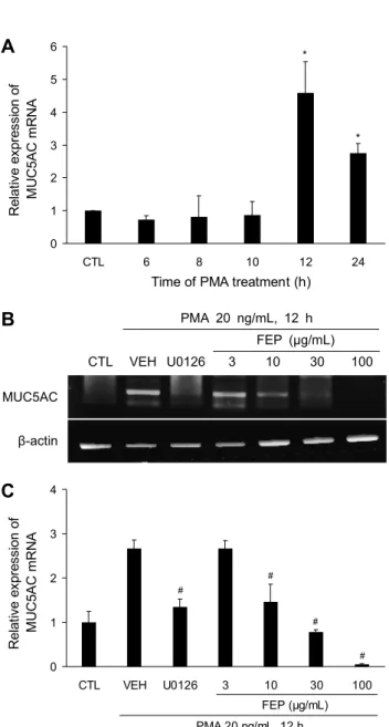
PMA에 의해 점액유전자 MUC5AC mRNA가 발현되는 조건을 구하기 위해 실험을 진행하였다. NCI-H292 세포에 서 PMA 20 ng/mL를 처리한 후 6, 8, 10, 12, 24시간 후에 MUC5AC mRNA 발현을 real time PCR 분석을 통해 측정 하였다. 실험 결과 PMA 처리 후 12시간이 경과하고 MUC 5AC mRNA의 발현을 정상군과 비교했을 때 가장 많이 증가 하는 것을 확인하였다(Fig. 2A) PMA에 의한 염증자극으로 인해 증가한 점액유전자 MUC5AC mRNA 발현에 대한 FEP 의 억제 효과를 조사하기 위해 FEP를 3, 10, 30, 100 μg/mL 의 농도로 NCI-H292 세포에서 30분 동안 전처리한 후 PMA 20 ng/mL를 12시간 동안 처리하였다. 그 결과 FEP 10 μg/mL 이상부터 점액유전자 MUC5AC mRNA 발현이
0 1 2 3 4 5 6
CTL 6 8 10 12 24
Time of PMA treatment (h) Relative expression of . MUC5AC mRNA .
*
*
A
0 1 2 3 4
CTL VEH U0126 3 10 30 100
PMA 20 ng/mL, 12 h Relative expression of . MUC5AC mRNA .
FEP (μg/mL)
#
#
#
#
C
PMA 20 ng/mL, 12 h FEP (μg/mL) CTL VEH U0126 3 10 30 100
MUC5AC
β-actin
A
B
C
Fig. 2. Effects of FEP on PMA-induced MUC5AC mRNA ex- pression in NCI-H292. (A) PMA up-regulates MUC5AC mRNA expression. Cells were treated with PMA (20 ng/mL) for 6, 8, 10, 12, and 24 h. MUC5AC mRNA expression was determined by qRT-PCR, *P<0.05 vs CTL. (B, C) Effect of FEP on MUC 5AC gene expression. NCI-H292 cells were pretreated with FEP (3, 10, 30, and 100 μg/mL) or U0126 for 30 min and stimulated with PMA (20 ng/mL) for 12 h. The levels of MUC5AC mRNA were (B) determined by RT-PCR analysis and (C) quantified by densitometry. Each bar represents the mean±SEM of at least 3 independent experiments, #P<0.05 vs VEH.
농도 의존적으로 억제되는 것을 확인하였다(Fig. 2B). 이를 통해 FEP가 NCI-H292 세포에서 염증자극에 의해 증가한 점액유전자 MUC5AC mRNA 발현 억제에 효능이 있다는 것을 확인하였다.
FEP의 ERK 활성화 억제 효능 측정
PMA는 특정 유형의 PKC isoform을 활성화하며 그 PKC
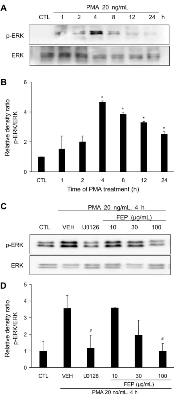
는 matrix metalloproteinases(MMPs)를 활성화하고 MMP 는 pro-EGFR ligands를 세포 표면에서 분리시켜 성숙한 EGFR ligands로 전환시킨다. EGFR ligands는 EGF 수용체 에 결합하고 intracellular tyrosine kinase를 인산화시키 며, mitogen-activated protein kinase kinase(MEK), ERK 의 활성화가 유발되고 전사인자인 specificity protein 1 (Sp1) 등이 활성화되어 MUC5AC gene promoter 상의 특 이결합 부위에 결합함으로써 MUC5AC 유전자의 전사와 뮤 신 단백질의 생성이 촉발되는 것으로 알려져 있다(Lee 등, 2002; Hewson 등, 2010). PMA에 의해 유도된 염증반응에 서 FEP가 증가한 점액성분인 MUC5AC를 조절하는 기전을 확인하기 위해 점액조절인자인 ERK의 활성화를 western blot을 이용하여 측정하였다. 그 결과 PMA 처리 후 4시간이 경과했을 때 ERK의 활성화(4.68±0.06)가 가장 높게 나타 나는 것을 확인할 수 있었다(Fig. 3A, 3B). PMA에 의해 유 도된 염증반응에서 증가한 점액조절인자인 ERK 활성화에 대한 FEP의 억제 효과를 조사하기 위해 FEP를 10, 30, 100 μg/mL의 농도로 세포에 30분 동안 전처리한 후, PMA 20 ng/mL를 4시간 동안 처리하였다. 그 결과 FEP 30 μg/mL 이상부터 대조군(3.56±0.77)과 비교했을 때 ERK의 활성화 가 억제되는 것으로 나타났고, 100 μg/mL의 농도(0.97±
0.47)에선 유의성 있게 억제되는 것을 확인하였다(Fig. 3C, 3D). 또한 Fig. 1B와 Fig. 2B의 결과에서 ERK 억제제인 U0126 처리에 의해 MUC5AC 단백질의 분비와 유전자 발 현이 억제되는 것을 확인하였다. 이는 FEP가 NCI-H292 세포에서 ERK의 활성화 억제를 통해 PMA에 의해 유도된 염증반응에서 증가한 점액성분인 MUC5AC 분비 및 발현 억제에 효능을 나타낼 것으로 사료된다.
FEP의 항산화 효능
페퍼민트는 많은 연구에서 강력한 항산화능을 가진다고 알려져 있으므로(Riachi와 Maria, 2015; Wu 등, 2019) FEP의 항산화능을 검증하기 위해서 DPPH 라디칼 소거능 을 측정하였다. DPPH법은 천연물의 항산화 활성을 측정하 기 위해 가장 많이 이용되고 있는 방법으로 항산화 능력이 있는 물질이 안정한 DPPH 라디칼에 전자를 제공하고 환원 시켜 보라색에서 노란색으로 탈색반응이 나타나는 원리를 이용하여 항산화 정도를 측정한다(Bondet 등, 1997). 양성 대조군으로 Trolox(CALBIOCHEM, Darmstadt, Germany) 1 mM을 사용했으며 80.2±2.1%의 항산화능을 나타냈다.
FEP 농도별로 DPPH 라디칼 소거능을 측정한 결과, FEP의 농도가 높아질수록 유의적으로 DPPH 라디칼 소거능이 증 가하였다. 10 mg/mL에서 51.01±3.01%, 30 mg/mL에서 61.9±4.4%의 높은 항산화능을 나타내었으며, FEP의 DPPH 라디칼 소거능의 EC50은 9.5 μg/mL로 나타났다(Fig. 4). 이 와 같은 결과로 볼 때 FEP는 천연 항산화 소재로서 가능성 이 있다고 판단된다.
0 20 40 60 80 100
1 3 10 30 100 Trolox
FEP (μg/mL)
DPPH scavenging activity (%) .
*
*
*
Fig. 4. DPPH radical scavenging activity (%) of FEP. DPPH radical scavenging activity of the FEP increased in a concen- tration-dependent manner. Data are presented as the mean±SEM (n=3).
0 2 4 6
CTL 1 2 4 8 12 24
Time of PMA treatment (h) Relative density ratio . p-ERK/ERK .
*
*
*
*
B
0 1 2 3 4 5
CTL VEH U0126 10 30 100
PMA 20 ng/mL, 4 h Relative density ratio . p-ERK/ERK .
#
#
FEP (μg/mL)
D
p-ERK
ERK
PMA 20 ng/mL
CTL 1 2 4 8 12 24 h
PMA 20 ng/mL, 4 h FEP (μg/mL) CTL VEH U0126 10 30 100
p-ERK
ERK
A
B
C
D
Fig. 3. Effects of FEP on PMA-induced phosphorylation of ERK in NCI-H292. (A, B) PMA increases the phosphorylation of ERK. Cells were treated with PMA (20 ng/mL) for the indicated time periods (1, 2, 4, 8, 12, and 24 h). The expression of protein signals were (A) analyzed via western blot and (B) quantified using densitometry. Values represent pERK/ERK ratio of 2 in- dependent experiments. *P<0.05 vs CTL. (C, D) FEP inhibits ERK phosphorylation. Cells were treated with peppermint (10, 30, and 100 μg/mL) or U0126 (20 μM) for 30 min, followed by PMA (20 ng/mL) stimulation for 4 h. The phosphorylation of ERK was (C) evaluated by western blot and (D) quantified using densitometry. Significant difference were indicated by
#P<0.05 vs VEH (n=3).
FEP의 세포 내 활성산소 생성 억제 효능 측정
ROS는 주로 세포의 미토콘드리아 대사의 부산물로 발생 하며 비・부비동염과 천식, 만성 폐쇄성 폐질환, 낭종성 섬유 증과 같은 염증성 호흡기 질환 악화의 중요인자 중 하나로 알려져 있다(Wiegman 등, 2015). 비극성분자인 DCF-DA 가 세포 내로 용이하게 들어간 후에 세포 내 esterase 효소 에 의해 acetate기가 떨어져 나가고 비형광성 물질인 DCFH 가 되며, 이 물질이 활성산소에 의해 산화되어 형광물질인 DCF로 바뀌는 원리를 이용하여 세포 내에 존재하는 활성산 소를 측정하였다(Okimoto 등, 2000). PMA는 세포 내 NADPH oxidase 활성을 통하여 ROS 생성을 유도한다고 알 려져 있다(Kirchner 등, 2012). NCI-H292 세포에서 PMA 자극에 의한 염증반응에 의해 ROS가 생성되는 조건을 구하 기 위하여 PMA 20 ng/mL를 처리 후 15, 30, 60, 120, 240 분이 경과했을 때 DCFH-DA 형광물질을 첨가한 뒤 con- focal fluorescence microscopy를 이용하여 ROS 생성량 을 측정하였다. 그 결과 정상군(3.93±0.9)과 비교했을 때 PMA 처리 후 60분이 경과 시 ROS 생성량(38.12±6.77)이 가장 높게 나타나는 것을 확인하였다(Fig. 5B).
FEP가 PMA로 유도한 산화적 스트레스 상태에서 NCI- H292 세포 내의 항산화 효과를 알아보기 위해 FEP를 농도 별로 처리하고, DCF-DA 염색 방법으로 세포 내 항산화 효 과를 측정했으며 측정한 결과는 Fig. 5와 같다. NCI-H292 세포에서 PMA를 20 ng/mL 농도로 1시간 처리했을 때 정상 군(5.12±4.3)에 비해 세포 내 ROS는 30.5±9.1까지 증가하 였다. ROS가 증가한 세포에 10, 30, 100 μg/mL FEP 처리 시 10 μg/mL(18.35±3.4) 이상부터 ROS 생성이 억제되는 것이 나타났고, 30, 100 μg/mL의 농도(5.09±1.9, 4.9±
2.6)에서는 정상군 수준까지 ROS 생성이 감소하는 것을 확 인하였다. 이를 통해서 FEP가 NCI-H292 세포에서 ROS 생성 억제를 통해 PMA에 의해 유도된 염증반응에서 증가한 점액성분인 MUC5AC 분비 및 발현 억제에 효능을 나타내
0 5 10 15 20 25 30 35 40 45
CTL VEH U0126 10 30 100 Trolox
PMA 20 ng/mL, 1 h
DCF-DA (relative fluorescence) .
FEP (μg/mL)
#
#
# #
PMA 20 ng/mL
0 15 30 60 120 240 min
0 5 10 15 20 25 30 35 40 45 50
0 50 100 150 200 250
Time of PMA treatment (min)
DCF-DA (relative fluorescence) .
PMA 20 ng/mL, 1 h FEP (μg/mL)
CTL VEH U0126 10 30 100 Trolox
A
B
C
D
Fig. 5. Effects of FEP on PMA-induced ROS production in NCI-H292. PMA generates ROS production. Cells were treated with PMA (20 ng/mL) at 0, 15, 30, 60, 120, and 240 min, followed by incubation with DCF-DA dye for 30 min. Changes in ROS production was (A) observed through laser confocal microscopy and (B) quantified using densitometry. Data are presented as the mean±SEM (n=3). FEP attenuates the generation of ROS. Cells were pretreated with peppermint (10, 30, and 100 μg/mL), U0126 (20 μM) or Trolox (1 mM) for 24 h, followed by stimulation with PMA (20 ng/mL) for 1 h. Cells were incubated with DCF-DA dye for 30 min. Changes in ROS production was (C) observed through laser confocal microscopy and (D) quantified using densitometry. #P<0.05 vs VEH.
는 것으로 사료된다. 요 약
본 연구에서는 다양한 치료적 효능이 보고된 페퍼민트의 효
능을 증진하기 위하여 유산균으로 발효하여 페퍼민트 추출 발효물(FEP)을 수득하였으며, FEP의 점액 과다분비에 대 한 조절 효능을 확인하고 그 기전을 규명하여 호흡기 개선에 활용될 수 있도록 정확한 용량과 효능 관계를 증명하고자 한다. 먼저 FEP는 PMA에 의해 증가한 MUC5AC의 유전자 발현 및 단백질 분비를 농도 의존적으로 억제하였으며 100 μg/mL의 농도에서 가장 효과적이었다. 이러한 억제기전은 MUC5AC 발현에 주로 관여하는 것으로 알려진 ERK/MAPK 신호전달 체계를 통해 이루어지는 것으로 추정할 수 있다.
또한 FEP의 항산화 효능을 관찰한 결과 농도 의존적인 DPPH 라디칼 소거능에 대한 효능과 ROS 억제 능력을 통해 FEP의 항산화능이 우수함을 확인하였다. 이러한 결과는 FEP가 PMA에 의해 증가한 MUC5AC의 유전자 발현을 억 제하고 ERK의 활성을 억제하여 활성산소 생성조절에 관여 한다. 이를 통해 FEP가 점액 과분비를 억제할 수 있는 새로 운 조절제로서의 활용 가능성이 있음을 시사한다. 또한 페퍼 민트의 호흡기 개선 기능을 확인하여 기능성 허브 소재로 개발한다면 현대인의 건강증진 및 기능성식품 산업발전 모 두에 기여할 수 있을 것으로 사료된다.
감사의 글
본 연구는 농림축산식품부 재원으로 포천허브 6차산업화 사 업단의 지원에 의하여 수행되었음.
REFERENCES
Ahn JH, Kim HG, Seo BH, Choi YS, Song SY, Bae CH, et al. Effect of multi-walled carbon nanotubes on MUC5AC and MUC5B expression in airway epithelial cells. Korean J Oto- rhinolaryngol-Head Neck Surg. 2015. 58:552-557.
Ben-Arye E, Dudai N, Eini A, Torem M, Schiff E, Rakover Y.
Treatment of upper respiratory tract infections in primary care:
a randomized study using aromatic herbs. Evid Based Com- plement Alternat Med. 2011. Article ID 690346. https://doi.
org/10.1155/2011/690346
Bondet V, Brand-Williams W, Berset C. Kinetics and mecha- nisms of antioxidant activity using the DPPH・ free radical method. LWT-Food Sci Technol. 1997. 30:609-615.
Chopra DP, Shoemaker RL, Taylor GW, Mathieu PA. Characteri- zation of epithelial cell cultures derived from human tracheal glands. In Vitro Cell Dev Biol. 1991. 27:13-20.
Heo HJ, Lee HJ, Kim YS, Kang SS, Son KH, Seok JH, et al.
Effects of baicalin and wogonin on mucin release from cultur- ed airway epithelial cells. Phytother Res. 2007. 21:1130-1134.
Hewson CA, Edbrooke MR, Johnston SL. PMA induces the MUC5AC respiratory mucin in human bronchial epithelial cells, via PKC, EGF/TGF-α, Ras/Raf, MEK, ERK and Sp1- dependent mechanisms. J Mol Biol. 2004. 344:683-695.
Hewson CA, Haas JJ, Bartlett NW, Message SD, Laza-Stanca V, Kebadze T, et al. Rhinovirus induces MUC5AC in a human infection model and in vitro via NF-κB and EGFR pathways.
Eur Respir J. 2010. 36:1425-1435.
Ishinaga H, Takeuchi K, Kishioka C, Suzuki S, Basbaum C, Majima Y. Pranlukast inhibits NF-κB activation and MUC2
gene expression in cultured human epithelial cells. Pharma- cology. 2005. 73:89-96.
Kim H, Choi YS, Lee JH, Park NK, Park CH, Lee YH, et al.
Effect of betulinic acid on MUC5AC and MUC5B expression in airway epithelial cells. Korean J Otorhinolaryngol-Head Neck Surg. 2014. 57:526-532.
Kim KC, Opaskar-Hincman H, Bhaskar KR. Secretions from primary hamster tracheal surface epithelial cells in culture:
mucin-like glycoproteins, proteoglycans, and lipids. Exp Lung Res. 1989. 15:299-314.
Kirchner T, Möller S, Klinger M, Solbach W, Laskay T, Behnen M. The impact of various reactive oxygen species on the for- mation of neutrophil extracellular traps. Mediators of inflamm.
2012. Article ID 849136. https://doi.org/10.1155/2012/849136 Kong BM, Park MJ, Min JW, Kim HB, Kim SH, Kim SY, et
al. Physico-chemical characteristics of white, fermented and red ginseng extracts. J Ginseng Res. 2008. 32:238-243.
Leach MJ, Page AT. Herbal medicine for insomnia: a systematic review and meta-analysis. Sleep Med Rev. 2015. 24:1-12.
Lee HW, Ahn DH, Crawley SC, Li JD, Gum JR Jr, Basbaum CB, et al. Phorbol 12-myristate 13-acetate up-regulates the transcription of MUC2 intestinal mucin via Ras, ERK, and NF-κB. J Biol Chem. 2002. 277:32624-32631.
Lin MY, Yen CL. Antioxidative ability of lactic acid bacteria.
J Agric Food Chem. 1999. 47:1460-1466.
Liu R, Guo X, Park Y, Huang X, Sinha R, Freedman ND, et al. Caffeine intake, smoking, and risk of Parkinson disease in men and women. Am J Epidemiol. 2012. 175:1200-1207.
McKay DL, Blumberg JB. A review of the bioactivity and poten- tial health benefits of peppermint tea (Mentha piperita L.).
Phytother Res. 2006. 20:619-633.
Okimoto Y, Watanabe A, Niki E, Yamashita T, Noguchi N. A novel fluorescent probe diphenyl-1-pyrenylphosphine to fol- low lipid peroxidation in cell membranes. FEBS Lett. 2000.
474:137-140.
Riachi LG, De Maria CAB. Peppermint antioxidants revisited.
Food Chem. 2015. 176:72-81.
Savikin K, Zdunić G, Menković N, Zivković J, Cujić N, Tereščenko M, et al. Ethnobotanical study on traditional use of medicinal plants in South-Western Serbia, Zlatibor district.
J Ethnopharmacol. 2013. 146:803-810.
Sekine K, Toida T, Saito M, Kuboyama M, Kawashima T, Hashimoto Y. A new morphologically characterized cell wall preparation (whole peptidoglycan) from Bifidobacterium in- fantis with a higher efficacy on the regression of an estab- lished tumor in mice. Cancer Res. 1985. 45:1300-1307.
Shao MXG, Ueki IF, Nadel JA. Tumor necrosis factor α-conver- ting enzyme mediates MUC5AC mucin expression in cultured human airway epithelial cells. Proc Natl Acad Sci U S A.
2003. 100:11618-11623.
Sharafi SM, Rasooli I, Owlia P, Taghizadeh M, Astaneh SDA.
Protective effects of bioactive phytochemicals from Mentha piperita with multiple health potentials. Pharmacogn Mag.
2010. 6:147-153.
Sikder A, Lee HJ, Ryu J, Park SH, Kim JO, Hong JH, et al.
Apigenin and wogonin regulate epidermal growth factor re- ceptor signaling pathway involved in MUC5AC mucin gene expression and production from cultured airway epithelial cells. Tuberc Respir Dis. 2014. 76:120-126.
Song EJ, Bae CH, Kim JY, Kim YW, Park SY, Song SY, et al.
Effect of epigallocatechin-3-gallate on PMA-induced MUC5B expression in human airway epithelial cells. Clin Exp Otorhi- nolaryngol. 2013. 6:237-242.
Song KS, Lee WJ, Chung KC, Koo JS, Yang EJ, Choi JY, et
al. Interleukin-1β and tumor necrosis factor-α induce MUC5AC overexpression through a mechanism involving ERK/p38 mi- togen-activated protein kinases-MSK1-CREB activation in hu- man airway epithelial cells. J Biol Chem. 2003. 278:23243- 23250.
Voynow JA, Rubin BK. Mucins, mucus, and sputum. Chest.
2009. 135:505-512.
Wiegman CH, Michaeloudes C, Haji G, Narang P, Clarke CJ, Russell KE, et al. Oxidative stress-induced mitochondrial dysfunction drives inflammation and airway smooth muscle remodeling in patients with chronic obstructive pulmonary disease. J Allergy Clin Immunol. 2015. 136:769-780.
Wu DY, Wu R, Reddy SP, Lee YC, Chang MMJ. Distinctive
epidermal growth factor receptor/extracellular regulated ki- nase-independent and -dependent signaling pathways in the induction of airway mucin 5B and mucin 5AC expression by phorbol 12-myristate 13-acetate. Am J Pathol. 2007. 170:
20-32.
Wu Z, Tan B, Liu Y, Dunn J, Guerola PM, Tortajada M, et al.
Chemical composition and antioxidant properties of essential oils from peppermint, native spearmint and scotch spearmint.
Molecules. 2019. 24:2825. doi: 10.3390/molecules24152825.
Zamin I, Majid A, Ali I, Khan TA, Ibrar M, Ali shah J, et al.
In-vitro antibacterial activity of Mentha piperita leaf extracts to some selective pathogenic bacterial strains. Int J Med Pharm Sci Res Rev. 2013. 103:1-13.