ISSN 2288-8101(Print) ISSN 2288-8586(Online)
Case Report
RECEIVED September 6, 2014, REVISED September 17, 2014, ACCEPTED October 16, 2014 Correspondence to Seung-O Ko
Department of Oral and Maxillofacial Surgery, School of Dentistry, Chonbuk National University 567 Baekje-daero, Deokjin-gu, Jeonju 561-756, Korea
Tel: 82-63-250-2113, Fax: 82-63-250-2089, E-mail: [email protected]
Copyright © 2014 by The Korean Association of Maxillofacial Plastic and Reconstructive Surgeons. All rights reserved.
CC
This is an open access article distributed under the terms of the Creative Commons Attribution Non-Commercial License (http://creativecommons.org/licenses/
by-nc/3.0) which permits unrestricted non-commercial use, distribution, and reproduction in any medium, provided the original work is properly cited.
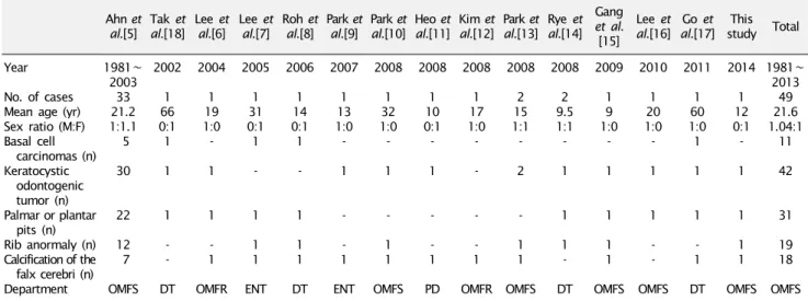
Nevoid Basal Cell Carcinoma Syndrome: A Case Report and Review of Korean Cases
Eun-Joo Jung 1 , Hyokeun Shin 1,2 , Jin-A Baek 1,2 , Dae-Ho Leem 1,2 , Seung-O Ko 1,2
1
Department of Oral and Maxillofacial Surgery, School of Dentistry, Chonbuk National University,
2
![Table 2. Relative frequencies of associated complications compared with other countries Evans et al.[22] Shanley et al.[26] Kimonis et al.[19] Lo Muzio et al.[27] Pruvost-Balland et al.[28] Ahn et al.[5] Shimada](https://thumb-ap.123doks.com/thumbv2/123dokinfo/5113047.330458/3.892.79.816.932.1127/relative-frequencies-associated-complications-compared-countries-shanley-kimonis.webp)