Received May 15, 2013, Revised June 10, 2013, Accepted for publication June 13, 2013
Corresponding author: Moon-Bum Kim, Department of Dermatology, Pusan National University Hospital, 179 Gudeok-ro, Seo-gu, Busan 602-739, Korea. Tel: 82-51-240-7338, Fax: 82-51-245-9467, E-mail:
This is an Open Access article distributed under the terms of the Creative Commons Attribution Non-Commercial License (http://
creativecommons.org/licenses/by-nc/3.0) which permits unrestricted non-commercial use, distribution, and reproduction in any medium, provided the original work is properly cited.
ORIGINAL ARTICLE
Basal Cell Carcinoma-Mimicking Lesions in Korean Clinical Settings
Hoon-Soo Kim1, Tae-Wook Kim, Je-Ho Mun, Margaret Song, Hyun-Chang Ko, Byung-Soo Kim1, Moon-Bum Kim1
Department of Dermatology, Pusan National University School of Medicine, Yangsan,
1Biomedical Research Institute, Pusan National University Hospital, Busan, Korea
Background: Basal cell carcinoma (BCC) is the most com- mon form of skin cancer and possesses various clinical features including translucency, ulceration, pigmentation, telangiectasia, and rolled borders. Accordingly, many cuta- neous lesions can mimic BCCs and differential diagnosis is difficult. Objective: To clarify the differences in clinical cha- racteristics between BCCs and BCC-mimicking lesions (BMLs), and to determine which clinical characteristics are helpful for an accurate clinical diagnosis of BCC. Methods:
We performed clinicopathologic analysis of cutaneous le- sions that received a clinical diagnosis of BCC. All lesions included in this study showed more than one of the following characteristics of BCCs: translucency, ulceration, flecked pigmentation, black or blue hue, telangiectasia, and rolled borders. We compared six clinical characteristics between the BCC group and the BML group. Results: Among 48 lesions in the BML group, there were 15 premalignant or malignant lesions and 33 benign lesions. Various derma- toses mimicking BCC that have not been reported in the dermatological literature were identified, including angio- sarcoma, vulvar intraepithelial neoplasm, foreign body gra- nuloma, intravascular papillary endothelial hyperplasia, sarcoidosis, and others. Compared to the BML group, the BCC group had a significantly higher frequency of translu-
cency (76.3% vs. 52.1%, p<0.001), ulceration or erosion (44.2% vs. 27.1%, p=0.022), black or blue hue (40.0% vs.
22.9%, p=0.020), and rolled borders (49.5% vs. 14.6%, p<
0.001). Cutaneous lesions with two or less clinical features of BCC were significantly more likely to be BMLs.
Conclusion: The results of this study could be helpful for the differential diagnosis of BCCs and BCC-mimicking cuta- neous lesions. (Ann Dermatol 26(4) 431∼436, 2014) -Keywords-
Basal cell carcinoma, Differential diagnosis, Mimicking
INTRODUCTION
Basal cell carcinoma (BCC) is the most common form of skin cancer, and its prevalence has been consistently increasing. BCCs have several textbook clinical charac- teristics such as translucency, ulceration, pigmentation, telangiectasia, and rolled borders. These characteristics are shared by many skin diseases and each subtype of BCCs should therefore be differentiated from a variety of other cutaneous disorders (Table 1)1. A variety of cutane- ous lesions can mimic the clinical features of BCCs, in- cluding adult-onset xanthogranuloma, rhabdomyomatous mesenchymal hamartoma, Darier’s disease, epidermal cysts, lymphoma, and several others (Table 2)2-15. There- fore, the differential diagnosis of BCC and BCC-mimicking lesions (BMLs) is complex, yet there have been no studies clarifying the differences in clinical characteristics bet- ween BCCs and BMLs, and which clinical characteristics are most helpful for making accurate clinical diagnoses of BCC. Accordingly, we conducted a comparative study bet- ween BCCs and BMLs.
Table 1. Differential diagnosis of basal cell carcinoma Clinical subtype Differential diagnosis Nodular Intradermal nevus
Squamous cell carcinoma Skin appendage tumors Seborrheic keratosis Dermatofibroma Pigmented Malignant melanoma
Skin appendage tumor Compound nevus Blue nevus
Morpheaform Scar
Morphea Trichoepithelioma Superficial Bowen’s disease
Paget’s disease Psoriasis Eczema Fibroepithelioma Skin tag
Fibroma
Papillomatous dermal nevus
Modified from Fitzpatrick's dermotology in general medicine. 8th ed.
New York: McGrow-Hill, 2012:1294-13031.
Table 2. Reported cases of other dermatoses mimicking basal cell carcinoma in searching PubMed/MEDLINE*
Case Diagnosis Diagnostic pitfalls
Bohn and Sanchez-Sosa2 Rhabdomyomatous mesenchymal hamartoma Focal ulceration Lovato et al.3 Adult onset xanthogranuloma Yellowish hue, telangiectasia
Russell et al.4 Darier disease Telangiectasia, translucency
Akinyemi et al.5 Diffuse large B-cell lymphoma Ulceration, rolled border
Ghaffar et al.6 Epidermoid cyst Telangiectasia, translucency
Hinz et al.7 Lymphoepithelioma-like carcinoma Central erosion, telangiectasia Lott et al.8 T-cell primary cutaneous anaplastic large cell lymphoma Ulceration, rolled border, translucency Hague and Ilchyshyn9 Allergic contact dermatitis due to nickel Ulceration, rolled border
Bechara et al.10 Pomade crust Ulceration
Askar et al.11 Syringocystadenoma papilliferum Ulceration, translucency Goto et al.12 Digital syringomatous carcinoma Ulceration, rolled border
Ingleton et al.13 Cutaneous cryptococcosis Erosion, rolled border
Tsao et al.14 Chronic varicella zoster infection Erosion, rolled border, translucency
Lobur et al.15 Irritant contact dermatitis Erosion, rolled border
*Studies published between 1983 and 2011 with the searching terms of “mimicking basal cell carcinoma.”
Table 3. Demographic data of both groups
BCCs BCC-mimicking lesions
Total number 608 48
Male : Female 1 : 1.2 1 : 1
Mean age (yr) 65.4 (18∼89) 64.9 (32∼87) Mean duration of disease (mo) 35.8 (4∼360) 34.6 (1∼240) Location of lesion
Head and neck 556 (91.4)00 46 (95.8)0
Trunk 40 (6.6)00 2 (4.2)0
Extremities 12 (2.0)00 0 (0.0)0
Values are presented as number, mean (range), or number (%).
BCC: basal cell carcinoma.
MATERIALS AND METHODS
Six clinical characteristics of BCCs
There are six textbook clinical characteristics of BCCs: tra- nslucency, ulceration, pigmentation, telangiectasia, and rolled borders1. In this study, “pigmentation” was divided into flecked pigmentation and black or blue hue since pigmented BCCs, which are frequent in Asia, possess various degrees of pigmentation16. We defined “flecked pigmentation” as multiple small spots of pigmentation, and “black or blue hue” as homogenous pigmentation.
More than one of these six clinical characteristics of BCCs was present in all cases reported as a BML, or in cases mistakenly reported as BCC, in the dermatological litera- ture (Table 2).
Subjects
We enrolled 656 patients with cutaneous lesions, of whi- ch the first clinical diagnosis was BCC at the Skin Cancer Clinic of the Department of Dermatology at Pusan Nati- onal University Hospital, from August 2002 to July 2011.
The study was approved by the ethics committee of PNUH (E-2013023). All lesions showed more than one of the six characteristics listed above. The number of patients in the BCC group was 608, and in the BML group was 48.
The demographic data are shown in Table 3.
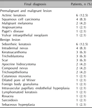
Table 4. Final diagnosis of basal cell carcinoma-mimicking lesions
Final diagnosis Patients, n (%) Premalignant and malignant lesion
Actinic keratosis
Squamous cell carcinoma Malignant melanoma Angiosarcoma Paget’s disease
Vulvar intraepithelial neoplasm
6 (12.5) 4 (8.3) 2 (4.2) 1 (2.1) 1 (2.1) 1 (2.1) Benign lesion
Seborrheic keratosis Intradermal nevus Keratoacanthoma Trichoblastoma Scar
Apocrine hidrocystoma Compound nevus Trichoepithelioma Cutaneous myxoma Dilated pore of Winer Foreign body granuloma
Intravascular papillary endothelial hyperplasia Lymphomatoid keratosis
Rosacea Sarcoidosis
6 (12.5) 4 (8.3) 3 (6.3) 3 (6.3) 3 (6.3) 2 (4.2) 2 (4.2) 2 (4.2) 1 (2.1) 1 (2.1) 1 (2.1) 1 (2.1) 1 (2.1) 1 (2.1) 1 (2.1)
Sebaceous hyperplasia 1 (2.1)
Fig. 1. Percentage according to number of clinical characteristics of basal cell carcinoma (BCC) in cases of BCCs and BCC-mi- micking lesions. *p<0.05.
Table 5. Comparison of six clinical characteristics between basal cell carcinomas (BCCs) and BCC-mimicking lesions*
Clinical features BCCs,
n (%) BCC-mimicking lesions, n (%) p-value Translucency 464 (76.3) 25 (52.1) <0.001 Telangiectasia 253 (41.6) 25 (52.1) <0.158 Flecked pigmentation 230 (37.9) 21 (43.8) <0.416 Ulceration or erosion 268 (44.2) 13 (27.1) <0.022 Black or blue hue 243 (40.0) 11 (22.9) <0.020 Rolled border 301 (49.5) 07 (14.6) <0.001
*Fisher’s exact test was utilized for statistical analysis. The level of significance in this study refers to a p-value of below 0.05.
Assessment
1) Final diagnoses of the BML group
After histopathologic evaluation, we analyzed which cuta- neous diseases can mimic BCC.
2) Comparison of the six characteristics between the BCC group and the BML group
On the basis of clinical photographs, we evaluated how often each characteristic (translucency, telangiectasia, fleck- ed pigmentation, ulceration or erosion, black or blue hue, and rolled borders) was found in the BCC group and the
BML group. All statistical analyses were performed by means of IBM SPSS Statistics 21.0 (IBM Co., Armonk, NY, USA).
To compare the six characteristics between the BCC group and the BML group, statistical analysis was performed using Fisher’s exact test. The level of significance in this study was set at a p-value of below 0.05.
RESULTS
Final diagnoses of the BML group (Table 4)
Among 48 BMLs, there were 15 premalignant or malignant lesions and 33 benign lesions. Cases of precancerous and malignant lesions included six cases of actinic keratosis;
four cases of squamous cell carcinoma; two cases of malignant melanoma; and one case each of angiosarcoma, Paget’s disease, and vulvar intraepithelial neoplasia. Benign disorders included six cases of seborrheic keratosis; four cases of intradermal nevus; three cases each of keratoacan- thomas, trichoblastoma, and scars; two cases each of trichoepithelioma, compound nevus, and apocrine hidro- cystoma; and one case each of cutaneous myxoma, dilated pore of Winer, foreign body granuloma, intravascular papillary endothelial hyperplasia, lymphomatoid keratosis, rosacea, sarcoidosis, and sebaceous hyperplasia.
Comparison of the six characteristics between the BCC group and the BML group
Compared to the BML group, the BCC group had a signi- ficantly higher frequency of translucency (76.3% vs.
52.1%, p<0.001), ulceration or erosion (44.2% vs.
27.1%, p=0.022), black or blue hue (40.0% vs. 22.9%, p=0.020), and rolled borders (49.5% vs. 14.6%, p<
0.001). In the case of telangiectasia (41.6% vs. 52.1%,
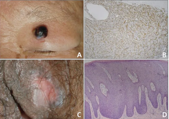
Fig. 2. Various malignant and premalignant cutaneous lesions mimicking basal cell carcinoma. (A, B) Angiosarcoma. Diagnostic pitfalls;
black hue (CD31 ×100). (C, D) Vulvar intraepithelial neoplasm. Diagnostic pitfalls; translucency, telangiectasia, erosion, blue hue, and rolled border (H&E, ×100).
p=0.158) and flecked pigmentation (37.9% vs. 43.8%, p=0.416), there was no statistically significant difference between the two groups (Table 5).
With respect to the number of clinical characteristics in each case, when the number present was two or less, the relevant lesions were highly likely to belong to the BML group (35.7% vs. 72.9%, p<0.001). When the number was three or more, they were highly likely to belong to the BCC group (64.3% vs. 27.1%, p<0.001; Fig. 1).
DISCUSSION
The clinical characteristics of BCC are commonly known to include translucency, ulceration, pigmentation, telan- giectasia, and a rolled border. Of these, ulceration, pig- mentation, and telangiectasia are commonly seen in daily dermatological practice. Therefore, various diseases, inclu- ding infectious skin disorders, can mimic BCC (Table 1, 2), and diagnostic pitfalls might exist between BCC and BMLs. However, there has been no systematic trial to
analyze and resolve these issues.
In this study, the BML group included various malignant and benign dermatoses. Among these, there were a variety of additional cutaneous disorders that have not yet been reported: angiosarcoma, vulvar intraepithelial neoplasm, foreign body granuloma, intravascular papillary endothe- lial hyperplasia, sarcoidosis, and others (Fig. 2, 3).
Among the six main clinical characteristics, translucency, ulceration or erosion, black or blue hue, and rolled bor- ders were found more frequently in the BCC group and this was statistically significant. In the case of telangiec- tasia and flecked pigmentation, there was no significant difference between the BCC group and the BML group.
Telangiectasia and flecked pigmentation could therefore be less reliable clinical characteristics for the diagnosis of BCCs in Korean clinical practice than the other four. Over the past two decades, laser ablation of benign skin lesions such as nevi or seborrheic keratosis on the face has gained wide popularity in Korea. Accordingly, some patients with BCCs are likely to be treated by laser ablation after being
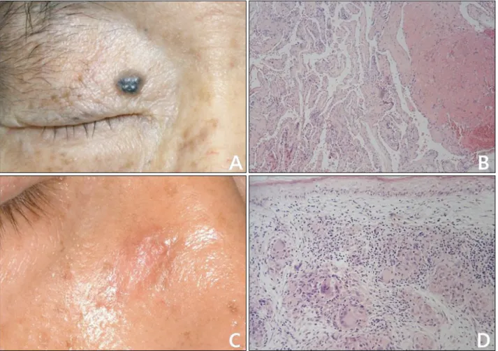
Fig. 3. Various benign cutaneous lesions mimicking basal cell carcinoma (A, B) Intravascular papillary endothelial hyperplasia. Diagnostic pitfalls; blue hue (H&E, ×40). (C, D) Sarcoidosis. Diagnostic pitfalls; translucency, rolled border (H&E, ×100).
diagnosed with these benign skin tumors17. In this respect, the results of this study may be helpful for the early detection of BCCs in Korea.
In addition, when a lesion shows two or fewer of the six textbook characteristics of BCCs, it is highly likely to be a BML. When three or more clinical characteristics are pre- sent, it is highly likely to be a BCC. Therefore, when one or two clinical characteristics of BCCs are observed at the time of cutaneous lesion examination, other cutaneous disorders should be considered before BCC.
In conclusion, we identified more cutaneous disorders capable of mimicking BCCs than have been previously reported in the literature. In cases of BCC in Korea, translucency, ulceration or erosion, black or blue hue, and rolled borders could be more reliable as diagnostic cli- nical characteristics than telangiectasia and flecked pig- mentation. If a cutaneous lesion suspected to be a BCC possesses three or more clinical characteristics of BCCs, including translucency, telangiectasias, flecked pigmen- tation, ulceration or erosion, black or blue hue, and rolled borders, it is significantly more likely to be a BCC. The
results of this study are thought to contribute to the accurate clinical diagnosis of BCC and provide more detailed information compared to dermoscopic findings alone.
ACKNOWLEDGMENT
This study was supported by a Medical Research Institute Grant (2011-08), Pusan National University Hospital.
REFERENCES
1. Carucci JA, Leffel DJ, Pettersen JS. Basal cell carcinoma. In:
Goldsmith LA, Katz SI, Gilchrest BA, Paller AS, Leffell DJ, Wolff K, editors. Fitzpatrick's dermotology in general medi- cine. 8th ed. New York: McGrow-Hill, 2012:1294-1303.
2. Bohn OL, Sanchez-Sosa S. Rhabdomyomatous mesenchymal hamartoma mimicking basal cell carcinoma. Am J Dermato- pathol 2009;31:309-310.
3. Lovato L, Salerni G, Puig S, Carrera C, Palou J, Malvehy J.
Adult xanthogranuloma mimicking basal cell carcinoma:
dermoscopy, reflectance confocal microscopy and patholo-
gical correlation. Dermatology 2010;220:66-70.
4. Russell DJ, Dutton JJ, Fowler AM. Darier disease mimicking Basal cell carcinoma of the eyelid. Ophthal Plast Reconstr Surg 2009;25:144-146.
5. Akinyemi E, Mai L, Matin A, Maini A. Diffuse large B-cell lymphoma mimicking advanced basal cell carcinoma. J Natl Med Assoc 2007;99:948-950.
6. Ghaffar SA, Clements SE, Lear JT. Epidermoid cysts mimi- cking recurrence of superficial basal cell carcinoma follo- wing photodynamic therapy. Clin Exp Dermatol 2007;32:
223-224.
7. Hinz T, Wiechert A, Bieber T, Bauer R, Schmid-Wendtner MH. Lymphoepithelioma-like carcinoma of the skin mimi- cking a basal cell carcinoma. Eur J Dermatol 2009;19:
179-180.
8. Lott DG, Akst LM, Greene D, Roberts JK. T-cell primary cutaneous anaplastic large cell lymphoma mimicking appea- rance of large basal cell carcinoma. Otolaryngol Head Neck Surg 2006;135:170-171.
9. Hague J, Ilchyshyn A. Nickel allergy mimicking basal cell carcinoma. Contact Dermatitis 2006;54:344-345.
10. Bechara FG, Rotterdam S, Hoffmann K, Altmeyer P, Stücker M, Jansen T. Pomade crust on the scalp mimicking recurrent basal cell carcinoma. Dermatol Nurs 2003;15:426-427.
11. Askar S, Kilinc N, Aytekin S. Syringocystadenoma papilli- ferum mimicking basal cell carcinoma on the lower eyelid: a case report. Acta Chir Plast 2002;44:117-119.
12. Goto M, Sonoda T, Shibuya H, Terashi H, Kai Y, Sato T, et al. Digital syringomatous carcinoma mimicking basal cell carcinoma. Br J Dermatol 2001;144:438-439.
13. Ingleton R, Koestenblatt E, Don P, Levy H, Szaniawski W, Weinberg JM. Cutaneous cryptococcosis mimicking basal cell carcinoma in a patient with AIDS. J Cutan Med Surg 1998;3:43-45.
14. Tsao H, Tahan SR, Johnson RA. Chronic varicella zoster infection mimicking a basal cell carcinoma in an AIDS patient. J Am Acad Dermatol 1997;36:831-833.
15. Lobur DM, Bailin PL, Taylor JS. Severe irritant dermatitis mimicking a basal cell carcinoma. Cleve Clin Q 1983;50:
465-467.
16. Ono T, Egawa K, Yamamoto S, Arao T. Pigmented basal cell carcinoma developing on the lower extremities--three cases masquerading as malignant melanoma. J Dermatol 1989;16:
325-329.
17. Jung DS, Cho HH, Ko HC, Bae YC, Oh CK, Kim MB, et al.
Recurrent basal cell carcinoma following ablative laser procedures. J Am Acad Dermatol 2011;64:723-729.