J Korean Ophthalmol Soc 2018;59(10):989-994 ISSN 0378-6471 (Print)⋅ISSN 2092-9374 (Online)
https://doi.org/10.3341/jkos.2018.59.10.989
Case Report
유리체 내 베바시주맙 및 애플리버셉트 주사를 통해 호전된 망막하출혈을 동반한 맥락막골종 1예
A Case of Choroidal Osteoma with Subretinal Hemorrhage Improved by Intravitreal Bevacizumab and Aflibercept Injections
김 참⋅최경식⋅선해정
Charm Kim, MD, Kyung Seek Choi, MD, Hae Jung Sun, MD
순천향대학교 의과대학 서울병원 안과학교실
Department of Ophthalmology, Seoul Hospital, Soonchunhyang University College of Medicine, Seoul, Korea
Purpose: To report a case of choroidal osteoma (CO) complicated by extensive subretinal hemorrhage treated with intravitreal bevacizumab and aflibercept injections.
Case summary: A 42-year-old female patient presented with decreased visual acuity and a temporal visual field defect in the left eye. The patient had a history of retinal hemorrhage in the left eye 3 years prior, which improved without any treatment. The pa- tient’s visual acuity had decreased to 0.6 at the initial visit. On fundus examination, orange-colored elevated lesions involving the superior peripapillary area with massive subretinal hemorrhage extending to the macular area were revealed. Optical coherence tomography, fluorescein angiography, and B-scan ultrasonography results indicated CO complicated by choroidal neo- vascularization (CNV). With multiple intravitreal injections of bevacizumab and aflibercept (bevacizumab ×1, aflibercept ×2), the patient’s visual acuity improved and the CNV lesion was kept stable without recurrence as of the 1-year follow-up visit.
Conclusions: Intravitreal bevacizumab and aflibercept injections can be helpful in the treatment of CO complicated by CNV, by improving visual acuity and the retinal anatomy.
J Korean Ophthalmol Soc 2018;59(10):989-994
Keywords: Aflibercept, Bevacizumab, Choroidal osteoma, Subretinal hemorrhage
■Received: 2018. 6. 7. ■ Revised: 2018. 7. 24.
■Accepted: 2018. 9. 27.
■Address reprint requests to Hae Jung Sun, MD
Department of Ophthalmology, Soonchunhyang University Seoul Hospital, #59 Daesagwan-ro, Yongsan-gu, Seoul 04401, Korea
Tel: 82-2-709-9354, Fax: 82-2-710-3196 E-mail: [email protected]
* The study was presented as an e-poster at the 117th Annual Meeting of the Korean Ophthalmological Society 2017.
* This study was supported by an Industry-academic cooperation of Soonchunhyang University 2018.
*Conflicts of Interest: The authors have no conflicts to disclose.
ⓒ2018 The Korean Ophthalmological Society
This is an Open Access article distributed under the terms of the Creative Commons Attribution Non-Commercial License (http://creativecommons.org/licenses/by-nc/3.0/) which permits unrestricted non-commercial use, distribution, and reproduction in any medium, provided the original work is properly cited.
맥락막골종은 주로 젊은 여성에서 시신경유두주위에 발 생하는 양성종양으로, 약 75%에서 단안에 발생하며 드물 게 양안에서 발생하기도 한다.1-4 시신경유두주위 또는 시신 경유두곁에 주로 위치하며 황반부까지 침범하기도 한다.3,5 증상은 무증상부터 변시증, 시야결손 등 합병증의 발생 정 도에 따라 다양하게 나타나며, 황백색 또는 황적색의 경계 가 명확한 지도 모양 또는 부채꼴 모양으로 융기된 특징적인 안저 소견과 초음파검사 및 전산화단층촬영상 석회화 골음영 을 확인하여 진단을 할 수 있다. 맥락막골종에서 맥락막신생 혈관이 합병되어 유리체 내 베바시주맙(Avastin®, Genetech, South San Francisco, CA, USA) 및 라니비주맙(Lucentis®, Novartis Pharmaceuticals Corp., East Hanover, NJ, USA)
A B
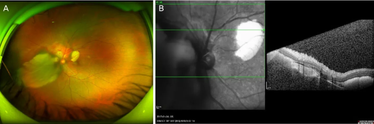
Figure 1. Ultrawide fundus photograph (UW FP) of the left eye. UWFP shows a well demarcated, orange-colored elevated lesion in-
volving superior peripapillary area with massive subretinal hemorrhage extending to the macular area (A). Optical coherence tomog- raphy shows the well-demarcated mass lesion at the choroidal space above the optic nerve head. Diffuse subretinal fluid with sub- retinal hemorrhage is seen (B).A B
Figure 2. Early fluorescein angiogram (FA) of the left eye. Early FA demonstrates hypofluorescence due to bloackage correspond-
ing to subretinal hemorrhage (A). Late FA of the left eye demonstrates hypofluorescence and hyperfluorescence with leakage at the superior disc area (B).주사를 시행 후 호전된 증례가 국외에서 다수 보고되었으 며,6-12 국내에서는 베바시주맙 및 라니비주맙 주사를 시행한 증례는 보고되었으나,13-15 애플리버셉트(Eylea®, Regeneron, Tarrytown, New York, NY, USA and Bayer HealthCare, Berlin, Germany) 주사 후 호전된 증례는 없었다. 저자들은 광범위한 망막하출혈을 동반한 맥락막골종 환자에서 유리 체 내 베바시주맙 및 애플리버셉트 주사를 시행한 후 증상 이 호전된 1예를 경험하여 이를 보고하고자 한다.
증례보고
42세 여자 환자가 좌안의 반복적인 망막하출혈을 주소로 타 병원에서 전원되었다. 환자는 3년 전에도 좌안에 망막하 출혈을 경험하였으며, 그 당시 특별한 치료 없이 증상 및 망막하출혈이 호전되었다고 하였다. 내원 3주 전 좌안에 시 력저하 및 이측시야가 가려 보이는 증상이 있어 타 병원에
서 좌안의 망막하출혈을 진단받고 경과관찰을 하던 중 1주 일 전 재출혈이 발생하면서 이측시야가 더욱 좁아짐을 호 소하여 본원으로 전원되었다. 환자는 특이 전신질환 및 과 거력, 안과적 수술력이나 가족력은 없었다. 내원 당시 좌안 의 나안시력 0.6, 최대교정시력은 0.7이었고 안압은 16 mmHg로 측정되었다. 세극등현미경검사에서 전안부에 특 이소견은 관찰되지 않았으나 안저검사에서 좌안의 시신경 유두 위쪽에 오렌지색 종괴로 보이는 병변과 이를 둘러싼 광범위한 망막하출혈이 관찰되었다(Fig. 1A). 빛간섭단층 촬영에서는 시신경유두 위쪽으로 경계가 분명한 고반사의 종괴와 주변의 광범위한 망막하액의 고임 및 망막하출혈이 관찰되었다(Fig. 1B). 형광안저혈관조영술에서 종괴 부위의 형광염색이 관찰되었으며, 후기 시신경유두 위쪽으로 약한 누출이 확인되었다(Fig. 2). B-scan 안초음파에서 좌안의 시 신경유두주위에 균질한 구조로 강한 반향을 보이면서 시신 경 뒤쪽으로 그림자 음영이 확인되었다(Fig. 3).
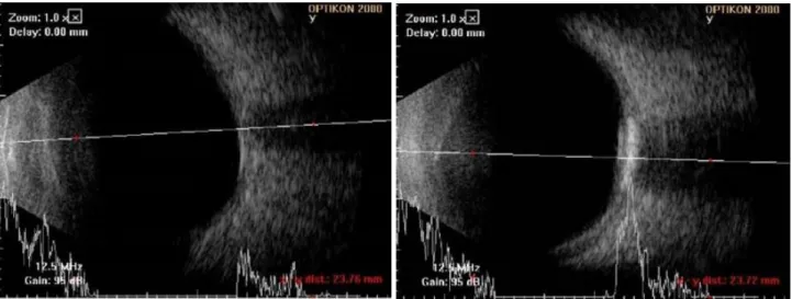
Figure 3. B-scan ultrasonography (USG). B-scan USG demonstrates slightly elevated, highly reflective choroidal mass with posteri-
or acoustic shadowing.형광안저혈관조영술에서 망막하출혈로 인해 누출이 명 확하게 보이지 않았으나, 안저검사 및 빛간섭단층촬영, 안 초음파 결과를 토대로 맥락막골종을 진단할 수 있었다. 맥 락막골종과 함께 맥락막신생혈관이 합병되어 반복적인 망 막하출혈이 발생하였던 것으로 추정하여 맥락막신생혈관 에 대한 치료를 시행하기로 결정하였다. 맥락막신생혈관에 대한 치료로 유리체 내의 항혈관내피성장인자 주사를 단독치 료로 시행하기로 결정하고, 유리체 내 베바시주맙(1.25 mg/
0.05 mL) 주사를 1차 시행하였다. 1개월 뒤 좌안의 최대교 정시력은 0.6으로 안저검사에서 망막하출혈의 범위가 감소 하였고(Fig. 4A), 빛간섭단층촬영에서 망막하액이 줄어들 고 시신경유두 위쪽으로 맥락막신생혈관 병변이 확인되었 다(Fig. 4B).
유리체 내 베바시주맙 주사를 시행하고 1개월 뒤 내원하 였을 때 환자가 주사 약제를 애플리버셉트로 교체하기를 희 망하여 2차 치료는 유리체 내 애플리버셉트(2.0 mg/0.05 mL) 주사를 시행하였다. 유리체 내 애플리버셉트 주사를 시행 하고 1개월 뒤 좌안의 최대교정시력은 1.0이었고, 안저검사 에서 망막하출혈의 범위가 더욱 감소하였으며(Fig. 4C), 빛 간섭단층촬영에서 망막하액은 관찰되지 않았고, 맥락막신 생혈관의 크기도 감소한 것을 확인하였다(Fig. 4D).
유리체 내 애플리버셉트 주사를 한 달 간격으로 한 번 더 시행하고 1개월 뒤 내원하였을 때 좌안의 최대교정시력은 1.0이었고 안저검사에서 망막하출혈 범위가 감소하였고 (Fig. 4E), 빛간섭단층촬영에서 맥락막신생혈관의 크기도 작아진 소견이 보였다(Fig. 4F). 유리체 내 애플리버셉트 주 사를 두 차례 시행한 후에 환자의 시력 및 안저 상태가 안 정되어 이후 경과관찰을 시행하였으며 치료 6개월 후 좌안
의 나안시력 0.9, 최대교정시력은 1.0으로 유지되었으며 안 저검사에서도 망막하출혈의 범위 및 크기가 감소하였다.
1년 뒤 내원하였을 때 좌안의 나안시력 0.9, 최대교정시 력은 1.0이었고 안저검사에서 망막하출혈이 있던 자리에 망막반흔이 생긴 것이 확인되었으며 그 범위 및 크기는 증 가하지 않았다(Fig. 4G). 빛간섭단층촬영에서도 망막하액 은 확인되지 않았고 망막하 융기된 조직의 크기는 안정적 이었으며, 맥락막신생혈관으로 의심되는 병변은 비활성화 된 것으로 판단되었다(Fig. 4H).
고 찰
맥락막골종은 비특이적으로 발생하고 맥락막 전층의 국 소적 골성변화를 보이는 드문 안구 내 양성 종양이다. 이 질환의 전형적인 임상 양상과 초음파 소견, 방사선학적 소 견, 형광안저혈관조영술 및 조직학적 소견은 1978년 Gass et al1과 Williams et al16이 처음 소개하였다. 병변은 주로 황백색 또는 황적색(오렌지색)의 경계가 명확한 지도 모양 또는 부채꼴 모양으로 융기된 특징적인 안저 소견을 보이 며, 종양의 표면은 불규칙적이고 갈색, 주황색 또는 회색의 색소침착을 보일 수 있다. 맥락막골종은 주로 젊은 여성에 서 발병하나 남성 또는 10세 이하의 어린이에서 보고되는 경우도 있으며, 75%에서는 단안에 발생하고 양안에 발생 하는 경우에는 양안의 발병 시기가 서로 다른 것으로 알려 져 있다.5,17 증상은 합병증의 발생 정도에 따라 무증상에서 부터 변시증, 시력저하, 시야결손 및 암점 등 다양하게 나 타날 수 있다.
맥락막신생혈관은 맥락막골종에서 가장 빈도가 높게 발
A B
C D
E F
G H
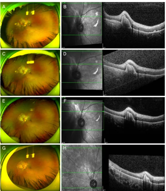
Figure 4. Ultrawide fundus photograph (UWFP) and optical coherence tomography (OCT) of the left eye. UW FP shows decreased
subretinal hemorrahge (SRH) at 1 month after intravitreal bevacizumab injection (A) and decreased subretinal fluid (SRF) with prominent subretinal elevated lesion is seen on OCT (B). 1 month after intravitreal aflibercept injection (1st), much decreased SRH (C) and decreased size of subretinal lesion with minimal SRF (D). 1 month after intravitreal aflibercept injection (2nd), SRH is near- ly absorbed (E) and height of subretinal lesion is more decreased on OCT (F). After 1 year from the first visit, chorioretinal scarring (G) and remnant but stable subretinal lesion is noticed (H).생하는 합병증으로, 맥락막신생혈관이 합병되는 경우 망막 하출혈, 맥락막신생혈관막 형성 및 망막색소상피박리 등으 로 시력 장애를 유발할 수 있으며, 맥락막골종 발병 후 10 년 이내에 31-47%의 환자에서 맥락막신생혈관이 발생한 다.18 맥락막골종 환자에서 2차적으로 발생한 맥락막신생혈 관의 치료에 대해서는 아직 논란이 많다. 맥락막골종 환자 에서 맥락막신생혈관이 동반되지 않은 망막하출혈이 발생 하였을 때에는 치료를 시행하지 않고 자발적으로 호전된 경우도 있었으나19, 맥락막신생혈관이 동반된 경우에는 회
복이 지연되거나 영구적인 시력 장애까지 유발할 수 있다.18 과거에는 레이저광응고술, 경동공온열치료, 광역학요법, 맥 락막골종절제술 등을 시행하였으나, 맥락막신생혈관을 효 과적으로 조절할 수 없었으며 시력예후도 불량하였다.
최근 다수의 국외 증례에 따르면, 맥락막골종 연관 맥락막 신생혈관 환자에서 유리체 내 항혈관내피성장인자 주사의 치료 효과가 입증되고 있으며, 특히 유리체 내 라니비주맙 (Lucentis®, Novartis Pharmaceuticals Corp., East Hanover, NJ, USA)과 베바시주맙(Avastin®, Genetech, South SanFrancisco,
CA, USA) 주사를 시행한 환자에서 망막구조의 개선 및 시 력 호전에 효과가 있음이 확인되었다.6-15 하지만 반복적인 주사 치료로 약제에 대한 내성 발생 시 항혈관내피성장인 자 치료의 반응성이 감소할 수 있으며, 한 연구 보고에 따 르면 맥락막골종 연관 맥락막신생혈관 환자에서 4명 중 1 명이 주사 후에도 지속적으로 망막하액이 존재했다고 보고 하고 있다.9 한 국외 증례에서는 유리체 내 라니비주맙 주 사와 저빈도 광역학요법을 함께 시행한 후 황반하 맥락막 신생혈관막이 감소하였다는 보고가 있었으나,20 또 다른 증 례에서는 맥락막신생혈관을 동반하지 않은 맥락막골종에 서 광역학요법을 시행한 후 망막하출혈이 발생하여 시력예 후가 불량하였다는 보고도 있었다.21
애플리버셉트(Eylea®, Regeneron, Tarrytown, New York, NY, USA and Bayer HealthCare, Berlin, Germany)는 인간 의 수용성 재조합 혈관내피성장인자 수용체로 면역글로불 린 G-1 (Immunoglobulin G1)의 불변 결정가능 조각(fragment crystallizable, Fc) 부위에 융합된 VEGF 수용체-A, B의 주 요 세포 외 vascular endothelial growth factor (VEGF) 결합 도메인을 포함하도록 설계되었다.22 VEGF-A만 억제하는 베 바시주맙과 라니비주맙과는 달리 애플리버셉트는 VEGF-A 뿐만 아니라 VEGF-B, 태반성장인자(placental growth fac- tor, PGF)에도 결합하여 더 높은 결합친화도를 보인다. 게 다가 분자 구조의 생물학적 특성상 베바시주맙, 라니비주 맙과 비교하여 더 오랜 기간 신생혈관 억제 효과를 보여 유 리체 내 주사 횟수를 줄일 수 있다. 최근 많은 증례에서 베 바시주맙이나 라니비주맙 등 고식적인 항혈관내피성장인 자 치료에 반응이 없는 황반변성으로 인해 치료되지 않고 반복되는 망막하액을 가진 환자에서 유리체 내 애플리버셉 트 2.0 mg을 주사 시 시력 호전 및 망막의 해부학적 호전을 보인 예가 다수 보고되었다.23-25
본 증례에서 맥락막신생혈관이 합병된 맥락막골종의 치 료로 유리체 내 베바시주맙 1.25 mg 주사 시행 후 시력 호 전 및 망막구조의 해부학적 개선이 있으며, 이후 환자가 주 사약제를 교체하기를 희망하였고 국외 증례를 토대로하여 유리체 내 애플리버셉트 2.0 mg 주사로 치료를 변경하고 두 차례 주사를 시행한 후 치료 전보다 더 나은 시력 호전 과 해부학적 개선을 보였다. 이러한 유리체 내 베바시주맙 및 애플리버셉트 주사 후 좋은 결과는 맥락막신생혈관이 합병된 맥락막골종 환자의 치료에 있어 치료제 선택의 폭 을 넓히는 데 도움을 줄 것이다.
저자들은 맥락막신생혈관이 합병된 맥락막골종 환자에 서 유리체 내 애플리버셉트 주사를 시행한 후 좋은 결과를 얻었기에 본 증례를 보고하는 바이다. 항혈관내피성장인자 약제에 따른 치료 반응의 차이가 있는지에 대해서는 향후
추가적인 비교연구가 필요할 것이나 맥락막신생혈관이 합 병된 맥락막골종 환자에서 유리체 내 애플리버셉트 주사로 시력의 호전 및 망막의 해부학적 구조 개선을 기대해 볼 수 있을 것이다.
REFERENCES
1) Gass JD, Guerry RK, Jack RL, Harris G. Choroidal osteoma. Arch Ophthalmol 1978;96:428-35.
2) Shields CL, Shields JA, Augsburger JJ. Choroidal osteoma. Surv Ophthalmol 1988;33:17-27.
3) Baum MD, Pilkerton AR, Berler DK, Kramer KK. Choroidal osteoma. Ann Ophthalmol 1979;11:1849-51.
4) Voluck MR, Say EA, Shields CL. Progressive growth of bilateral choroidal osteomas in a child. J Pediatr Ophthalmol Strabismus 2011;48 Online:e66-8.
5) Gass JD. New observations concerning choroidal osteomas. Int Ophthalmol 1979;1:71-84.
6) Narayanan R, Shah VA. Intravitreal bevacizumab in the manage- ment of choroidal neovascular membrane secondary to choroidal osteoma. Eur J Ophthalmol 2008;18:466-8.
7) Pandey N, Guruprasad A. Choroidal osteoma with choroidal ne- ovascular membrane: successful treatment with intravitreal bevacizumab. Clin Ophthalmol 2010;4:1081-4.
8) Wu ZH, Wong MY, Lai TY. Long-term follow-up of intravitreal ra- nibizumab for the treatment of choroidal neovascularization due to choroidal osteoma. Case Rep Ophthalmol 2012;3:200-4.
9) Khan MA, DeCroos FC, Storey PP, et al. Outcomes of anti-vas- cular endothelial growth factor therapy in the management of cho- roidal neovascularization associated with choroidal osteoma.
Retina 2014;34:1750-6.
10) Najafabadi FF, Hendimarjan SM, Zarrin Y, Najafabadi MF.
Intravitreal bevacizumab for management of choroidal osteoma without choroidal neovascularization. J Ophthalmic Vis Res 2015;
10:484-6.
11) Papastefanou VP, Pefkianaki M, Al Harby L, et al. Intravitreal bev- acizumab monotherapy for choroidal neovascularisation secon- dary to choroidal osteoma. Eye (Lond) 2016;30:843-9.
12) Zafar S, Burq MA, Ifhtikar M, Ali A. Intravitreal ranibizumab for treatment of choroidal neovascularization secondary to a bilateral choroidal osteoma. Am J Ophthalmol Case Rep 2016;4:7-10.
13) Song MH, Roh YJ. Intravitreal ranibizumab in a patient with cho- roidal neovascularization secondary to choroidal osteoma. Eye (Lond) 2009;23:1745-6.
14) Song WK, Koh HJ, Kwon OW, et al. Intravitreal bevacizumab for choroidal neovascularization secondary to choroidal osteoma.
Acta Ophthalmol 2009;87:100-1.
15) Song JH, Bae JH, Rho MI, Lee SC. Intravitreal bevacizumab in the management of subretinal fluid associated with choroidal osteoma.
Retina 2010;30:945-51.
16) Williams AT, Font RL, Van Dyk HJ, Riekhof FT. Osseous chori- stoma of the choroid simulating a choroidal melanoma. association with a positive 32P test. Arch Ophthalmol 1978;96:1874-7.
17) Aylward GW, Chang TS, Pautler SE, Gass JD. A long-term fol- low-up of choroidal osteoma. Arch Ophthalmol 1998;116:1337-41.
= 국문초록 =
유리체 내 베바시주맙 및 애플리버셉트 주사를 통해 호전된 망막하출혈을 동반한 맥락막골종 1예
목적: 광범위한 망막하출혈을 동반한 맥락막골종 환자에서 유리체 내 베바시주맙 및 애플리버셉트 주사를 시행한 후 시력과 망막의 해부학적 구조가 개선된 환자 1예를 보고하고자 한다.
증례요약: 42세 여자 환자가 내원 1개월 전부터 시작된 좌안의 시력저하 및 이측의 시야가림을 주소로 내원하였다. 환자는 3년 전에도 좌안에 망막출혈이 있었으나 특별한 치료 없이 증상 및 망막출혈이 호전되었다고 하였다. 내원 당시 환자의 시력은 0.6이었고, 안저검 사에서 좌안에 황반부를 침범하는 거대 망막하출혈을 동반한 오렌지색의 융기된 종괴가 시신경 위쪽에서 관찰되었다. 빛간섭단층촬 영과 형광안저혈관조영술, B-scan 초음파검사를 이용하여 맥락막신생혈관이 합병된 맥락막골종을 진단하였다. 맥락막신생혈관의 치 료를 위해 유리체 내 베바시주맙과 애플리버셉트 주사를 각각 한 차례와 두 차례 시행하였으며, 치료 후 환자의 시력 및 망막의 해부 학적 구조가 개선되었고 이후 1년간 안정적으로 유지되었다.
결론: 맥락막신생혈관이 합병된 맥락막골종 환자에서 시력 호전 및 망막의 해부학적 구조 개선을 위해 유리체 내 베바시주맙 및 애플 리버셉트 주사를 시도해 볼 수 있다.
<대한안과학회지 2018;59(10):989-994>
김 참 / Charm Kim
순천향대학교 의과대학 서울병원 안과학교실 Department of Ophthalmology, Seoul Hospital, Soonchunhyang University College of Medicine 18) Shields CL, Perez B, Materin MA, et al. Optical coherence tomog-
raphy of choroidal osteoma in 22 cases: evidence for photoreceptor atrophy over the decalcified portion of the tumor. Ophthalmology 2007;114:e53-8.
19) Koylu MT, Gokce G, Uysal Y, Durukan AH. Spontaneous reso- lution of subretinal hemorrhage secondary to choroidal osteoma un- associated with choroidal neovascularization. Case Rep Ophthalmol Med 2014;2014:823953.
20) Morris RJ, Prabhu VV, Shah PK, Narendran V. Combination ther- apy of low-fluence photodynamic therapy and intravitreal ranibi- zumab for choroidal neovascular membrane in choroidal osteoma.
Indian J Ophthalmol 2011;59:394-6.
21) Palamar M, Uretmen O, Gunduz K. Transient subretinal hemor- rhage after photodynamic therapy of subfoveal choroidal osteoma.
Retin Cases Brief Rep 2012;6:166-8.
22) Heier JS, Brown DM, Chong V, et al. Intravitreal aflibercept (VEGF trap-eye) in wet age-related macular degeneration. Ophthalmology 2012;119:2537-48.
23) Bakall B, Folk JC, Boldt HC, et al. Aflibercept therapy for exuda- tive age-related macular degeneration resistant to bevacizumab and ranibizumab. Am J Ophthalmol 2013;156:15-22.e1.
24) Cho H, Shah CP, Weber M, Heier JS. Aflibercept for exudative AMD with persistent fluid on ranibizumab and/or bevacizumab. Br J Ophthalmol 2013;97:1032-5.
25) Kumar N, Marsiglia M, Mrejen S, et al. Visual and anatomical out- comes of intravitreal aflibercept in eyes with persistent subfoveal flu- id despite previous treatments with ranibizumab in patients with neo- vascular age-related macular degeneration. Retina 2013;33:1605-12.