© 2019 The Korean Ophthalmological Society
This is an Open Access article distributed under the terms of the Creative Commons Attribution Non-Commercial License (http://creativecommons.org/licenses /by-nc/3.0/) which permits unrestricted non-commercial use, distribution, and reproduction in any medium, provided the original work is properly cited.
Original Article
Intravitreal Dexamethasone Implantation in Intravitreal Bevacizumab Treatment-resistant Pseudophakic Cystoid Macular Edema
Ayse Gul Kocak Altintas1, Cagri Ilhan2
1Department of Ophthalmology, Ankara Ulucanlar Eye Education and Research Hospital, University of Health Sciences, Ankara, Turkey
2Department of Ophthalmology, Hatay State Hospital, Hatay, Turkey
Pseudophakic cystoid macular edema (PCME) (Ir- vine-Gass syndrome) is a common cause of a painless de-
crease in visual acuity (VA) following cataract surgery [1].
PCME can occur even after uneventful surgery, and the pathogenesis is based predominantly on inflammation, vascular instability, vitreomacular traction, and light toxic- ity [2-4]. Some pathologies such as vasculopathy, hyper- tension, and uveitis are predisposing factors for the devel- opment of PCME, and the incidence of PCME is higher in patients with these diseases [5-7]. Optical coherence to-
Received: January 3, 2019 Final revision: February 9, 2019 Accepted: February 19, 2019
Corresponding Author: Ayse Gul Kocak Altintas, MD. Ankara Ulucan- lar Eye Education and Research Hospital, University of Health Sciences, Ulucanlar Cad. No. 59 06230, Altindag, Ankara, Turkey. Tel: 90-312- 3126261, Fax: 90-312-3124827, E-mail: [email protected]
Purpose: To evaluate the changes in visual acuity (VA) and central macular thickness (CMT) after intravitreal dexamethasone (IVD) implantation in intravitreal bevacizumab (IVB) treatment-resistant cases with pseu- dophakic cystoid macular edema (PCME).
Methods: This study included 10 PCME cases who underwent uneventful phacoemulsification and intraoc- ular lens implantation with similar methods and six PCME cases referred to our hospital for treatment of low VA after cataract surgery. Due to the persistence of PCME, both topical steroid and anti-inflammatory medication were administered first, followed by IVB injection. IVD implantation was performed for all IVB treatment-resistant cases. VA and CMT values were compared before and at three months after the first IVD implantation.
Results: The mean VA values before and at 3 months after the first IVD implantation were 0.69 ± 0.19 loga- rithm of the minimum angle of resolution (logMAR) (1.50 to 0.10 logMAR) and 0.26 ± 0.07 logMAR (1.00 to 0.00 logMAR), respectively (p < 0.001). The mean CMT was 476.13 ± 135.13 mm (314 to 750 mm) and 294.06 ± 15.26 mm (222 to 480 mm), respectively (p < 0.001). The mean number of implanted IVD was 1.44 ± 0.89 (1 to 4) and the mean follow-up time was 7.4 ± 4.6 months (6 to 24 months). After IVD implantation therapy, the mean VA and CMT values were 0.19 ± 0.05 logMAR (0.70 to 0.00 logMAR) and 268.38 ± 31.35 mm (217 to 351 mm), respectively.
Conclusions: To the best of our knowledge, this is the first report to show the efficacy of IVD implantation even after repeated IVB injections in treatment-resistant PCME. IVD implantation is both a safe and effective method for decreasing PCME after both uneventful and complicated cataract surgery.
Key Words: Bevacizumab, Dexamethasone, Macular edema, Pseudophakic cystoid macular edema
mography (OCT) is widely used for diagnosing PCME as it clearly demonstrates the presence and nature of perifo- veal cystic spaces [8].
PCME is usually self-limiting and spontaneous resolu- tion can occur within a few months in many cases [9].
However, in some cases, PCME persists for more than six months and is then considered chronic [10,11]. Although medical therapies are usually applied, there is still no widely accepted consensus on the efficacy of various ther- apeutic options. Topical non-steroidal anti-inflammatory drugs and topical/periocular corticosteroids may be effica- cious in reducing PCME. Although the dominant patho- genesis in the development of PCME is inflammation, in- travitreal bevacizumab (IVB) and other anti-vascular endothelial growth factor agents are important treatment options that can inhibit the development of PCME, as vas- cular endothelial growth factor leads to an increase in vas- cular permeability [12]. Sustained-release intravitreal dexamethasone (IVD) implants can be effective in treat- ment-resistant cases through their highly potent anti-in- flammatory and anti-edema effects [13,14].
In this study, we evaluated the functional and anatomic response to IVD implantation by comparing VA and cen- tral macular thickness (CMT) before and after IVD im- plantation therapy in IVB treatment-resistant cases with PCME.
Materials and Methods
This retrospective, case series study was conducted by the department of ophthalmology of a referral university hospital, after obtaining approval from the Ministry of Health Review Board (75642246-518.01-78994). A written infromed consent was obtained from each patient. All pro- cedures were performed in accordance with the ethical standards of the Helsinki Declaration for human subjects.
Study subjects
We retrospectively examined the medical documents of IVB (Avastin; Genentech, South San Francisco, CA, USA) treatment-resistant adult patients with PCME after phacoemulsification and intraocular lens (IOL) implanta- tion between 2012 and 2017. Detailed medical history, ocu- lar examination, ophthalmological ancillary tests, and rou-
tine laboratory results were present in the medical records.
Patients with a history of any type of uveitis, anterior or posterior segment surgery, retinal artery or vein occlusion, patients with preoperative vitreomacular interface diseas- es, those taking topical prostaglandin analog medication for glaucoma, those with complications after cataract sur- gery (such as endophthalmitis or retained cortical materi- al), diabetic retinopathy, hypertensive retinopathy, and eyes with choroidal neovascular membrane were excluded from this study.
Phacoemulsification procedure
In 10 cases, phacoemulsification and IOL implantation were performed with similar methods by a single experi- enced surgeon (AKA). Six additional cases were referred to our hospital for treatment of low VA after cataract sur- gery.
In the 10 cases treated at our hospital, topical tropi- camide 1% and proparacaine 0.5% was administered for pupil dilation and anesthesia. A triplanar main incision and side port incisions were made then 0.1 mL adrenalin 0.001% and 0.1 mL aritmal 2% were injected. Ophthalmic viscoelastic devices were used with the soft-shell method.
Phacoemulsification was performed with the Constellation Vision System (Alcon, Fort Worth, TX, USA). The divide and conquer technique was used for nucleus fragmentation and phacoemulsification. Low vacuum and ultrasonic pow- er parameters were set for nucleofractis and irrigation/as- piration. All 10 patients had uneventful surgery and poste- rior chamber in the bag AcrySof IQ monofocal IOLs (Alcon) were implanted. After intracameral antibiotic and subconjunctival steroid injection, surgery was finalized.
Topical moxifloxacin (1 week 3 drops per day) and topical dexamethasone 0.1% (1 month, 3 drops per day) were pre- scribed as standard postoperative care. No postoperative complications, such as corneal edema, anterior chamber reaction, or increased intraocular pressure, developed in any patients.
In four of the six patients referred to our hospital, cata- ract surgeries were complicated by posterior capsular rup- ture, and a 3-piece posterior chamber ciliary sulcus fixated IOL was implanted (Tecnis 3-Piece; Abbot Medical Optics, Santa Ana, CA, USA). Posterior chamber in the bag Acry- Sof IQ monofocal IOL (Alcon) was successfully implanted in the other two patients.
Post-surgical evaluation
The six patients referred from other hospitals and the 10 patients operated on in our hospital who had VA that only slightly increased in the early postoperative period or de- creased one month postoperatively after a significant early increment underwent a detailed ophthalmological evalua- tion to investigate the cause of low VA. PCME was diag- nosed based on symptoms including painless visual loss or absence of visual improvement after surgery, ocular exam- inations including best corrected VA, slit-lamp biomicros- copy and fundoscopy after pupillary dilation, and spec- tral-domain OCT (Spectralis; Heidelberg Engineering, Heidelberg, Germany) evaluation, which revealed the in- crease of CMT without associated permanent fibrotic or atrophic changes. PCME had persisted for a minimum of three months, and during this period, all patients were ad- ministered topical dexamethasone 0.1% and ketorolac tro- methamine 0.4% for a minimum of 1 month. At least one IVB injection was administered to topical anti-inflamma- tory treatment-resistant patients. At 1-month post-IVB in- jection, the patients were re-evaluated and IVB injections were repeated for the patients with an increase in VA (at least 0.1 increase with the Snellen chart) or a decrease in CMT (at least 100 mm with OCT) but who had not reached normal VA and CMT. After one or more IVB injections, patients who had not obtained any significant VA increase or CMT decrease were considered as IVB treatment-resis- tant patients. Finally, the 16 IVB treatment-resistant pa- tients with PCME underwent IVD (Ozurdex; Allergan, Ir- vine, CA, USA) implantation. At three months after IVD implantation, the patients were re-evaluated and repeated IVD implantations at three-month intervals were adminis- tered for patients who did not have normal VA and CMT.
IVD implantation therapy was finalized when normal VA and CMT were obtained or when it was decided that no additional benefit could be gained from further implanta- tions.
Each patient was evaluated regularly with OCT in addi- tion to monthly clinical examinations. The morphology of macular changes such as cystic macular edema or diffuse macular thickening was evaluated using the same OCT device and the same observer. CMT was measured in the same area each time with the aid of an eye tracker system and differences in CMT at the same point of the fovea were calculated with the progression mode of the OCT de-
vices in each evaluation period. An example of the pro- gression in CMT examined by OCT is shown in Fig. 1.
Statistical analyses
Statistical analyses were performed using the IBM SPSS Statistics ver. 24.0 (IBM Corp., Armonk, NY, USA).
The results are presented as mean ± standard deviation.
The conformity of numerical data to a normal distribution was evaluated using the Kolmogorov-Smirnov test and the numerical data did not fit a normal distribution, thus the Wilcoxon test was used for statistical analysis of indepen- dent variables. A value of p < 0.05 was considered statisti- cally significant.
Results
The female : male ratio was 7 : 9 in this study. Patient mean age was 66.2 ± 3.9 years (range, 57 to 73 years). Four patients had type 2 diabetes mellitus without diabetic reti- nopathy and were using insulin. Five patients had systemic hypertension and were taking oral anti-hypertensive medi- cation.
The mean duration between cataract surgery and the first IVB injection was 6.3 ± 2.1 months (3 to 11 months).
The mean number of IVB injections was 2.19 ± 1.43 (1 to 5). The mean interval between the last IVB injection and the first IVD implantation was 3.6 ± 1.1 months (1 to 6 months).
The mean number of IVD implantations was 1.44 ± 0.89 (1 to 4). In patients who underwent multiple IVD implan- tations, the mean interval between two IVD implantations was 4.4 ± 1.5 months (3 to 6 months). The mean follow-up time after the first IVD implantation was 7.4 ± 4.6 months (6 to 24 months). All patients who needed repeat IVD im- plantations had complicated cataract surgery with sulcus IOL implantation. All of these patients also needed more IVB injections compared with patients who underwent un- eventful surgery. One of the referred patients had three IVD implantations and required keratoplasty for pseu- dophakic bullous keratopathy. One patient who had four IVD implantations had vitreous in the anterior chamber due to complicated cataract surgery and this was treated with anterior vitrectomy and anterior chamber restoration after an unsuccessful attempt of Nd:YAG laser vitreolysis.
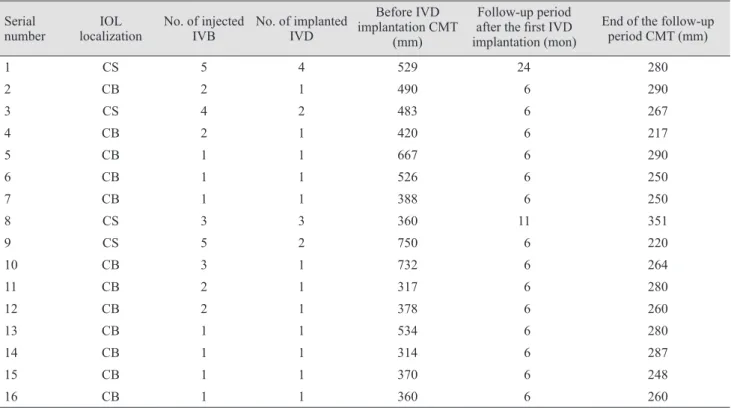
No vitreomacular interface abnormalities were seen in the follow-up period and no patients required vitreoretinal surgery. The treatment was finalized early after the 3rd IVD implantation in one patient (serial number 8) due to permanent low VA and an outer nuclear layer defect. The clinical features, number of intravitreal injections or im- plantations, pre-IVD implantation and final CMTs, and pa- tient follow-up periods are summarized in Table 1.
Before IVD implantation, the mean VA and CMT were 0.69 ± 0.19 logarithm of the minimum angle of resolution (logMAR) (1.50 to 0.10 logMAR) and 476.13 ± 135.13 mm (314 to 750 mm), respectively. At three months after the first IVD implantation, mean VA and CMT were 0.26 ± 0.07 logMAR (1.00 to 0.00 logMAR) and 294.06 ± 15.26 mm (222 to 480 mm), respectively. The differences be- tween the values before and at 3 months after the IVD im- Fig. 1. An example of the progression in central macular thickness examined by optical coherence tomography (OCT). ILM = internal limiting membrane; BM = Bruch membrane; IR = infrared; HS = high speed; ART = automatic realtime tracking; Q = quality factor.
0 200 400 600 800 1000
Thickness (μm)
0.0 1.0 2.0 3.0 4.0 5.0 Position (mm)
OCT 20° (5.8 mm) ART (10) Q: 18 [HS]
IR 30° [HS]
0 200 400 600 800 1000
Thickness (μm)
0.0 1.0 2.0 3.0 4.0 5.0 Position (mm)
OCT 20° (5.8 mm) ART (10) Q: 28 [ HS]
IR 30° ART [HS]
plantation values were statistically significant (p < 0.001 for both). The time-based changes in VA and CMT with IVD implantation are summarized in Table 2. At the end of the follow-up period, the mean VA and CMT values in the whole group were 0.19 ± 0.05 logMAR (0.70 to 0.00 logMAR) and 268.38 ± 31.35 mm (217 to 351 mm), respec- tively. The mean CMT decrement in this series was 207.81
± 151.13 mm (9 to 530 mm). The CMT values before IVD implantation and at the end of the follow-up periods are shown in Fig. 2.
Discussion
While the occurrence of PCME has declined with the use of modern surgical techniques, recently developed sur- gical materials and IOL design, PCME can still occur even following uneventful cataract surgery [15]. Furthermore, the incidence of clinically significant PCME remains around 0.6% to 3.6%, while higher rates have been deter- mined with OCT evidence of CME [5,10,16].
Although the pathogenesis of PCME has been reported to be multifactorial, the activation of inflammatory path- ways seems to play a critical role in its onset and continua- tion. Surgical mechanical trauma triggers a cascade of in- f lammatory events and increases the synthesis of
Table 2. Time-based changes in VA and CMT with IVD implantation
Before IVD implantation 3 Months after the first IVD implantation p-value
VA (logMAR) 0.69 ± 0.19 0.26 ± 0.07 <0.001
CMT (mm) 476.13 ± 135.13 294.06 ± 15.26 <0.001
VA = visual acuity; CMT = central macular thickness; IVD = intravitreal dexamethasone; logMAR = logarithm of the minimum angle of resolution.
Table 1. Summary of patient treatment-related conditions Serial
number IOL
localization No. of injected
IVB No. of implanted IVD
Before IVD implantation CMT
(mm)
Follow-up period after the first IVD implantation (mon)
End of the follow-up period CMT (mm)
1 CS 5 4 529 24 280
2 CB 2 1 490 6 290
3 CS 4 2 483 6 267
4 CB 2 1 420 6 217
5 CB 1 1 667 6 290
6 CB 1 1 526 6 250
7 CB 1 1 388 6 250
8 CS 3 3 360 11 351
9 CS 5 2 750 6 220
10 CB 3 1 732 6 264
11 CB 2 1 317 6 280
12 CB 2 1 378 6 260
13 CB 1 1 534 6 280
14 CB 1 1 314 6 287
15 CB 1 1 370 6 248
16 CB 1 1 360 6 260
IOL = intraocular lens; IVB = intravitreal bevacizumab; IVD = intravitreal dexamethasone; CMT = central macular thickness; CS = cili- ary sulcus; CB = capsular bag.
inflammatory mediators such as prostaglandins and cyto- kines. Inflammatory mediators cause the breakdown of the blood-retinal barrier that leads to the accumulation of ex- tracellular intra-retinal fluid, resulting in macular thicken- ing and creating cystic spaces [17]. In the current series, the persistence and re-occurrence of macular edema eval- uated on OCT was observed more frequently in patients who had complicated surgery than in subjects with un- eventful surgery. The number of IVB injections was high- er in eyes that had complicated cataract surgery, and re- peated IVD implantation was only performed in eyes that had complicated surgery with sulcus IOL implantation.
These results provide indirect evidence that surgical com- plications are important risk factors for the development of treatment-resistant PCME.
Several treatment methods have been applied for PCME, depending on the etiology. Based on the key role of prostaglandins and leukotriene-mediated inflammation in PCME, conventional treatments for PCME have includ- ed steroid and non-steroidal anti-inflammatory drugs [18].
Brynskov et al. [19] reported successful treatment with IVD implantation in a subtenon triamcinolone treat- ment-resistant PCME case. Dang et al. [20] compared the efficacy of intravitreal triamcinolone injection and IVD implantation. The authors reported that both treatments similarly restored VA and CMT, but IVD implantation showed longer efficacy and was well tolerated. Therefore, IVD implantation was suggested as a new treatment option in PCME cases [20]. Garcia et al. [21] applied IVD implan- tation in a series of six patients and favorable anatomic outcomes via OCT were confirmed. Kakkassery et al. [22]
showed the efficacy of IVD implantation in PCME resis-
tant to both several topical and oral medication combina- tions, including topical diclofenac, topical and oral pred- nisolone, and oral acetazolamide. Mayer et al. [23]
observed that CMT decreased from 520.8 ± 71.4 to 232.7 ± 26.6 μm after IVD implantation for treatment of PCME in 23 patients after uneventful cataract surgery. The decrease in CMT in the current series (mean values, from 476.13 to 207.81 μm) was similar to those findings. However, the previous study reported nine recurrences with a peak at three months after implantation that required a second IVD implantation [23]. Although only patients with un- eventful cataract surgery were evaluated in the previous study, the recurrence rate was higher than that of the cur- rent series. This result showed that risk factors other than complicated surgery may also be important for the recur- rence of PCME. Dutra Medeiros et al. [24] reported that the mean CMT decreased from 542.22 to 143.89 μm at 6 months after a single IVD implantation. The mean decre- ment in CMT was lower than the current series (207.81 mm). This result demonstrated that single IVD implanta- tion was not sufficient in treatment-resistant cases and re- peated injections were required, as in the current series.
Another hypothesis that could explain the increase in endothelial permeability is the vascular endothelial growth factor-associated breakdown of the blood-retinal barrier [25]. Many ophthalmologists use IVB in treatment-naïve or topical anti-inflammatory treatment-resistant cases of PCME. Arevalo et al. [26] used the IVB treatment option in 36 eyes with PCME. After a 12-month follow-up period, a significant improvement was obtained on OCT with a mean of 2.7 IVB injections [24]. Similar findings have been reported by other authors [27,28]. In contrast, Spitzer et al. [29] showed that a 1.25 mg IVB injection caused a slight decrease in CMT in a series of 16 eyes with refracto- ry PCME. In our clinical practice, we observed that PCME can be treated with IVB, although there is no single treat- ment regimen that is suitable for every patient. In this study, we observed that some patients who received a mean of 2.19 repeat IVB injections, similar to the Arevalo et al. series [26], still had persistent macular edema even without any significant ocular and systemic risk factors other than complicated surgery in some. Therefore, only cases resistant to both topical and IVB treatment were evaluated to investigate a better option for these patients.
The main limitation of this study was the lack of a con- trol group for comparison with the IVD-treated group.
Central macular thickness
( μm
)
0 100 200 300 400 500 600 700 800
Before intravitreal dexamethasone implantation End of the follow-up period
Fig. 2. Central macular thickness values before intravitreal dexa- methasone implantation and at the end of the follow-up period.
This approach could clearly show the efficacy of IVD ther- apy on patients with IVB treatment-resistant PCME. Fur- thermore, the mean follow-up time after implantation of IVD was 7.4 months, which may be considered a short pe- riod. This study also only included 16 patients and this small number restricts results generalization. The current study sample was not fully homogeneous because it in- cluded patients with complicated and uncomplicated sur- geries. In addition, the patients had not received the same types of treatments before IVD implantation. The retro- spective nature of the study is another important limita- tion.
To the best of our knowledge, this is the first report to show the efficacy of IVD implants even after repeated IVB injections in treatment-resistant PCME. Although there is no widely accepted treatment algorithm for recal- citrant PCME cases, the current findings suggest that pro- longed activity of IVD implantation is both a safe and ef- fective method for the resolution of macular edema resistant to other possible treatments after both uneventful and complicated cataract surgery. Further studies with a large number of patients are needed for the development of new treatment algorithms in patients with recalcitrant macular edema resulting from Irvine-Gass syndrome.
Conflict of Interest
No potential conflict of interest relevant to this article was reported.
References
1. Yonekawa Y, Kim IK. Pseudophakic cystoid macular ede- ma. Curr Opin Ophthalmol 2012;23:26-32.
2. Gass JD, Norton EW. Cystoid macular edema and papill- edema following cataract extraction: a fluorescein fundo- scopic and angiographic study. Arch Ophthalmol 1966;76:646-61.
3. Schubert HD. Cystoid macular edema: the apparent role of mechanical factors. Prog Clin Biol Res 1989;312:277-91.
4. Ursell PG, Spalton DJ, Whitcup SM, Nussenblatt RB.
Cystoid macular edema after phacoemulsification: relation- ship to blood-aqueous barrier damage and visual acuity. J Cataract Refract Surg 1999;25:1492-7.
5. Henderson BA, Kim JY, Ament CS, et al. Clinical pseu- dophakic cystoid macular edema. Risk factors for develop- ment and duration after treatment. J Cataract Refract Surg 2007;33:1550-8.
6. Eriksson U, Alm A, Bjarnhall G, et al. Macular edema and visual outcome following cataract surgery in patients with diabetic retinopathy and controls. Graefes Arch Clin Exp Ophthalmol 2011;249:349-59.
7. Chu CJ, Johnston RL, Buscombe C, et al. Risk factors and incidence of macular edema after cataract surgery: a data- base study of 81984 eyes. Ophthalmology 2016;123:316-23.
8. Sacconi R, Corbelli E, Carnevali A, et al. Optical coher- ence tomography angiography in pseudophakic cystoid macular oedema compared to diabetic macular oedema:
qualitative and quantitative evaluation of retinal vascula- ture. Br J Ophthalmol 2018;102:1684-90.
9. Benitah NR, Arroyo JG. Pseudophakic cystoid macular edema. Int Ophthalmol Clin 2010;50:139-53.
10. Vukicevic M, Gin T, Al-Qureshi S. Prevalence of optical coherence tomography-diagnosed postoperative cystoid macular oedema in patients following uncomplicated pha- co-emulsification cataract surgery. Clin Exp Ophthalmol 2012;40:282-7.
11. Altintas AG, Coban P, Arifoglu HB, et al. Comparison of phaco parameters effect on macular thickness changes af- ter uneventful phacosurgery in diabetic and non-diabetic patients. Int Eye Sci 2016;16:201-6.
12. Lin CJ, Tsai YY. Use of aflibercept for the management of refractory pseudophakic macular edema in Irvine-Gass syndrome and literature review. Retin Cases Brief Rep 2018;12:59-62.
13. Kiernan DF, Hariprasad SM. Controversies in the manage- ment of Irvine-Gass syndrome. Ophthalmic Surg Lasers Imaging Retina 2013;44:522-7.
14. Randazzo A, Vinciguerra P. Chronic macular edema medi- cal treatment in Irvine-Gass syndrome: case report. Eur J Ophthalmol 2010;20:462-5.
15. Ilhan C. Current developments in monofocal intraocular lens technology. Int J Ophthalmic Res 2017;3:239-42.
16. Lobo CL, Faria PM, Soares MA, et al. Macular alterations after small-incision cataract surgery. J Cataract Refract Surg 2004;30:752-60.
17. Hudes GR, Li WY, Rockey JH, White P. Prostacyclin is the major prostaglandin synthesized by bovine retinal cap- illary pericytes in culture. Invest Ophthalmol Vis Sci 1988;29:1511-6.
18. Shelsta HN, Jampol LM. Pharmacologic therapy of pseu- dophakic cystoid macular edema: 2010 update. Retina 2011;31:4-12.
19. Brynskov T, Laugesen CS, Halborg J, et al. Longstanding refractory pseudophakic cystoid macular edema resolved using intravitreal 0.7 mg dexamethasone implants. Clin Ophthalmol 2013;7:1171-4.
20. Dang Y, Mu Y, Li L, et al. Comparison of dexamethasone intravitreal implant and intravitreal triamcinolone aceton- ide for the treatment of pseudophakic cystoid macular ede- ma in diabetic patients. Drug Des Devel Ther 2014;8:1441-9.
21. Garcia JM, Isaac DL, Avila MP. Dexamethasone 0.7 mg implants in the management of pseudophakic cystoid mac- ular edema. Arq Bras Oftalmol 2016;79:113-5.
22. Kakkassery V, Schultz T, Wunderlich MI, et al. Evaluation of predictive factors for successful intravitreal dexametha- sone in pseudophakic cystoid macular edema. J Ophthal- mol 2017;2017:4625730.
23. Mayer WJ, Kurz S, Wolf A, et al. Dexamethasone implant as an effective treatment option for macular edema due to Ir- vine-Gass syndrome. J Cataract Refract Surg 2015;41:1954- 61.
24. Dutra Medeiros M, Navarro R, Garcia-Arumi J, et al. Dexa- methasone intravitreal implant for treatment of patients with recalcitrant macular edema resulting from Irvine-Gass syndrome. Invest Ophthalmol Vis Sci 2013;54:3320-4.
25. Tolentino MJ, McLeod DS, Taomoto M, et al. Pathologic features of vascular endothelial growth factor-induced reti- nopathy in the nonhuman primate. Am J Ophthalmol 2002;133:373-85.
26. Arevalo JF, Maia M, Garcia-Amaris RA, et al. Intravitreal bevacizumab for refractory pseudophakic cystoid macular edema: the Pan-American Collaborative Retina Study Group results. Ophthalmology 2009;116:1481-7.
27. Mason JO 3rd, Albert MA Jr, Vail R. Intravitreal bevaci- zumab (Avastin) for refractory pseudophakic cystoid mac- ular edema. Retina 2006;26:356-7.
28. Barone A, Prascina F, Russo V, et al. Successful treatment of pseudophakic cystoid macular edema with intravitreal bevacizumab. J Cataract Refract Surg 2008;34:1210-2.
29. Spitzer MS, Ziemssen F, Yoeruek E, et al. Efficacy of intra- vitreal bevacizumab in treating postoperative pseudopha- kic cystoid macular edema. J Cataract Refract Surg 2008;34:70-5.